
Payload Information
General Information of This Payload (ID: PAY0QLDVX)
| Name |
Monomethyl auristatin F
|
|||||
|---|---|---|---|---|---|---|
| Synonyms |
MMAF; 745017-94-1; Monomethyl Auristatin F; Monomethylauristatin F; (2S)-2-[[(2R,3R)-3-methoxy-3-[(2S)-1-[(3R,4S,5S)-3-methoxy-5-methyl-4-[methyl-[(2S)-3-methyl-2-[[(2S)-3-methyl-2-(methylamino)butanoyl]amino]butanoyl]amino]heptanoyl]pyrrolidin-2-yl]-2-methylpropanoyl]amino]-3-phenylpropanoic acid; MonomethylauristatinF; SCHEMBL3208014; CHEMBL3359822; MonoMethyl auristatin F (MMAF); MFRNYXJJRJQHNW-DEMKXPNLSA-N; DTXSID601031563; AMY30768; EX-A1046; MFCD25976742; MONOMETHYLAURISTATIN PHENYLALANINE; AC-30596; BP-22316; HY-15579; ((2R,3R)-3-((S)-1-((3R,4S,5S)-4-((S)-N,3-dimethyl-2-((S)-3-methyl-2-(methylamino)butanamido)butanamido)-3-methoxy-5-methylheptanoyl)pyrrolidin-2-yl)-3-methoxy-2-methylpropanoyl)-L-phenylalanine; (2S)-2-[(2R)-2-[(R)-[(2S)-1-[(3R,4S,5S)-4-[(2S)-N,3-dimethyl-2-[(2S)-3-methyl-2-(methylamino)butanamido]butanamido]-3-methoxy-5-methylheptanoyl]pyrrolidin-2-yl](methoxy)methyl]propanamido]-3-phenylpropanoic acid; (S)-2-((2R,3R)-3-((S)-1-((3R,4S,5S)-4-((S)-N,3-Dimethyl-2-((S)-3-methyl-2-(methylamino)butanamido)butanamido)-3-methoxy-5-methylheptanoyl)pyrrolidin-2-yl)-3-methoxy-2-methylpropanamido)-3-phenylpropanoic acid; ;(2~{S})-2-[[(2~{R},3~{R})-3-methoxy-3-[(2~{S})-1-[(3~{R},4~{S},5~{S})-3-methoxy-5-methyl-4-[methyl-[(2~{S})-3-methyl-2-[[(2~{S})-3-methyl-2-(methylamino)butanoyl]amino]butanoyl]amino]heptanoyl]pyrrolidin-2-yl]-2-methyl-propanoyl]amino]-3-phenyl-propanoic acid; L-PHENYLALANINE, N-METHYL-L-VALYL-L-VALYL-(3R,4S,5S)-3-METHOXY-5-METHYL-4-(METHYLAMINO)HEPTANOYL-(.ALPHA.R,.BETA.R,2S)-.BETA.-METHOXY-.ALPHA.-METHYL-2-PYRROLIDINEPROPANOYL-
Click to Show/Hide
|
|||||
| Target | Microtubule (MT) | |||||
| Structure |
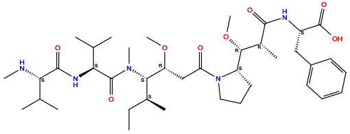
|
|||||
| Formula | C39H65N5O8 |
|||||
| Isosmiles | CC[C@H](C)[C@@H]([C@@H](CC(=O)N1CCC[C@H]1[C@@H]([C@@H](C)C(=O)N[C@@H](CC2=CC=CC=C2)C(=O)O)OC)OC)N(C)C(=O)[C@H](C(C)C)NC(=O)[C@H](C(C)C)NC |
|||||
| PubChem CID | ||||||
| InChI |
InChI=1S/C39H65N5O8/c1-12-25(6)34(43(9)38(48)33(24(4)5)42-37(47)32(40-8)23(2)3)30(51-10)22-31(45)44-20-16-19-29(44)35(52-11)26(7)36(46)41-28(39(49)50)21-27-17-14-13-15-18-27/h13-15,17-18,23-26,28-30,32-35,40H,12,16,19-22H2,1-11H3,(H,41,46)(H,42,47)(H,49,50)/t25-,26+,28-,29-,30+,32-,33-,34-,35+/m0/s1
|
|||||
| InChIKey |
MFRNYXJJRJQHNW-DEMKXPNLSA-N
|
|||||
| IUPAC Name |
(2S)-2-[[(2R,3R)-3-methoxy-3-[(2S)-1-[(3R,4S,5S)-3-methoxy-5-methyl-4-[methyl-[(2S)-3-methyl-2-[[(2S)-3-methyl-2-(methylamino)butanoyl]amino]butanoyl]amino]heptanoyl]pyrrolidin-2-yl]-2-methylpropanoyl]amino]-3-phenylpropanoic acid
|
|||||
| Pharmaceutical Properties | Molecule Weight |
732 |
Polar area |
167 |
||
Complexity |
1160 |
xlogp Value |
2.1 |
|||
Heavy Count |
52 |
Rot Bonds |
21 |
|||
Hbond acc |
9 |
Hbond Donor |
4 |
|||
The activity data of This Payload
| Standard Type | Value | Units | Cell line | Disease Model | Cell line ID | Reference |
|---|---|---|---|---|---|---|
| Half Maximal Inhibitory Concentration (IC50) | 10 | nM |
HT-29 cells
|
Colon adenocarcinoma
|
[1] | |
| Half Maximal Inhibitory Concentration (IC50) | 105 | nM |
H3396 cells
|
Breast carcinoma
|
[1] | |
| Half Maximal Inhibitory Concentration (IC50) | 137 | nM |
HL-60 cells
|
Adult acute myeloid leukemia
|
[1] | |
| Half Maximal Inhibitory Concentration (IC50) | 2880 | nM |
HCT 116 cells
|
Colon carcinoma
|
[1] | |
| Half Maximal Inhibitory Concentration (IC50) | 5.3 | nM |
SK-BR-3 cells
|
Breast adenocarcinoma
|
[2] | |
| Half Maximal Inhibitory Concentration (IC50) | 7 | nM |
Hep-G2 cells
|
Hepatoblastoma
|
[3] | |
| Half Maximal Inhibitory Concentration (IC50) | 8.8 | nM |
BT474-M1 cells
|
Invasive breast carcinoma
|
[2] |
Each Antibody-drug Conjugate Related to This Payload
Full Information of The Activity Data of The ADC(s) Related to This Payload
caxmotabart entudotin [Phase 3]
Identified from the Human Clinical Data
| Experiment 1 Reporting the Activity Date of This ADC | [4] | ||||
| Efficacy Data | Objective Response Rate (ORR) |
66.70
42.90 % |
|||
| Patients Enrolled |
Patients with HER2-expresssing advanced solid tumors who had failed prior standard of care therapies.
|
||||
| Administration Dosage |
FS-1502 was given IV once in 21-day or 28-day cycle at doses of 0.10-3.50 mg/kg.
|
||||
Belantamab mafodotin [Approved in 2020 (withdrawn in 2022, approved again in 2025)]
Identified from the Human Clinical Data
| Experiment 1 Reporting the Activity Date of This ADC | [5] | ||||
| Efficacy Data | Objective Response Rate (ORR) |
56.30%
|
High BCMA expression (BCMA +++) | ||
| Patients Enrolled |
Eligible adult (18 years of age) patients for part 2 had histologically or cytologically confirmed MM, Eastern Cooperative Oncology Group performance status 0 or 1, prior therapy with alkylators, proteasome inhibitors and immunomodulators, and were refractory to the last line of treatment.
|
||||
| Administration Dosage |
GSK2857916 3.4 mg/kg was administered through 1-h intravenous infusions once every 3 weeks, for a maximum of 16 cycles.
|
||||
| Related Clinical Trial | |||||
| NCT Number | NCT02064387 | Phase Status | Phase 1 | ||
| Clinical Description |
A Phase I open-label, dose escalation study to investigate the safety, pharmacokinetics, pharmacodynamics, immunogenicity and clinical activity of the antibody drug conjugate GSK2857916 in subjects with relapsed/refractory multiple myeloma and other advanced hematologic malignancies expressing BCMA.
|
||||
| Primary Endpoint |
Objective response rate=60.00% (95% CI 42.10-76.10),comprising 3 (9.00%) complete responses and 14.00 (40%) partial responses.
|
||||
| Other Endpoint |
The median progression-free survival was 12.00 months and the median duration of response was 14.30 months.
|
||||
| Experiment 2 Reporting the Activity Date of This ADC | [6] | ||||
| Efficacy Data | Objective Response Rate (ORR) |
60%
|
|||
| Patients Enrolled |
Histologically or cytologically confirmed MM, a European Cooperative Oncology Group performance status of 0 or 1, prior therapy with alkylators, PI and IMiD, had undergone stem cell transplant (if eligible) and refractory to the last line of treatment (defined as progressive disease on or within 60 days of completion of the last therapy) that included stem cell transplant and those patients with a history of autologous stem cell transplant must have received the transplant >100 days prior to study enrolment and have no active infection.
Click to Show/Hide
|
||||
| Administration Dosage |
Doses ranging between 0.03 mg/kg and 4.60 mg/kg was administered as a 1-hour intravenous infusion every 3 weeks for a maximum of 16 cycles.
|
||||
| Related Clinical Trial | |||||
| NCT Number | NCT02064387 | Phase Status | Phase 1 | ||
| Clinical Description |
A phase 1 open-label, dose escalation study to investigate the safety, pharmacokinetics, pharmacodynamics, immunogenicity and clinical activity of the antibody drug conjugate GSK2857916 in subjects with relapsed/refractory multiple myeloma and other advanced hematologic malignancies expressing BCMA.
|
||||
| Primary Endpoint |
The primary endpoints of the trial were to determine the safety, tolerability, maximum tolerated dose (MTD) and RP2D and schedule of GSK2857916. Median PFS (post hoc analysis) was 7.90 months (95% CI: 3.1-not estimable),Overall response rate at 3.40 mg/kg in Part 2 was 60.00% (21/35; 95% confidence interval: 42.10%-76.10%).
|
||||
| Other Endpoint |
PK profile (single dose area under the curve, maximum serum concentration [Cmax], time to Cmax , clearance, steady-state volume of distribution [Vss], half-life [t]; repeat dose Cmax and trough plasma concentration), the incidence of anti-drug antibodies, and clinical activity measured as overall response rate (ORR), defined as the percentage of subjects achieving confirmed partial response or better (PR) and clinical benefit rate, defined as the percentages of subjects with minimal response or better (MR).
Click to Show/Hide
|
||||
| Experiment 3 Reporting the Activity Date of This ADC | [7] | ||||
| Related Clinical Trial | |||||
| NCT Number | NCT03769506 | Phase Status | Phase 3 | ||
| Clinical Description |
A phase 3, randomized, double-arm, open-label, controlled trial of ASP-1929 photoimmunotherapy versus physician's choice standard of care for the treatment of locoregional, recurrent head and neck squamous cell carcinoma in patients who have failed or progressed on or after at least two lines of therapy, of which at least one line must be systemic therapy.
Click to Show/Hide
|
||||
Revealed Based on the Cell Line Data
| Experiment 1 Reporting the Activity Date of This ADC | [8] | ||||
| Efficacy Data | Half Maximal Effective Concentration (EC50) |
30.6 ug/mL
|
Moderate BCMA expression (BCMA++) | ||
| Method Description |
Cells (5 x 10⁵ cells/mL) were left untreated or exposed to the indicated treatments for the indicated time. Cells were counted on a Vi-Cell-XR Cell Viability Analyzer (Beckman Coulter).
|
||||
| In Vitro Model | Thymoma | EL4 cells (BCMA expression) | CVCL_0255 | ||
AGS-16C3F [Phase 2 (discontinued)]
Identified from the Human Clinical Data
| Experiment 1 Reporting the Activity Date of This ADC | [9] | ||||
| Efficacy Data | stable disease (SD) |
50%
|
|||
| Patients Enrolled |
Exclusion criteria comprise uncontrolled CNS metastases, investigational drug use within 4 weeks prior, AGS-16C3F hypersensitivity, thromboembolic events (≤3 months), severe cardiac conditions (e.g., CHF Class III/IV), major surgery within 4 weeks, pregnancy/lactation, HIV/hepatitis B/C positivity, active infections requiring systemic treatment, or recent eye surgery/cataracts affecting vision.
Click to Show/Hide
|
||||
| Administration Dosage |
ADCs were given q3w until PD or unacceptable toxicity.AGS-16M8F and AGS-16C3F studies treated 26 and 34 subjects in dose range 0.6 - 4.8 and 1.8 - 4.8 mg/kg, respectively.
|
||||
| Related Clinical Trial | |||||
| NCT Number | NCT01672775 | Phase Status | PHASE1 | ||
| Clinical Description |
A Phase 1, Open Label, Multi-center Study to Assess the Safety, Pharmacokinetics and Effectiveness of AGS-16C3F Monotherapy in Subjects With Renal Cell Carcinoma (RCC) of Clear Cell or Papillary Histology
|
||||
| Primary Endpoint |
The study evaluates the incidence of adverse events over 24 months and assesses pharmacokinetics (TAb, ADC, MMAF) including Ceoi/Cmax, Ctrough, Tmax, AUCτ, t1/2, CL, Vss at specified time points up to Day 92. Secondary endpoints include antidrug antibody formation, tumor response (ORR, DCR), and bone scan changes during the study period.
|
||||
| Other Endpoint |
Eligible participants include metastatic RCC patients (clear cell/non-clear cell or papillary histology) with prior anti-VEGFR therapy (clear cell) or ENPP3+ status (non-clear cell/papillary). Key requirements: measurable disease (RECIST 1.1), ECOG 0-1, adequate hematologic (ANC ≥1.5x10⁹/L, platelet ≥100x109/L, Hb ≥9 g/dL), renal (creatinine ≤1.5xULN or GFR >50 mL/min), and hepatic function (AST/ALT ≤2.5xULN or ≤5xULN with metastases; bilirubin ≤1.5xULN). Contraception is mandated for participants of childbearing potential.
Click to Show/Hide
|
||||
| Experiment 2 Reporting the Activity Date of This ADC | [10] | ||||
| Efficacy Data | Progression Free Survival |
3.5 months
|
|||
| Patients Enrolled |
Exclusion criteria comprised prior axitinib/AGS-16C3F treatment, untreated or unstable brain metastases (>3 months post-radiation/surgery if treated), uncontrolled hypertension (>150/90), GI disorders affecting absorption, active ocular conditions (e.g., infections, corneal ulcers, glaucoma), strong CYP3A4/5 inhibitors/inducers use within 14 days, thromboembolic events (≤4 weeks unless anticoagulated), bleeding disorders (≤2 months), severe cardiac disease (NYHA Class III/IV, MI ≤6 months), major surgery ≤4 weeks, pregnancy/lactation, unresolved infections, or inability to comply with study protocols.
Click to Show/Hide
|
||||
| Administration Dosage |
Participants received 1.8 milligram per kilogram (mg/kg) of AGS-16C3F once every three weeks by single intravenous (IV) infusion.
|
||||
| Related Clinical Trial | |||||
| NCT Number | NCT02639182 | Phase Status | PHASE2 | ||
| Clinical Description |
A Multi-Center, Open Label, Randomized Phase 2 Study of AGS-16C3F vs. Axitinib in Metastatic Renal Cell Carcinoma
|
||||
| Primary Endpoint |
The study assessed Progression-Free Survival (PFS) per RECIST v1.1 by investigator (up to 53 months) and central radiology review (up to 40 months), along with Objective Response Rate (ORR), Duration of Response (DOR), Overall Survival (OS), and Disease Control Rate (DCR). Pharmacokinetic parameters (Cmax, Ctrough, Tmax, AUC0- 21, t1/ 2) for ADC, TAb, and Cys-mcMMAF were evaluated over 21-day cycles. Adverse events (AEs) were monitored for 53 months, with serious AEs requiring intervention or hospitalization.
Click to Show/Hide
|
||||
| Experiment 3 Reporting the Activity Date of This ADC | [9] | ||||
| Efficacy Data | Partial Response (PR) |
8.80%
|
|||
| Patients Enrolled |
Exclusion criteria comprise uncontrolled CNS metastases, investigational drug use within 4 weeks prior, AGS-16C3F hypersensitivity, thromboembolic events (≤3 months), severe cardiac conditions (e.g., CHF Class III/IV), major surgery within 4 weeks, pregnancy/lactation, HIV/hepatitis B/C positivity, active infections requiring systemic treatment, or recent eye surgery/cataracts affecting vision.
Click to Show/Hide
|
||||
| Administration Dosage |
ADCs were given q3w until PD or unacceptable toxicity.AGS-16M8F and AGS-16C3F studies treated 26 and 34 subjects in dose range 0.6 - 4.8 and 1.8 - 4.8 mg/kg, respectively.
|
||||
| Related Clinical Trial | |||||
| NCT Number | NCT01672775 | Phase Status | PHASE1 | ||
| Clinical Description |
A Phase 1, Open Label, Multi-center Study to Assess the Safety, Pharmacokinetics and Effectiveness of AGS-16C3F Monotherapy in Subjects With Renal Cell Carcinoma (RCC) of Clear Cell or Papillary Histology
|
||||
| Primary Endpoint |
The study evaluates the incidence of adverse events over 24 months and assesses pharmacokinetics (TAb, ADC, MMAF) including Ceoi/Cmax, Ctrough, Tmax, AUCτ, t1/2, CL, Vss at specified time points up to Day 92. Secondary endpoints include antidrug antibody formation, tumor response (ORR, DCR), and bone scan changes during the study period.
|
||||
| Other Endpoint |
Eligible participants include metastatic RCC patients (clear cell/non-clear cell or papillary histology) with prior anti-VEGFR therapy (clear cell) or ENPP3+ status (non-clear cell/papillary). Key requirements: measurable disease (RECIST 1.1), ECOG 0-1, adequate hematologic (ANC ≥1.5x10⁹/L, platelet ≥100x109/L, Hb ≥9 g/dL), renal (creatinine ≤1.5xULN or GFR >50 mL/min), and hepatic function (AST/ALT ≤2.5xULN or ≤5xULN with metastases; bilirubin ≤1.5xULN). Contraception is mandated for participants of childbearing potential.
Click to Show/Hide
|
||||
| Experiment 4 Reporting the Activity Date of This ADC | [10] | ||||
| Efficacy Data | Objective Response Rate (ORR) |
7.50%
|
|||
| Patients Enrolled |
Exclusion criteria comprised prior axitinib/AGS-16C3F treatment, untreated or unstable brain metastases (>3 months post-radiation/surgery if treated), uncontrolled hypertension (>150/90), GI disorders affecting absorption, active ocular conditions (e.g., infections, corneal ulcers, glaucoma), strong CYP3A4/5 inhibitors/inducers use within 14 days, thromboembolic events (≤4 weeks unless anticoagulated), bleeding disorders (≤2 months), severe cardiac disease (NYHA Class III/IV, MI ≤6 months), major surgery ≤4 weeks, pregnancy/lactation, unresolved infections, or inability to comply with study protocols.
Click to Show/Hide
|
||||
| Administration Dosage |
Participants received 1.8 milligram per kilogram (mg/kg) of AGS-16C3F once every three weeks by single intravenous (IV) infusion.
|
||||
| Related Clinical Trial | |||||
| NCT Number | NCT02639182 | Phase Status | PHASE2 | ||
| Clinical Description |
A Multi-Center, Open Label, Randomized Phase 2 Study of AGS-16C3F vs. Axitinib in Metastatic Renal Cell Carcinoma
|
||||
| Primary Endpoint |
The study assessed Progression-Free Survival (PFS) per RECIST v1.1 by investigator (up to 53 months) and central radiology review (up to 40 months), along with Objective Response Rate (ORR), Duration of Response (DOR), Overall Survival (OS), and Disease Control Rate (DCR). Pharmacokinetic parameters (Cmax, Ctrough, Tmax, AUC0- 21, t1/ 2) for ADC, TAb, and Cys-mcMMAF were evaluated over 21-day cycles. Adverse events (AEs) were monitored for 53 months, with serious AEs requiring intervention or hospitalization.
Click to Show/Hide
|
||||
| Experiment 5 Reporting the Activity Date of This ADC | [10] | ||||
| Efficacy Data | Disease control rate (DCR) |
13.40%
|
|||
| Patients Enrolled |
Exclusion criteria comprised prior axitinib/AGS-16C3F treatment, untreated or unstable brain metastases (>3 months post-radiation/surgery if treated), uncontrolled hypertension (>150/90), GI disorders affecting absorption, active ocular conditions (e.g., infections, corneal ulcers, glaucoma), strong CYP3A4/5 inhibitors/inducers use within 14 days, thromboembolic events (≤4 weeks unless anticoagulated), bleeding disorders (≤2 months), severe cardiac disease (NYHA Class III/IV, MI ≤6 months), major surgery ≤4 weeks, pregnancy/lactation, unresolved infections, or inability to comply with study protocols.
Click to Show/Hide
|
||||
| Administration Dosage |
Participants received 1.8 milligram per kilogram (mg/kg) of AGS-16C3F once every three weeks by single intravenous (IV) infusion.
|
||||
| Related Clinical Trial | |||||
| NCT Number | NCT02639182 | Phase Status | PHASE2 | ||
| Clinical Description |
A Multi-Center, Open Label, Randomized Phase 2 Study of AGS-16C3F vs. Axitinib in Metastatic Renal Cell Carcinoma
|
||||
| Primary Endpoint |
The study assessed Progression-Free Survival (PFS) per RECIST v1.1 by investigator (up to 53 months) and central radiology review (up to 40 months), along with Objective Response Rate (ORR), Duration of Response (DOR), Overall Survival (OS), and Disease Control Rate (DCR). Pharmacokinetic parameters (Cmax, Ctrough, Tmax, AUC0- 21, t1/ 2) for ADC, TAb, and Cys-mcMMAF were evaluated over 21-day cycles. Adverse events (AEs) were monitored for 53 months, with serious AEs requiring intervention or hospitalization.
Click to Show/Hide
|
||||
| Experiment 6 Reporting the Activity Date of This ADC | [11] | ||||
| Efficacy Data | Partial Response (PR) |
23.08%
|
High ENPP3 expression (ENPP3+++) | ||
| Patients Enrolled |
Metastatic renal cell carcinoma (MRCC), Eastern Cooperative Oncology Group (ECOG) performance status 1, adequate organ and bone marrow function.
|
||||
| Administration Dosage |
AGS-16M8F was administered intravenously every 3 weeks at 5 dose levels ranging from 0.60 to 4.80 mg/kg until unacceptable toxicity or progression. A second study with AGS-16C3F started with the AGS-16M8F bridging dose of 4.80 mg/kg given every 3 weeks.
|
||||
| Related Clinical Trial | |||||
| NCT Number | NCT01672775 | Phase Status | Phase 1 | ||
| Clinical Description |
A phase 1, open label, multi-center study to assess the safety, pharmacokinetics and effectiveness of AGS-16C3F monotherapy in subjects with renal cell carcinoma (RCC) of clear cell or papillary histology.
|
||||
| Primary Endpoint |
In the AGS-16C3F study (n = 34),the MTD was 3.60 mg/kg,but this was not tolerated. The 1.80 mg/kg dose was determined to be safe and was associated antitumor response.
|
||||
| Other Endpoint |
3 subjects at 1.80 mg/kg achieved durable PR (3/13, 23.08%). The disease control rate at 1.80 mg/kg was 92.30% (N=12/13). The disease control rate for the entire study was 58.82% (N=20/34).
|
||||
| Experiment 7 Reporting the Activity Date of This ADC | [12] | ||||
| Efficacy Data | Objective Response Rate (ORR) |
7.50
18.20 % |
Moderate ENPP3 expression (ENPP3++) | ||
| Patients Enrolled |
Advanced renal cell carcinoma (RCC).
|
||||
| Administration Dosage |
Intravenous AGS-16C3F 1.80 mg/kg every 3 weeks or oral axitinib 5 mg twice daily (starting dose).
|
||||
| Related Clinical Trial | |||||
| NCT Number | NCT02639182 | Phase Status | Phase 2 | ||
| Clinical Description |
A multi-center, open label, randomized phase 2 study of AGS-16C3F vs. axitinib in metastatic renal cell carcinoma.
|
||||
| Primary Endpoint |
Median PFS=2.90 months (95% CI,2.00-4.00) for AGS16C3F,Median PFS=5.7 months (95% CI,5.30-9.10) for axitinib.
|
||||
| Other Endpoint |
Disease Control Rate (DCR)=13.40% (95% CI,6.3-24.0) for AGS16C3F, Disease Control Rate (DCR)=22.70% (95% CI,13.30-34.70) for axitinib. Median duration of Response (mDoR)=6.80 months (95% CI,3.80-18.40) for AGS16C3F, Median duration of Response (mDoR)=6.7 months (95% CI,1.80-9.20) for axitinib. Objective Response Rate (ORR)=7.50% (95% CI,2.50-16.60) for AGS16C3F, Objective Response Rate (ORR)=18.20% (95% CI,9.80-29.60) for axitinib. Median Overall Survival (mOS)=13.10 months for AGS16C3F, Median Overall Survival (mOS)=15.40 months for axitinib.
Click to Show/Hide
|
||||
Discovered Using Cell Line-derived Xenograft Model
| Experiment 1 Reporting the Activity Date of This ADC | [13] | ||||
| Efficacy Data | Half Maximal Inhibitory Concentration (IC50) |
1.1 nM
|
|||
| Method Description |
Cells were incubated in triplicate in medium containing AGS-16C3F or cHmLYS-1c3.G2k-mcMMAF (0 [Control],0.001,0.003,0.008,0.02,0.07,0.21,0.62,1.82,5.57,16.67,50,150,450,and 1350 nM) in a 5% CO2 incubator at 37°C for 96 hours. IC50 values at day 5 for AGS-16C3F were calculated for each cell line.
|
||||
| In Vitro Model | Normal | ROSA KIT D816V cells | CVCL_5G50 | ||
| Experiment 2 Reporting the Activity Date of This ADC | [13] | ||||
| Efficacy Data | Half Maximal Inhibitory Concentration (IC50) |
2.73 nM
|
Moderate ENPP3 expression (ENPP3++) | ||
| Method Description |
Cells were incubated in triplicate in medium containing AGS-16C3F or cHmLYS-1c3.G2k-mcMMAF (0 [Control],0.001,0.003,0.008,0.02,0.07,0.21,0.62,1.82,5.57,16.67,50,150,450,and 1350 nM) in a 5% CO2 incubator at 37°C for 96 hours. IC50 values at day 5 for AGS-16C3F were calculated for each cell line.
|
||||
| In Vitro Model | Mast-cell sarcoma | ROSA KIT D816V Gluc cells | Homo sapiens | ||
| Experiment 3 Reporting the Activity Date of This ADC | [13] | ||||
| Efficacy Data | Half Maximal Inhibitory Concentration (IC50) |
109.9 nM
|
High ENPP3 expression (ENPP3+++) | ||
| Method Description |
Cells were incubated in triplicate in medium containing AGS-16C3F or cHmLYS-1c3.G2k-mcMMAF (0 [Control],0.001,0.003,0.008,0.02,0.07,0.21,0.62,1.82,5.57,16.67,50,150,450,and 1350 nM) in a 5% CO2 incubator at 37°C for 96 hours. IC50 values at day 5 for AGS-16C3F were calculated for each cell line.
|
||||
| In Vitro Model | Mast cell leukemia | HMC-1.1 cells | CVCL_H206 | ||
| Experiment 4 Reporting the Activity Date of This ADC | [13] | ||||
| Efficacy Data | Half Maximal Inhibitory Concentration (IC50) |
146.5 nM
|
|||
| Method Description |
Cells were incubated in triplicate in medium containing AGS-16C3F or cHmLYS-1c3.G2k-mcMMAF (0 [Control],0.001,0.003,0.008,0.02,0.07,0.21,0.62,1.82,5.57,16.67,50,150,450,and 1350 nM) in a 5% CO2 incubator at 37°C for 96 hours. IC50 values at day 5 for AGS-16C3F were calculated for each cell line.
|
||||
| In Vitro Model | Mast cell leukemia | HMC-1.2 cells | CVCL_H205 | ||
Depatuxizumab mafodotin [Phase 3 (discontinued)]
Identified from the Human Clinical Data
| Experiment 1 Reporting the Activity Date of This ADC | [14] | ||||
| Efficacy Data | Progression Free Survival |
2.1 months
|
|||
| Patients Enrolled |
Japanese participants with WHO grade III or IV malignant glioma
|
||||
| Administration Dosage |
ABT-414 administered every other weeks monotherapy
|
||||
| Related Clinical Trial | |||||
| NCT Number | NCT02590263 | Phase Status | PHASE1|||PHASE2 | ||
| Clinical Description |
A Non-Randomized, Open-Label, Multi-Center Phase 1/2 Study Evaluating the Safety, Pharmacokinetics and Efficacy of ABT-414 in Japanese Subjects With Malignant Glioma
|
||||
| Primary Endpoint |
For all patients in the 2L Depatux-M + CT arm, the 6-month PFS estimate by central review was 25.6% (95% CI 11.4-42.6) with a median PFS of 2.1 months (95% CI 1.9-3.9; Figure 3 A, B). The 6-month OS estimate was 89.7% (95% CI 71.3-96.5), and the median OS was 14.7 months (95% CI 10.7-15.4; Figure 4). ORR, analyzed in patients with at least one measurable disease at baseline, was 21.7% (5/23) by central review with all responses being PR and the median DoR was 5.5 months (95% CI 1.9-NE; Table 6). Seven patients were considered to have 6-month PFS by investigator review, but not by central review.
Click to Show/Hide
|
||||
| Other Endpoint |
Objective Response Rate [Time Frame: At each visit for approximately 1 year], Overall Survival [Time Frame: At each visit for approximately 1 year], Duration of Overall Response [Time Frame: At each visit for approximately 1 year]
|
||||
| Experiment 2 Reporting the Activity Date of This ADC | [14] | ||||
| Efficacy Data | Overall suvival (OS) |
14.7 months
|
|||
| Patients Enrolled |
Japanese participants with WHO grade III or IV malignant glioma
|
||||
| Administration Dosage |
ABT-414 administered every other weeks monotherapy
|
||||
| Related Clinical Trial | |||||
| NCT Number | NCT02590263 | Phase Status | PHASE1|||PHASE2 | ||
| Clinical Description |
A Non-Randomized, Open-Label, Multi-Center Phase 1/2 Study Evaluating the Safety, Pharmacokinetics and Efficacy of ABT-414 in Japanese Subjects With Malignant Glioma
|
||||
| Primary Endpoint |
For all patients in the 2L Depatux-M + CT arm, the 6-month PFS estimate by central review was 25.6% (95% CI 11.4-42.6) with a median PFS of 2.1 months (95% CI 1.9-3.9; Figure 3 A, B). The 6-month OS estimate was 89.7% (95% CI 71.3-96.5), and the median OS was 14.7 months (95% CI 10.7-15.4; Figure 4). ORR, analyzed in patients with at least one measurable disease at baseline, was 21.7% (5/23) by central review with all responses being PR and the median DoR was 5.5 months (95% CI 1.9-NE; Table 6). Seven patients were considered to have 6-month PFS by investigator review, but not by central review.
Click to Show/Hide
|
||||
| Other Endpoint |
Objective Response Rate [Time Frame: At each visit for approximately 1 year], Overall Survival [Time Frame: At each visit for approximately 1 year], Duration of Overall Response [Time Frame: At each visit for approximately 1 year]
|
||||
| Experiment 3 Reporting the Activity Date of This ADC | [15] | ||||
| Efficacy Data | Overall suvival (OS) |
15.5 months
|
|||
| Patients Enrolled |
Must have a clinical diagnosis of glioblastoma (GBM).
|
||||
| Administration Dosage |
Depatuxizumab mafodotin is given on Day 1 of Week 1, 3 and 5 along with the standard therapy of TMZ and radiation during the chemoradiation phase. Depatuxizumab mafodotin is given on Day 1 and 15 of each cycle along with TMZ (Days 1-5 of each cycle) per standard of care during the adjuvant phase.
|
||||
| Related Clinical Trial | |||||
| NCT Number | NCT02573324 | Phase Status | PHASE3 | ||
| Clinical Description |
A Randomized, Placebo Controlled Phase 3 Study of ABT-414 With Concurrent Chemoradiation and Adjuvant Temozolomide in Subjects With Newly Diagnosed Glioblastoma (GBM) With Epidermal Growth Factor Receptor (EGFR) Amplification (Intellance1)
|
||||
| Primary Endpoint |
Overall Survival (OS) [Time Frame: Overall median duration of follow-up was 15.5 months (range: 0.1, 35.6).]
|
||||
| Other Endpoint |
OS for the O6-methylguaninemethlytransferese (MGMT) Unmethylated Group [Time Frame: Overall median duration of follow-up was 15.5 months (range: 0.1, 35.6).]
|
||||
| Experiment 4 Reporting the Activity Date of This ADC | [14] | ||||
| Efficacy Data | During of response (DoR) |
5.5 months
|
|||
| Patients Enrolled |
Japanese participants with WHO grade III or IV malignant glioma
|
||||
| Administration Dosage |
ABT-414 administered every other weeks monotherapy
|
||||
| Related Clinical Trial | |||||
| NCT Number | NCT02590263 | Phase Status | PHASE1|||PHASE2 | ||
| Clinical Description |
A Non-Randomized, Open-Label, Multi-Center Phase 1/2 Study Evaluating the Safety, Pharmacokinetics and Efficacy of ABT-414 in Japanese Subjects With Malignant Glioma
|
||||
| Primary Endpoint |
For all patients in the 2L Depatux-M + CT arm, the 6-month PFS estimate by central review was 25.6% (95% CI 11.4-42.6) with a median PFS of 2.1 months (95% CI 1.9-3.9; Figure 3 A, B). The 6-month OS estimate was 89.7% (95% CI 71.3-96.5), and the median OS was 14.7 months (95% CI 10.7-15.4; Figure 4). ORR, analyzed in patients with at least one measurable disease at baseline, was 21.7% (5/23) by central review with all responses being PR and the median DoR was 5.5 months (95% CI 1.9-NE; Table 6). Seven patients were considered to have 6-month PFS by investigator review, but not by central review.
Click to Show/Hide
|
||||
| Other Endpoint |
Objective Response Rate [Time Frame: At each visit for approximately 1 year], Overall Survival [Time Frame: At each visit for approximately 1 year], Duration of Overall Response [Time Frame: At each visit for approximately 1 year]
|
||||
| Experiment 5 Reporting the Activity Date of This ADC | [16] | ||||
| Patients Enrolled |
Subjects must have a solid tumor type likely to over-express Epidermal Growth Factor Receptor (EGFR) (Phase 1)
|
||||
| Administration Dosage |
Data from patients who received ABT-414 monotherapy at a dose of 1-4 mg/kg once every 3 weeks or 1 or 1.5 mg/kg weekly for 2 out of every 3 weeks (alternate schedule) by intravenous infusion were included in the analysis of triplicate 12-lead ECGs obtained before dosing and through 168 h after dosing.
|
||||
| Related Clinical Trial | |||||
| NCT Number | NCT01741727 | Phase Status | PHASE1 | ||
| Clinical Description |
A Phase 1/2 Study Evaluating the Safety, Pharmacokinetics and Efficacy of ABT-414 in Subjects With Advanced Solid Tumors Likely to Over-Express the Epidermal Growth Factor Receptor (EGFR)
|
||||
| Primary Endpoint |
Phase 1 - Safety (Number of subjects with adverse events and/or dose limiting toxicities) [Time Frame: Every 1-3 weeks for an average of 20 weeks]
|
||||
| Other Endpoint |
Phase 2- Safety (Scheduled study visits occurring on average every 3 weeks) [Time Frame: Followed on average every 3 weeks for approximately 20 weeks]
|
||||
| Experiment 6 Reporting the Activity Date of This ADC | [17] | ||||
| Patients Enrolled |
Histologically confirmed de novo (primary) glioblastoma with unequivocal tumor progression or recurrence.
|
||||
| Administration Dosage |
intravenous administration (1.25 mg/kg or 1.0 mg/kg body weight) over 30 to 40 minutes once every 2 weeks until one of the treatment withdrawal criteria was met. The dose was 1.25 mg/kg in the original protocol (Version 1) and Version 2, Amendment 1, and was lowered to 1.0 mg/kg in protocol Version 3, Amendment 2. Pediatric participants: Intravenous administration (1.0 mg/kg body weight for those who were 6 to 17 years old at the date of first dose, or 1.3 mg/kg for those who were 0 to 5 years old) over 30 to 40 minutes or as directed by the guidelines once every 2 weeks until one of the treatment withdrawal criteria was met, for a maximum of one year. If used in combination with temozolomide, depatuxizumab mafodotin was dosed on Day 1 and Day 15 of the TMZ cycle (assuming a standard regimen of 200 mg/m^2/day for 5 days of each 28-day cycle; for other TMZ schedules, timing of the depatuxizumab mafodotin dosing schedule were to be discussed with the medical monitor).
Click to Show/Hide
|
||||
| Related Clinical Trial | |||||
| NCT Number | NCT02343406 | Phase Status | PHASE2 | ||
| Clinical Description |
INTELLANCE-2: ABT-414 Alone or ABT-414 Plus Temozolomide Versus Lomustine or Temozolomide for Recurrent Glioblastoma: A Randomized Phase 2 Study of the EORTC Brain Tumor Group
|
||||
| Primary Endpoint |
Overall Survival (OS); Progression-Free Survival (PFS)
|
||||
| Other Endpoint |
Objective Response Rate (ORR); Overall Survival in the Subgroup With Epidermal Growth Factor Receptor (EGFRvIII) Mutation
|
||||
| Experiment 7 Reporting the Activity Date of This ADC | [18] | ||||
| Patients Enrolled |
Newly diagnosed glioblastoma (GBM) histologically proven, World Health Organization (WHO) grade IV GBM or WHO grade IV gliosarcoma
|
||||
| Administration Dosage |
During the Chemoradiation Phase, participants were to receive depatuxizumab mafodotin at 2.0 mg/kg IV infusion over 30 - 40 minutes once every 2 weeks (Day 1 of Weeks 1, 3, and 5 of the 6-week regimen). During the Adjuvant Therapy Phase, participants were to receive depatuxizumab mafodotin at 1.25 mg/kg on Day 1 (± 2 days) and Day 15 (± 2 days) of each 28-day cycle as a 30 - 40 minute infusion for 12 cycles.
Click to Show/Hide
|
||||
| Related Clinical Trial | |||||
| NCT Number | NCT03419403 | Phase Status | PHASE3 | ||
| Clinical Description |
Phase 3b Study for Management of Ocular Side Effects in Subjects With EGFR-amplified Glioblastoma Receiving Depatuxizumab Mafodotin (ABT-414)
|
||||
| Primary Endpoint |
Percentage of Participants Who Required a Change in Ocular Side Effect (OSE) Management [Time Frame: Within 8 weeks after the initial dose of depatuxizumab mafodotin]
|
||||
| Other Endpoint |
Maximum Change From Baseline on the Logarithm of the Minimum Angle of Resolution (LogMAR) Scale; Time to Bandage Contact Lens (BCL) Intervention; Number of Participants With Depatuxizumab Mafodotin Dose Modifications Due to Ocular Side Effects (OSE)
|
||||
| Experiment 8 Reporting the Activity Date of This ADC | [19] | ||||
| Patients Enrolled |
Glioblastoma Multiforme (GBM)
|
||||
| Administration Dosage |
ABT-414 will be administered by intravenous infusion
|
||||
| Related Clinical Trial | |||||
| NCT Number | NCT01800695 | Phase Status | PHASE1 | ||
| Clinical Description |
A Phase 1 Study Evaluating the Safety and Pharmacokinetics of ABT-414 for Subjects With Glioblastoma Multiforme
|
||||
| Primary Endpoint |
Number and percentage of participants with adverse events
|
||||
| Other Endpoint |
Biomarker EGFR expression, Progression Free Survival, Overall Survival
|
||||
| Experiment 9 Reporting the Activity Date of This ADC | [20] | ||||
| Patients Enrolled |
Participant must have epidermal growth factor receptor (EGFR) amplification or EGFRvIII mutation.
|
||||
| Related Clinical Trial | |||||
| NCT Number | NCT03123952 | Phase Status | N.A. | ||
| Clinical Description |
This is an expanded access program (EAP) for eligible participants. This program is designed to provide access to ABT-414 prior to approval by the local regulatory agency. Availability will depend on territory eligibility. Participating sites will be added as they apply for and are approved for the EAP. A medical doctor must decide whether the potential benefit outweighs the risk of receiving an investigational therapy based on the individual patient's medical history and program eligibility criteria.
Click to Show/Hide
|
||||
| Experiment 10 Reporting the Activity Date of This ADC | [21] | ||||
| Efficacy Data | Median progression-free survival (mPFS) |
8.0 (depatux-m group); 6.3 (placebo group) Months
|
High EGFR expression (EGFR +++) | ||
| Patients Enrolled |
EGFR-amp newly diagnosed GBM were randomized 1:1 to radiotherapy, temozolomide, and depatux-m/placebo.
|
||||
| Administration Dosage |
Depatux-m was dosed at 2.0 mg/kg during RT, then 1.25 mg/kg thereafter on days 1 and 15/28, 19,21 and allowed to continue until disease progression.
|
||||
| Experiment 11 Reporting the Activity Date of This ADC | [21] | ||||
| Efficacy Data | Median Overall Survival (mOS) |
18.9 (depatux-m group); 18.7(placebo group) Months
|
High EGFR expression (EGFR +++) | ||
| Patients Enrolled |
EGFR-amp newly diagnosed GBM were randomized 1:1 to radiotherapy, temozolomide, and depatux-m/placebo.
|
||||
| Administration Dosage |
Depatux-m was dosed at 2.0 mg/kg during RT, then 1.25 mg/kg thereafter on days 1 and 15/2819, 21 and allowed to continue until disease progression.
|
||||
Revealed Based on the Cell Line Data
| Experiment 1 Reporting the Activity Date of This ADC | [22] | ||||
| Efficacy Data | Tumor Growth Inhibition value (TGI) | ≈ 87.50% | Positive EGFR expression (EGFR+++/++) | ||
| Method Description |
To establish xenografts, 2 x 10⁶ MSTO-211H cells mixed with 75-uL Matrigel were injected subcutaneously in the right flank of 5 to 6-week-old female BALB/c nu/nu miceFor the MSTO-211H study, mice received either ABT-414, ABBV-221 or ADC control (3 mg/kg) every 4 days.
|
||||
| In Vivo Model | MSTO-211H CDX model | ||||
| In Vitro Model | Pleural biphasic mesothelioma | MSTO-211H cells | CVCL_1430 | ||
| Experiment 2 Reporting the Activity Date of This ADC | [22] | ||||
| Efficacy Data | Half Maximal Inhibitory Concentration (IC50) |
10.00 - 35.00 ug/mL
|
Positive EGFR expression (EGFR+++/++) | ||
| Method Description |
Cells lines were plated at 1,000-3,000 cells per well in complete growth medium containing 10% FCS in 96-well plates and allowed to adhere overnight.
|
||||
| In Vitro Model | Pleural mesothelioma | NCI-H2052 cells | CVCL_1518 | ||
| Experiment 3 Reporting the Activity Date of This ADC | [22] | ||||
| Efficacy Data | Half Maximal Inhibitory Concentration (IC50) |
10.00 - 35.00 ug/mL
|
Positive EGFR expression (EGFR+++/++) | ||
| Method Description |
Cells lines were plated at 1,000-3,000 cells per well in complete growth medium containing 10% FCS in 96-well plates and allowed to adhere overnight.
|
||||
| In Vitro Model | Pleural mesothelioma | NCI-H2052 cells | CVCL_1518 | ||
| Experiment 4 Reporting the Activity Date of This ADC | [22] | ||||
| Efficacy Data | Half Maximal Inhibitory Concentration (IC50) |
10.00 - 35.00 ug/mL
|
Positive EGFR expression (EGFR+++/++) | ||
| Method Description |
Cells lines were plated at 1,000-3,000 cells per well in complete growth medium containing 10% FCS in 96-well plates and allowed to adhere overnight.
|
||||
| In Vitro Model | Pleural mesothelioma | NCI-H28 cells | CVCL_1555 | ||
| Experiment 5 Reporting the Activity Date of This ADC | [22] | ||||
| Efficacy Data | Half Maximal Inhibitory Concentration (IC50) |
10.00 - 35.00 ug/mL
|
Positive EGFR expression (EGFR+++/++) | ||
| Method Description |
Cells lines were plated at 1,000-3,000 cells per well in complete growth medium containing 10% FCS in 96-well plates and allowed to adhere overnight.
|
||||
| In Vitro Model | Pleural biphasic mesothelioma | MSTO-211H cells | CVCL_1430 | ||
PF-06263507 [Phase 1 (discontinued)]
Identified from the Human Clinical Data
| Experiment 1 Reporting the Activity Date of This ADC | [23] | ||||
| Efficacy Data | stable disease (SD) |
7.60%
|
|||
| Patients Enrolled |
Eligible participants had advanced/metastatic solid tumors refractory to standard therapy, ECOG 0-1, and adequate organ function. Exclusion criteria included uncontrolled brain metastases, recent major surgery/anticancer therapy, and active infections.
|
||||
| Administration Dosage |
Drug: PF-06263507 Part 1 - PF-06263507 will be administered intravenously in 21-day cycles in cohorts of 2 or more patients starting at a dose of 0.05 mg/kg. Increases in dose will continue until MTD is determined. Drug: PF-06263507 Part 2 - Patients with select tumor types will be treated at the MTD or Recommended Phase 2 dose selected in Part 1.
|
||||
| Related Clinical Trial | |||||
| NCT Number | NCT01891669 | Phase Status | PHASE1 | ||
| Clinical Description |
A PHASE 1, DOSE ESCALATION STUDY OF PF-06263507 IN PATIENTS WITH ADVANCED SOLID TUMORS
|
||||
| Primary Endpoint |
The study assessed dose-limiting toxicities (DLTs) occurring within the first treatment cycle (21 days), including severe neutropenia, febrile neutropenia, thrombocytopenia with bleeding, non-hematologic toxicities, or cardiac troponin abnormalities. AEs were graded per CTCAE v4.0, and persistent toxicity delaying treatment by >2 weeks was considered a DLT.
Click to Show/Hide
|
||||
| Other Endpoint |
Treatment-emergent adverse events (TEAEs) and treatment-related AEs were monitored from baseline through treatment cycles and follow-up, with severity graded per NCI CTCAE v4.0. Hematologic, chemistry, and urine protein abnormalities were tracked, along with vital sign changes meeting predefined criteria. Anti-drug antibodies and pharmacokinetic parameters (Tmax for PF-06263507 and metabolites) were also evaluated.
Click to Show/Hide
|
||||
Discovered Using Cell Line-derived Xenograft Model
| Experiment 1 Reporting the Activity Date of This ADC | [33] | ||||
| Efficacy Data | Tumor Growth Inhibition value (TGI) | ≈ 71.70% | Low 5T4 expression (5T4+) | ||
| Method Description |
ASN004 was evaluated for efficacy in human tumor mouse xenograft models,derived from four different human tumor cell types,having a wide range of 5T4 expression levels. ASN004 was further evaluated in a tumor xenograft model derived from the H1975 human lung carcinoma cell line [5T4+; 15, 800 binding sites per cell]. Subcutaneous tumor xenografts were developed in nude mice with established mean tumor volumes of 150 mm3. The dose of PF-06263507 was 3 mg/kg Q4D 4.
Click to Show/Hide
|
||||
| In Vivo Model | Lung cancer CDX model | ||||
| In Vitro Model | Lung cancer | Lung cancer cells | Homo sapiens | ||
| Experiment 2 Reporting the Activity Date of This ADC | [33] | ||||
| Efficacy Data | Tumor Growth Inhibition value (TGI) | ≈ 100% | Low 5T4 expression (5T4+) | ||
| Method Description |
ASN004 was evaluated for efficacy in human tumor mouse xenograft models,derived from four different human tumor cell types,having a wide range of 5T4 expression levels. ASN004 was further evaluated in a tumor xenograft model derived from the H1975 human lung carcinoma cell line [5T4+; 15, 800 binding sites per cell]. Subcutaneous tumor xenografts were developed in nude mice with established mean tumor volumes of 150 mm3. The dose of PF-06263507 was 10 mg/kg Q4D 4.
Click to Show/Hide
|
||||
| In Vivo Model | Lung cancer CDX model | ||||
| In Vitro Model | Lung cancer | Lung cancer cells | Homo sapiens | ||
MEDI-547 [Phase 1 (discontinued)]
Identified from the Human Clinical Data
| Experiment 1 Reporting the Activity Date of This ADC | [24] | ||||
| Efficacy Data | stable disease (SD) |
16.70%
|
|||
| Patients Enrolled |
Eligible patients must have relapsed/refractory solid tumors (ovarian, prostate, NSCLC, etc.) with prior histologic confirmation, ECOG 0-2, adequate organ function, and measurable disease (for dose expansion). Exclusions include uncontrolled CNS metastases, active infections, significant cardiac history, recent stroke/TIA, uncontrolled hypertension, anticoagulant use, and pregnancy/lactation. Prior therapies must be completed ≥30 days before enrollment.
Click to Show/Hide
|
||||
| Administration Dosage |
All 6 patients were exposed to MEDI-547 at a dose of 0.08 mg/kg once every 3 weeks.
|
||||
| Related Clinical Trial | |||||
| NCT Number | NCT00796055 | Phase Status | PHASE1 | ||
| Clinical Description |
A Phase 1, Open-Label Study of MEDI-547 to Evaluate the Safety, Tolerability, Pharmacokinetics, and Biologic Activity of Intravenous Administration in Subjects With Relapsed or Refractory Solid Tumors Associated With EphA2 Expression
|
||||
| Primary Endpoint |
The safety and tolerability of MEDI-547 will be evaluated through monitoring adverse events (AEs), serious AEs (SAEs), lab abnormalities, physical exam changes, treatment discontinuations due to toxicity, and drug-related deaths during treatment and up to 30 days post-treatment.
|
||||
| Other Endpoint |
Antitumor activity will be assessed via objective response rate (ORR), time to response (TTR), duration of response (DR), time to progression (TTP), progression-free survival (PFS), and overall survival (OS), with measurements continuing until 30 days after the last MEDI-547 dose.
|
||||
| Experiment 2 Reporting the Activity Date of This ADC | [24] | ||||
| Efficacy Data | progressive disease (PD) |
83.30%
|
|||
| Patients Enrolled |
Eligible patients must have relapsed/refractory solid tumors (ovarian, prostate, NSCLC, etc.) with prior histologic confirmation, ECOG 0-2, adequate organ function, and measurable disease (for dose expansion). Exclusions include uncontrolled CNS metastases, active infections, significant cardiac history, recent stroke/TIA, uncontrolled hypertension, anticoagulant use, and pregnancy/lactation. Prior therapies must be completed ≥30 days before enrollment.
Click to Show/Hide
|
||||
| Administration Dosage |
All 6 patients were exposed to MEDI-547 at a dose of 0.08 mg/kg once every 3 weeks.
|
||||
| Related Clinical Trial | |||||
| NCT Number | NCT00796055 | Phase Status | PHASE1 | ||
| Clinical Description |
A Phase 1, Open-Label Study of MEDI-547 to Evaluate the Safety, Tolerability, Pharmacokinetics, and Biologic Activity of Intravenous Administration in Subjects With Relapsed or Refractory Solid Tumors Associated With EphA2 Expression
|
||||
| Primary Endpoint |
The safety and tolerability of MEDI-547 will be evaluated through monitoring adverse events (AEs), serious AEs (SAEs), lab abnormalities, physical exam changes, treatment discontinuations due to toxicity, and drug-related deaths during treatment and up to 30 days post-treatment.
|
||||
| Other Endpoint |
Antitumor activity will be assessed via objective response rate (ORR), time to response (TTR), duration of response (DR), time to progression (TTP), progression-free survival (PFS), and overall survival (OS), with measurements continuing until 30 days after the last MEDI-547 dose.
|
||||
| Experiment 3 Reporting the Activity Date of This ADC | [31] | ||||
| Efficacy Data | Objective Response Rate (ORR) |
0%
|
|||
| Patients Enrolled |
Malignant solid tumor thought to be associated with increased expression of EphA2 (endometrial, breast, ovarian, prostate, non-small cell lung, colon, esophageal, gastric, and bladder cancers, renal cell carcinoma, melanoma), relapsed or refractory to standard therapy, and an Eastern Cooperative Oncology Group (ECOG) performance status score of 0-2.
Click to Show/Hide
|
||||
| Administration Dosage |
0.08 mg/kg, 1-h intravenous (IV) infusion once q3wks or qwk for 3 consecutive weeks until unacceptable toxicity, progressive disease.
|
||||
| Related Clinical Trial | |||||
| NCT Number | NCT00796055 | Phase Status | Phase 1 | ||
| Clinical Description |
A phase 1, open-label study of MEDI-547 to evaluate the safety, tolerability, pharmacokinetics, and biologic activity of intravenous administration in subjects with relapsed or refractory solid tumors associated with epha2 expression.
|
||||
| Primary Endpoint |
Best response included progressive disease (n=5, 83.33%) and stable disease (n=1, 16.67%), No complete or partial tumor responses.
|
||||
| Other Endpoint |
MTD could not be selected.
|
||||
Discovered Using Cell Line-derived Xenograft Model
| Experiment 1 Reporting the Activity Date of This ADC | [32] | ||||
| Efficacy Data | Tumor Growth Inhibition value (TGI) | ≈ 48% | Negative EPHA2 expression (EPHA2-) | ||
| Method Description |
Mice injected with EphA2-negative SPEC-2 cell was assigned to one of four groups (n = 10 mice per group),MEDI-547,3 mg/kg weekly.
|
||||
| In Vivo Model | Endometrial cancer CDX model | ||||
| In Vitro Model | Endometrial cancer | Endometrial cancer cells | Homo sapiens | ||
| Experiment 2 Reporting the Activity Date of This ADC | [32] | ||||
| Efficacy Data | Tumor Growth Inhibition value (TGI) | ≈ 86.67% | Positive EPHA2 expression (EPHA2+++/++) | ||
| Method Description |
Mice injected with either Hec-1A or Ishikawa were assigned to one of four groups (n = 10 mice per group),MEDI-547,3 mg/kg weekly.
|
||||
| In Vivo Model | Endometrial cancer CDX model | ||||
| In Vitro Model | Endometrial adenocarcinoma | HEC-1-A cells | CVCL_0293 | ||
| Experiment 3 Reporting the Activity Date of This ADC | [32] | ||||
| Efficacy Data | Tumor Growth Inhibition value (TGI) | ≈ 92% | Positive EPHA2 expression (EPHA2+++/++) | ||
| Method Description |
Mice injected with either Hec-1A or Ishikawa were assigned to one of four groups (n = 10 mice per group),MEDI-547,3 mg/kg weekly.
|
||||
| In Vivo Model | Endometrial cancer CDX model | ||||
| In Vitro Model | Endometrial adenocarcinoma | Ishikawa cells | CVCL_2529 | ||
Vorsetuzumab mafodotin [Phase 1 (discontinued)]
Identified from the Human Clinical Data
| Experiment 1 Reporting the Activity Date of This ADC | [25] | ||||
| Efficacy Data | stable disease (SD) |
58%
|
|||
| Patients Enrolled |
Eligible participants require confirmed NHL/RCC with CD70+ status, ≥1 prior systemic therapy failure, measurable lesions (NHL>1.5cm/RCC≥10mm). Key exclusions are prior allogeneic transplant, active secondary malignancy (<3y remission), or previous anti-CD70 therapy.
|
||||
| Administration Dosage |
SGN-75 was administered as an intravenous (IV) infusion without premedication every 3 weeks (Q3Wk), or on Days 1, 8, and 15 of 28-day cycles (weekly). Dose levels assessed were 0.3, 1.0, 1.5, 2.0, 3.0, and 4.5 mg/kg administered Q3Wk and 0.3 and 0.6 mg/kg administered weekly.
|
||||
| Related Clinical Trial | |||||
| NCT Number | NCT01015911 | Phase Status | PHASE1 | ||
| Clinical Description |
A Phase 1, Open-label, Dose-escalation Study to Evaluate the Safety and Tolerability of SGN-75 in Patients With CD70-positive Relapsed or Refractory Non-Hodgkin Lymphoma or Metastatic Renal Cell Carcinoma
|
||||
| Primary Endpoint |
The study evaluates adverse events and lab abnormalities as primary safety outcomes, monitored until 1 month post-treatment.
|
||||
| Other Endpoint |
Secondary objectives include clinical response (assessed every 2 months), duration parameters (every 3 months), pharmacokinetics of SGN-75, and immunogenicity (both tracked through 1 month post-treatment).
|
||||
| Experiment 2 Reporting the Activity Date of This ADC | [30] | ||||
| Patients Enrolled |
Eligible patients must have CD70+ metastatic renal cell carcinoma with ≥1 prior TKI, measurable disease, ECOG 0-1, and adequate organ function. Exclusions involve prior anti-CD70 therapy or >1 mTOR inhibitor regimen.
|
||||
| Administration Dosage |
1-2 mg/kg IV every 21 days
|
||||
| Related Clinical Trial | |||||
| NCT Number | NCT01677390 | Phase Status | PHASE1 | ||
| Clinical Description |
A Phase 1b, Open-label, Dose-escalation Study to Evaluate the Safety and Tolerability of SGN-75 in Combination With Everolimus in Patients With CD70-positive Metastatic Renal Cell Carcinoma
|
||||
| Primary Endpoint |
The primary outcome measures include incidence of adverse events and lab abnormalities assessed up to 1 month post last dose, with clinical response evaluated per RECIST 1.1.
|
||||
| Other Endpoint |
Secondary endpoints cover progression-free survival (expected ~6 months), overall survival (expected ~1 year), SGN-75/metabolite blood levels monitored across cycles, and anti-therapeutic antibody incidence.
|
||||
Denintuzumab mafodotin [Phase 2 (discontinued)]
Identified from the Human Clinical Data
| Experiment 1 Reporting the Activity Date of This ADC | [26] | ||||
| Efficacy Data | Partial Response (PR) |
3%
|
|||
| Patients Enrolled |
Eligible patients must be relapsed/refractory to prior therapies with disease confirmation (B-ALL, Burkitt's or B-lymphoblastic lymphoma) and measurable disease, excluding recent transplant recipients (<60 days) or those with active GVHD/immunosuppression.
|
||||
| Administration Dosage |
SGN-CD19A (IV) once (Day 1) or twice (Days 1 and 8) every 21 days; dose range: 0.3-6 mg/kg
|
||||
| Related Clinical Trial | |||||
| NCT Number | NCT01786096 | Phase Status | PHASE1 | ||
| Clinical Description |
A Phase 1, Open-Label, Dose-Escalation Study of SGN-CD19A in Patients With B-Lineage Acute Lymphoblastic Leukemia and Highly Aggressive Lymphomas
|
||||
| Primary Endpoint |
The study evaluates adverse events and laboratory abnormalities occurring within 1 month post last dose as primary safety endpoints.
|
||||
| Other Endpoint |
Key secondary endpoints include objective response per modified AML or lymphoma criteria, duration of response (expected 3 months), overall survival (expected 6 months), pharmacokinetics of SGN-CD19A measured at specific timepoints, and immunogenicity assessed through antitherapeutic antibodies.
|
||||
| Experiment 2 Reporting the Activity Date of This ADC | [27] | ||||
| Efficacy Data | Objective Response Rate (ORR) |
33%
|
|||
| Patients Enrolled |
Patients eligible for enrollment must have confirmed B-cell malignancies (e.g., MCL, DLBCL, Burkitt lymphoma) and be relapsed/refractory post at least 1 prior therapy (intensive salvage required for DLBCL/Grade 3 FL), with ECOG 0-1 and measurable disease, excluding those with prior allogeneic SCT.
|
||||
| Administration Dosage |
Denintuzumab mafodotin was administered IV every 3 weeks (q3wk; 0.5-6 mg/kg) for dose escalation and every 6 weeks (q6wk; 3 mg/kg) in a subsequent expansion cohort.
|
||||
| Related Clinical Trial | |||||
| NCT Number | NCT01786135 | Phase Status | PHASE1 | ||
| Clinical Description |
A Phase 1, Open-Label, Dose-Escalation Study of SGN-CD19A in Patients With Relapsed or Refractory B-Lineage Non-Hodgkin Lymphoma
|
||||
| Primary Endpoint |
The primary endpoints assess safety, including adverse events and laboratory abnormalities within 1 month post last dose.
|
||||
| Other Endpoint |
Secondary objectives focus on efficacy outcomes, including objective response per Cheson 2007 (assessed at ~6 weeks post last dose), duration of response (~6 months), overall survival (~1 year), pharmacokinetics of SGN-CD19A, and immunogenicity (antitherapeutic antibodies).
|
||||
| Experiment 3 Reporting the Activity Date of This ADC | [28] | ||||
| Patients Enrolled |
Secondary outcomes include adverse events (AEs) and lab abnormalities graded by NCI CTCAE v4.03, Objective Response Rate (ORR), duration of Complete Response (CR) and Objective Response (OR) up to 27.9 months, Progression-Free Survival (PFS) and Overall Survival (OS) tracked up to 30 months, alongside PBSC mobilization success and autologous stem cell transplant (ASCT) rates post-treatment.
Click to Show/Hide
|
||||
| Related Clinical Trial | |||||
| NCT Number | NCT02592876 | Phase Status | PHASE2 | ||
| Clinical Description |
A Randomized, Open-Label Phase 2 Study of Denintuzumab Mafodotin (SGN-CD19A) Plus Rituximab, Ifosfamide, Carboplatin, and Etoposide (19A+RICE) Chemotherapy vs. RICE in the Treatment of Patients With Relapsed or Refractory Diffuse Large B-Cell Lymphoma (DLBCL) Who Are Candidates for Autologous Stem Cell Transplant
|
||||
| Primary Endpoint |
The primary endpoint is the Complete Remission Rate assessed via PET and CT scans (or CT alone) over 4 months, measuring the number of patients achieving complete metabolic or radiologic responses as evaluated by an independent review facility.
|
||||
| Other Endpoint |
Secondary outcomes include adverse events (AEs) and lab abnormalities graded by NCI CTCAE v4.03, Objective Response Rate (ORR), duration of Complete Response (CR) and Objective Response (OR) up to 27.9 months, Progression-Free Survival (PFS) and Overall Survival (OS) tracked up to 30 months, alongside PBSC mobilization success and autologous stem cell transplant (ASCT) rates post-treatment.
Click to Show/Hide
|
||||
| Experiment 4 Reporting the Activity Date of This ADC | [29] | ||||
| Patients Enrolled |
Eligible patients had untreated systemic DLBCL (de novo or transformed) or Grade 3b FL with high-intermediate/high-risk disease, measurable FDG-avid lesions, ECOG ≤2, and adequate baseline labs. Exclusions included prior lymphoma treatment, untreated CNS involvement, active cancers not in 3-year remission, PML history, or significant ocular conditions.
Click to Show/Hide
|
||||
| Administration Dosage |
SGN-CD19A at 3 mg/kg will be administered every 6 weeks via intravenous (IV) infusion, up to a maximum of three (3) doses, on Day 1 of Cycles 1, 3, and 5 of 21-day cycles
|
||||
| Related Clinical Trial | |||||
| NCT Number | NCT02855359 | Phase Status | PHASE2 | ||
| Clinical Description |
An Open Label Phase 2 Study of Denintuzumab Mafodotin (SGN-CD19A) in Combination With RCHOP (Rituximab, Cyclophosphamide, Doxorubicin, Vincristine, and Prednisone) or RCHP (Rituximab, Cyclophosphamide, Doxorubicin, and Prednisone) Compared With RCHOP Alone as Frontline Therapy in Patients With Diffuse Large B-cell Lymphoma (DLBCL) or Follicular Lymphoma (FL) Grade 3b
Click to Show/Hide
|
||||
| Primary Endpoint |
The study did not advance to Part B, so the Complete Response Rate (CR) was not assessed. Adverse events and laboratory abnormalities (Grade 1+) were recorded in Part A only, with safety data collected over 54.7 weeks and lab assessments up to 183 days.
|
||||
| Other Endpoint |
As the study did not proceed to Part B, none of the efficacy endpoints (Event-Free Survival, Progression-Free Survival, Overall Survival, Objective Response Rate, or Duration of Response) were evaluated, with no comparative data between study arms available.
|
||||
GPC-1-ADC [Investigative]
Discovered Using Patient-derived Xenograft Model
| Experiment 1 Reporting the Activity Date of This ADC | [34] | ||||
| Efficacy Data | Tumor Growth Inhibition value (TGI) | ≈ 22.63% | Positive GPC-1 expression (GPC-1 +++/++) | ||
| Method Description |
Tumour volumes reached >100 mm3, the mice were randomly divided into five groups (six per group). PBS, control ADC (10 mg/kg) or GPC-1-ADC (1 mg/kg) was injected from caudal veins every 4 days until four doses had been administered.
|
||||
| In Vivo Model | Pancreatic cancer PDX model | ||||
| Experiment 2 Reporting the Activity Date of This ADC | [34] | ||||
| Efficacy Data | Tumor Growth Inhibition value (TGI) | ≈ 29.66% | Positive GPC-1 expression (GPC-1 +++/++) | ||
| Method Description |
Tumour volumes reached >100 mm3, the mice were randomly divided into five groups (six per group). PBS, control ADC (10 mg/kg) or GPC-1-ADC (3 mg/kg) was injected from caudal veins every 4 days until four doses had been administered.
|
||||
| In Vivo Model | Pancreatic cancer PDX model | ||||
| Experiment 3 Reporting the Activity Date of This ADC | [34] | ||||
| Efficacy Data | Tumor Growth Inhibition value (TGI) | ≈ 69.22% | Positive GPC-1 expression (GPC-1 +++/++) | ||
| Method Description |
Tumour volumes reached >100 mm3, the mice were randomly divided into five groups (six per group). PBS, control ADC (10 mg/kg) or GPC-1-ADC (10 mg/kg) was injected from caudal veins every 4 days until four doses had been administered.
|
||||
| In Vivo Model | Pancreatic cancer PDX model | ||||
Discovered Using Cell Line-derived Xenograft Model
| Experiment 1 Reporting the Activity Date of This ADC | [34] | ||||
| Efficacy Data | Tumor Growth Inhibition value (TGI) | ≈ 71.97% | Positive GPC-1 expression (GPC-1 +++/++) | ||
| Method Description |
Tumour-bearing mice were intravenously administered PBS, control ADC or GPC-1-ADC (1 mg/kg) on days 0, 4, 8 and 12.
|
||||
| In Vivo Model | BxPC-3 CDX model | ||||
| In Vitro Model | Pancreatic ductal adenocarcinoma | BxPC-3 cells | CVCL_0186 | ||
| Experiment 2 Reporting the Activity Date of This ADC | [34] | ||||
| Efficacy Data | Tumor Growth Inhibition value (TGI) | ≈ 85% | Positive GPC-1 expression (GPC-1 +++/++) | ||
| Method Description |
Tumour-bearing mice were intravenously administered PBS, control ADC or GPC-1-ADC (3 mg/kg) on days 0, 4, 8 and 12.
|
||||
| In Vivo Model | BxPC-3 CDX model | ||||
| In Vitro Model | Pancreatic ductal adenocarcinoma | BxPC-3 cells | CVCL_0186 | ||
| Experiment 3 Reporting the Activity Date of This ADC | [34] | ||||
| Efficacy Data | Tumor Growth Inhibition value (TGI) | ≈ 96% | Positive GPC-1 expression (GPC-1 +++/++) | ||
| Method Description |
Tumour-bearing mice were intravenously administered PBS, control ADC (10 mg/kg ) or GPC-1-ADC (10 mg/kg) on days 0, 4, 8 and 12, once every 4 days for a total of four doses.
|
||||
| In Vivo Model | BxPC-3 CDX model | ||||
| In Vitro Model | Pancreatic ductal adenocarcinoma | BxPC-3 cells | CVCL_0186 | ||
Revealed Based on the Cell Line Data
| Experiment 1 Reporting the Activity Date of This ADC | [34] | ||||
| Efficacy Data | Half Maximal Inhibitory Concentration (IC50) |
0.06 nM
|
Positive GPC-1 expression (GPC-1 +++/++) | ||
| Method Description |
IC50 of MMAF and anti-glypican-1 ADC in human pancreatic cancer cell lines.
|
||||
| In Vitro Model | Pancreatic ductal adenocarcinoma | BxPC-3 cells | CVCL_0186 | ||
| Experiment 2 Reporting the Activity Date of This ADC | [34] | ||||
| Efficacy Data | Half Maximal Inhibitory Concentration (IC50) |
0.24 nM
|
Positive GPC-1 expression (GPC-1 +++/++) | ||
| Method Description |
IC50 of MMAF and anti-glypican-1 ADC in human pancreatic cancer cell lines.
|
||||
| In Vitro Model | Pancreatic ductal adenocarcinoma | T3M-4 cells | CVCL_4056 | ||
| Experiment 3 Reporting the Activity Date of This ADC | [34] | ||||
| Efficacy Data | Half Maximal Inhibitory Concentration (IC50) | > 100 nM | Negative GPC-1 expression (GPC-1 -) | ||
| Method Description |
IC50 of MMAF and anti-glypican-1 ADC in human pancreatic cancer cell lines.
|
||||
| In Vitro Model | Pancreatic ductal adenocarcinoma | SUIT-2 cells | CVCL_3172 | ||
ASG-15MF [Investigative]
Discovered Using Patient-derived Xenograft Model
| Experiment 1 Reporting the Activity Date of This ADC | [35] | ||||
| Efficacy Data | Tumor Growth Inhibition value (TGI) | ≈ 62.90% | High SLITRK6 expression (SLITRK6+++; IHC H-score=250) | ||
| Method Description |
PDX models were established by subcutaneous implantation of xenograft fragments (AG-B7 or AG-B8) in the flanks of SCID mice. When the tumor volume reached approximately 200 mm3,ASG-15MF was dosed at 0.25 mg/kg,2x per week i.v in AG-B8 PDX model. The last dose was given on day 14.
|
||||
| In Vivo Model | Bladder cancer PDX model (PDX: AG-B8) | ||||
| Experiment 2 Reporting the Activity Date of This ADC | [35] | ||||
| Efficacy Data | Tumor Growth Inhibition value (TGI) | ≈ 63.40% | High SLITRK6 expression (SLITRK6+++; IHC H-score=230) | ||
| Method Description |
PDX models were established by subcutaneous implantation of xenograft fragments (AG-B7 or AG-B8) in the flanks of SCID mice. When the tumor volume reached approximately 200 mm3,ASG-15MF was dosed at 0.5 mg/kg,2x per week i.v in AG-B7 PDX model. The last dose was given on day 21.
|
||||
| In Vivo Model | Bladder cancer PDX model (PDX: AG-B7) | ||||
| Experiment 3 Reporting the Activity Date of This ADC | [35] | ||||
| Efficacy Data | Tumor Growth Inhibition value (TGI) | ≈ 80% | High SLITRK6 expression (SLITRK6+++; IHC H-score=250) | ||
| Method Description |
PDX models were established by subcutaneous implantation of xenograft fragments (AG-B7 or AG-B8) in the flanks of SCID mice. When the tumor volume reached approximately 200 mm3,ASG-15MF was dosed at 0.5 mg/kg,2x per week i.v in AG-B8 PDX model. The last dose was given on day 14.
|
||||
| In Vivo Model | Bladder cancer PDX model (PDX: AG-B8) | ||||
Discovered Using Cell Line-derived Xenograft Model
| Experiment 1 Reporting the Activity Date of This ADC | [35] | ||||
| Efficacy Data | Tumor Growth Inhibition value (TGI) | ≈ 35.20% | Moderate SLITRK6 expression (SLITRK6++; IHC H-score=185) | ||
| Method Description |
CDX models were established by subcutaneous injection of between 2 and 10 million SW780, RT4 (ATCC) or NCI-H322M (NCI) cells in SCID mice. ASG-15MF was administered twice weekly at 3 mg/kg (n = 6) starting when the tumor volume reached approximately 200 mm3.
|
||||
| In Vivo Model | NCI-322M CDX model | ||||
| In Vitro Model | Lung cancer | NCI-322M cells | Homo sapiens | ||
| Experiment 2 Reporting the Activity Date of This ADC | [35] | ||||
| Efficacy Data | Tumor Growth Inhibition value (TGI) | ≈ 81.30% | High SLITRK6 expression (SLITRK6+++; IHC H-score=280) | ||
| Method Description |
CDX models were established by subcutaneous injection of between 2 and 10 million SW780, RT4 (ATCC) or NCI-H322M (NCI) cells in SCID mice. When the tumor volume reached approximately 230 mm3, a single dose of ASG-15MF, 5 mg/kg intravenously, was administered intravenously (iv) to the mice.
|
||||
| In Vivo Model | Bladder cancer CDX model | ||||
| In Vitro Model | Bladder cancer | Bladder cancer cells | Homo sapiens | ||
HuM25-mcMMAF-E2 [Investigative]
Discovered Using Cell Line-derived Xenograft Model
| Experiment 1 Reporting the Activity Date of This ADC | [36] | ||||
| Efficacy Data | Tumor Growth Inhibition value (TGI) |
0%
|
Positive LRRC15 expression (LRRC15 +++/++) | ||
| Method Description |
LRRC15 expression on stromal cells asassessed by IHC in an untreated xenograft tumor of 200-800 mm in volume, representative for eachxenograft model. In vivo activity of huM25-MCMMAF-E2 (12 mg/kg) was demonstrated in PANC-1 xenografts.
|
||||
| In Vivo Model | PANC-1 CDX model | ||||
| In Vitro Model | Pancreatic ductal adenocarcinoma | PANC-1 cells | CVCL_0480 | ||
| Experiment 2 Reporting the Activity Date of This ADC | [36] | ||||
| Efficacy Data | Tumor Growth Inhibition value (TGI) |
8.47%
|
High LRRC15 expression (LRRC15+++; IHC 3+) | ||
| Method Description |
LRRC15 expression on stromal cells asassessed by IHC in an untreated xenograft tumor of 200-800 mm in volume, representative for eachxenograft model. In vivo activity of huM25-MCMMAF-E2 (6 mg/kg) was demonstrated in EBC-1 xenografts.
|
||||
| In Vivo Model | EBC-1 CDX model | ||||
| In Vitro Model | Lung squamous cell carcinoma | EBC-1 cells | CVCL_2891 | ||
Revealed Based on the Cell Line Data
| Experiment 1 Reporting the Activity Date of This ADC | [36] | ||||
| Efficacy Data | Half Maximal Inhibitory Concentration (IC50) |
0.01 - 0.10 nM
|
Positive LRRC15 expression (LRRC15 +++/++) | ||
| Method Description |
Cancer cell lines were incubated with compounds for 72 h. IC50 values were determined by quantitating viable cells using a CellTiter-Glo luminescent assay.
|
||||
| In Vitro Model | Colon carcinoma | HCT 116 cells | CVCL_0291 | ||
GPC-1 ADC 1 [Investigative]
Discovered Using Cell Line-derived Xenograft Model
| Experiment 1 Reporting the Activity Date of This ADC | [37] | ||||
| Efficacy Data | Tumor Growth Inhibition value (TGI) | ≈ 21.71% | Positive GPC1 expression (GPC1 +++/++) | ||
| Method Description |
Antitumor efficacy of GPC1-ADC in HeLa (n = 6/group) xenograft models. When the mean tumor size of each cancer type reached approximately 130 mm3 , PBS, control-ADC or GPC1-ADC (1 mg/kg) was intravenously injected every four days for a total of 4 times.
|
||||
| In Vivo Model | HeLa CDX model | ||||
| In Vitro Model | Endocervical adenocarcinoma | HeLa cells | CVCL_0030 | ||
| Experiment 2 Reporting the Activity Date of This ADC | [37] | ||||
| Efficacy Data | Tumor Growth Inhibition value (TGI) | ≈ 28.13% | Positive GPC1 expression (GPC1 +++/++) | ||
| Method Description |
Antitumor efficacy of GPC1-ADC in ME180 (n = 7/group) xenograft models. When the mean tumor size of each cancer type reached approximately 130 mm3 , PBS, control-ADC or GPC1-ADC (1 mg/kg) was intravenously injected every four days for a total of 4 times.
|
||||
| In Vivo Model | ME180 CDX model | ||||
| In Vitro Model | Cervical squamous cell carcinoma | ME-180 cells | CVCL_1401 | ||
| Experiment 3 Reporting the Activity Date of This ADC | [37] | ||||
| Efficacy Data | Tumor Growth Inhibition value (TGI) | ≈ 33.65% | Positive GPC1 expression (GPC1 +++/++) | ||
| Method Description |
Antitumor efficacy of GPC1-ADC in ME180 (n = 7/group) xenograft models. When the mean tumor size of each cancer type reached approximately 130 mm3 , PBS, control-ADC or GPC1-ADC (3 mg/kg) was intravenously injected every four days for a total of 4 times.
|
||||
| In Vivo Model | ME180 CDX model | ||||
| In Vitro Model | Cervical squamous cell carcinoma | ME-180 cells | CVCL_1401 | ||
| Experiment 4 Reporting the Activity Date of This ADC | [37] | ||||
| Efficacy Data | Tumor Growth Inhibition value (TGI) | ≈ 43.94% | Positive GPC1 expression (GPC1 +++/++) | ||
| Method Description |
Antitumor efficacy of GPC1-ADC in HeLa (n = 6/group) xenograft models. When the mean tumor size of each cancer type reached approximately 130 mm3 , PBS, control-ADC or GPC1-ADC (3 mg/kg) was intravenously injected every four days for a total of 4 times.
|
||||
| In Vivo Model | HeLa CDX model | ||||
| In Vitro Model | Endocervical adenocarcinoma | HeLa cells | CVCL_0030 | ||
| Experiment 5 Reporting the Activity Date of This ADC | [37] | ||||
| Efficacy Data | Tumor Growth Inhibition value (TGI) | ≈ 69.65% | Positive GPC1 expression (GPC1 +++/++) | ||
| Method Description |
Antitumor efficacy of GPC1-ADC in HeLa (n = 6/group) xenograft models. When the mean tumor size of each cancer type reached approximately 130 mm3 , PBS, control-ADC or GPC1-ADC (10 mg/kg) was intravenously injected every four days for a total of 4 times.
|
||||
| In Vivo Model | HeLa CDX model | ||||
| In Vitro Model | Endocervical adenocarcinoma | HeLa cells | CVCL_0030 | ||
| Experiment 6 Reporting the Activity Date of This ADC | [37] | ||||
| Efficacy Data | Tumor Growth Inhibition value (TGI) | ≈ 99.49% | Positive GPC1 expression (GPC1 +++/++) | ||
| Method Description |
Antitumor efficacy of GPC1-ADC in ME180 (n = 7/group) xenograft models. When the mean tumor size of each cancer type reached approximately 130 mm3 , PBS, control-ADC or GPC1-ADC (10 mg/kg) was intravenously injected every four days for a total of 4 times.
|
||||
| In Vivo Model | ME180 CDX model | ||||
| In Vitro Model | Cervical squamous cell carcinoma | ME-180 cells | CVCL_1401 | ||
Revealed Based on the Cell Line Data
| Experiment 1 Reporting the Activity Date of This ADC | [37] | ||||
| Efficacy Data | Half Maximal Inhibitory Concentration (IC50) |
0.04 nM
|
Positive GPC1 expression (GPC1 +++/++) | ||
| Method Description |
HeLa, ME180, and RMG-1 cells were treated with anti-GPC1 monoclonal antibody (clone 01a033) or control IgG antibody for 144 h.
|
||||
| In Vitro Model | Endocervical adenocarcinoma | HeLa cells | CVCL_0030 | ||
| Experiment 2 Reporting the Activity Date of This ADC | [37] | ||||
| Efficacy Data | Half Maximal Inhibitory Concentration (IC50) |
0.09 nM
|
Positive GPC1 expression (GPC1 +++/++) | ||
| Method Description |
HeLa, ME180, and RMG-1 cells were treated with anti-GPC1 monoclonal antibody (clone 01a033) or control IgG antibody for 144 h.
|
||||
| In Vitro Model | Cervical squamous cell carcinoma | ME-180 cells | CVCL_1401 | ||
| Experiment 3 Reporting the Activity Date of This ADC | [37] | ||||
| Efficacy Data | Half Maximal Inhibitory Concentration (IC50) | > 100 nM | Negative GPC1 expression (GPC1 -) | ||
| Method Description |
HeLa, ME180, and RMG-1 cells were treated with anti-GPC1 monoclonal antibody (clone 01a033) or control IgG antibody for 144 h.
|
||||
| In Vitro Model | Ovarian clear cell adenocarcinoma | RMG-I cells | CVCL_1662 | ||
Anti-BCMA J6M0-mcMMAF [Investigative]
Discovered Using Cell Line-derived Xenograft Model
| Experiment 1 Reporting the Activity Date of This ADC | [38] | ||||
| Efficacy Data | Tumor Growth Inhibition value (TGI) | ≈ 24.38% | High BCMA expression (BCMA+++) | ||
| Method Description |
Mice were treated with either a single intravenous dose of the ADCs at 0.3 mg/kg or dosed intravenously with J6M0-mc-MMAFweekly at a dose of 0.3 mg/kg for 2 weeks.
|
||||
| In Vivo Model | NCI-H929 CDX model | ||||
| In Vitro Model | Plasma cell myeloma | NCI-H929 cells | CVCL_1600 | ||
| Experiment 2 Reporting the Activity Date of This ADC | [38] | ||||
| Efficacy Data | Tumor Growth Inhibition value (TGI) | ≈ 65.09% | Moderate BCMA expression (BCMA++) | ||
| Method Description |
Mice were treated with either a single intravenous dose of ADCs at 1 mg/kg, or dosed intravenously with J6M0-mc-MMAF ADC weekly at a dose of 1 mg/kg for 4 weeks. Control mice were left untreated.
|
||||
| In Vivo Model | MM.1S CDX model | ||||
| In Vitro Model | Plasma cell myeloma | MM1.S cells | CVCL_8792 | ||
| Experiment 3 Reporting the Activity Date of This ADC | [38] | ||||
| Efficacy Data | Tumor Growth Inhibition value (TGI) | ≈ 66.25% | Moderate BCMA expression (BCMA++) | ||
| Method Description |
Mice were treated with either a single intravenous dose of ADCs at 1 mg/kg, or dosed intravenously with J6M0-mc-MMAF ADC weekly at a dose of 1 mg/kg for 3 weeks. Control mice were left untreated.
|
||||
| In Vivo Model | JJN-3 CDX model | ||||
| In Vitro Model | Plasma cell myeloma | JJN-3 cells | CVCL_2078 | ||
| Experiment 4 Reporting the Activity Date of This ADC | [38] | ||||
| Efficacy Data | Tumor Growth Inhibition value (TGI) | ≈ 84.86% | High BCMA expression (BCMA+++) | ||
| Method Description |
Mice were treated with either a single intravenous dose of ADCs at 1 mg/kg, or dosed intravenously with J6M0-mc-MMAF ADC weekly at a dose of 3 mg/kg for 4 weeks. Control mice were left untreated.
|
||||
| In Vivo Model | MM.1R CDX model | ||||
| In Vitro Model | Plasma cell myeloma | MM1.R cells | CVCL_8794 | ||
Revealed Based on the Cell Line Data
| Experiment 1 Reporting the Activity Date of This ADC | [38] | ||||
| Efficacy Data | Half Maximal Inhibitory Concentration (IC50) |
29.28 nM
|
Moderate BCMA expression (BCMA++) | ||
| Method Description |
The ability of the ADCs to kill multiple myeloma cells in vitro inthe presence of soluble BCMA (sBCMA, 0 ng/ml) as compared to the 09-SG3249 ADC was evaluated in MM.1S cells, except that tested cell lines also were treated with BCMA-containing conditioned media collected from Ad293 cells expressing human BCMA.
|
||||
| In Vitro Model | Plasma cell myeloma | MM1.S cells | CVCL_8792 | ||
| Experiment 2 Reporting the Activity Date of This ADC | [38] | ||||
| Efficacy Data | Half Maximal Inhibitory Concentration (IC50) |
55.55 nM
|
Moderate BCMA expression (BCMA++) | ||
| Method Description |
The ability of the ADCs to kill multiple myeloma cells in vitro inthe presence of soluble BCMA (sBCMA, 75 ng/ml) as compared to the 09-SG3249 ADC was evaluated in MM.1S cells, except that tested cell lines also were treated with BCMA-containing conditioned media collected from Ad293 cells expressing human BCMA.
|
||||
| In Vitro Model | Plasma cell myeloma | MM1.S cells | CVCL_8792 | ||
| Experiment 3 Reporting the Activity Date of This ADC | [38] | ||||
| Efficacy Data | Half Maximal Inhibitory Concentration (IC50) |
159.7 nM
|
Moderate BCMA expression (BCMA++) | ||
| Method Description |
The ability of the ADCs to kill multiple myeloma cells in vitro inthe presence of soluble BCMA (sBCMA, 270 ng/ml) as compared to the 09-SG3249 ADC was evaluated in MM.1S cells, except that tested cell lines also were treated with BCMA-containing conditioned media collected from Ad293 cells expressing human BCMA.
|
||||
| In Vitro Model | Plasma cell myeloma | MM1.S cells | CVCL_8792 | ||
| Experiment 4 Reporting the Activity Date of This ADC | [38] | ||||
| Efficacy Data | Half Maximal Inhibitory Concentration (IC50) |
1.46 uM
|
Moderate BCMA expression (BCMA++) | ||
| Method Description |
The ability of the ADCs to kill multiple myeloma cells in vitro inthe presence of soluble BCMA (sBCMA, 720 ng/ml) as compared to the 09-SG3249 ADC was evaluated in MM.1S cells, except that tested cell lines also were treated with BCMA-containing conditioned media collected from Ad293 cells expressing human BCMA.
|
||||
| In Vitro Model | Plasma cell myeloma | MM1.S cells | CVCL_8792 | ||
Ab3-mcMMAF [Investigative]
Discovered Using Cell Line-derived Xenograft Model
| Experiment 1 Reporting the Activity Date of This ADC | [39] | ||||
| Efficacy Data | Tumor Growth Inhibition value (TGI) | ≈ 30.23% | Positive EGFR expression (EGFR+++/++) | ||
| Method Description |
Cells were subcutaneously inoculated at a dose of 1,000,000 cells to the right flank region of each female nude mouse (Day 0). On the day 7 of grouping, the antibody-drug conjugate was intravenously administered at doses of 3 mg/kg to thetail of each mouse.
|
||||
| In Vivo Model | EBC-1 CDX model | ||||
| In Vitro Model | Lung squamous cell carcinoma | EBC-1 cells | CVCL_2891 | ||
HA15-1C25F [Investigative]
Discovered Using Cell Line-derived Xenograft Model
| Experiment 1 Reporting the Activity Date of This ADC | [35] | ||||
| Efficacy Data | Tumor Growth Inhibition value (TGI) | ≈ 35.90% | High SLITRK6 expression (SLITRK6+++; IHC H-score=280) | ||
| Method Description |
CDX models were established by subcutaneous injection of between 2 and 10 million SW780, RT4 (ATCC) or NCI-H322M (NCI) cells in SCID mice. When the tumor volume reached approximately 230 mm3, a single dose of Ha15-1c25F, 5 mg/kg intravenously, was administered intravenously (iv) to the mice.
|
||||
| In Vivo Model | Bladder cancer CDX model | ||||
| In Vitro Model | Bladder cancer | Bladder cancer cells | Homo sapiens | ||
HA15-1ABE16F [Investigative]
Discovered Using Cell Line-derived Xenograft Model
| Experiment 1 Reporting the Activity Date of This ADC | [35] | ||||
| Efficacy Data | Tumor Growth Inhibition value (TGI) | ≈ 44.70% | High SLITRK6 expression (SLITRK6+++; IHC H-score=280) | ||
| Method Description |
CDX models were established by subcutaneous injection of between 2 and 10 million SW780, RT4 (ATCC) or NCI-H322M (NCI) cells in SCID mice. When the tumor volume reached approximately 230 mm3, a single dose of Ha15-1abe16F, 5 mg/kg intravenously, was administered intravenously (iv) to the mice.
|
||||
| In Vivo Model | Bladder cancer CDX model | ||||
| In Vitro Model | Bladder cancer | Bladder cancer cells | Homo sapiens | ||
WO2017089890A1 ADC34 [Investigative]
Discovered Using Cell Line-derived Xenograft Model
| Experiment 1 Reporting the Activity Date of This ADC | [40] | ||||
| Efficacy Data | Tumor Growth Inhibition value (TGI) | ≈ 58.68% | Moderate HER2 expression (HER2++) | ||
| Method Description |
JIMT-1 Cells of 5,000,000 suspended in 50 uL cold-saline were implanted intoright hind leg of balb/c-nude mouse. When the tumor volume reaches to about 200 mm3, mice having average valuewere selected and grouped according to tumor volume. Then, mice were treated with PBs (vehicle control), or ADCs (2 mg/kg, single).
|
||||
| In Vivo Model | JIMT-1 CDX model | ||||
| In Vitro Model | Breast ductal carcinoma | JIMT-1 cells | CVCL_2077 | ||
| Experiment 2 Reporting the Activity Date of This ADC | [40] | ||||
| Efficacy Data | Tumor Growth Inhibition value (TGI) | ≈ 87.39% | Moderate HER2 expression (HER2++) | ||
| Method Description |
JIMT-1 Cells of 5,000,000 suspended in 50 uL cold-saline were implanted intoright hind leg of balb/c-nude mouse. When the tumor volume reaches to about 200 mm3, mice having average valuewere selected and grouped according to tumor volume. Then, mice were treated with PBs (vehicle control), or ADCs (5 mg/kg, single).
|
||||
| In Vivo Model | JIMT-1 CDX model | ||||
| In Vitro Model | Breast ductal carcinoma | JIMT-1 cells | CVCL_2077 | ||
Revealed Based on the Cell Line Data
| Experiment 1 Reporting the Activity Date of This ADC | [40] | ||||
| Efficacy Data | Half Maximal Inhibitory Concentration (IC50) |
0.03 nM
|
High HER2 expression (HER2 +++) | ||
| Method Description |
Anti-proliferation activities of the antibodies, drugs, and conjugates with regard tothe cancer cell lines were measured. The cells were plated in 96-well, tissue culture platesat 10,000 cells per well. After 24 hour incubation, the antibodies, drugs, and conjugateswere added in various concentrations. The number of viable cells after 72 hours werecounted using SRB assay.
Click to Show/Hide
|
||||
| In Vitro Model | Breast adenocarcinoma | SK-BR-3 cells | CVCL_0033 | ||
| Experiment 2 Reporting the Activity Date of This ADC | [40] | ||||
| Efficacy Data | Half Maximal Inhibitory Concentration (IC50) |
0.09 nM
|
Moderate HER2 expression (HER2++) | ||
| Method Description |
Anti-proliferation activities of the antibodies, drugs, and conjugates with regard tothe cancer cell lines were measured. The cells were plated in 96-well, tissue culture platesat 10,000 cells per well. After 24 hour incubation, the antibodies, drugs, and conjugateswere added in various concentrations. The number of viable cells after 72 hours werecounted using SRB assay.
Click to Show/Hide
|
||||
| In Vitro Model | Breast ductal carcinoma | JIMT-1 cells | CVCL_2077 | ||
| Experiment 3 Reporting the Activity Date of This ADC | [40] | ||||
| Efficacy Data | Half Maximal Inhibitory Concentration (IC50) |
0.1 nM
|
Moderate HER2 expression (HER2++) | ||
| Method Description |
Anti-proliferation activities of the antibodies, drugs, and conjugates with regard tothe cancer cell lines were measured. The cells were plated in 96-well, tissue culture platesat 10,000 cells per well. After 24 hour incubation, the antibodies, drugs, and conjugateswere added in various concentrations. The number of viable cells after 72 hours werecounted using SRB assay.
Click to Show/Hide
|
||||
| In Vitro Model | Breast ductal carcinoma | JIMT-1 cells | CVCL_2077 | ||
| Experiment 4 Reporting the Activity Date of This ADC | [40] | ||||
| Efficacy Data | Half Maximal Inhibitory Concentration (IC50) | > 33.3 nM | Negative HER2 expression (HER2-) | ||
| Method Description |
Anti-proliferation activities of the antibodies, drugs, and conjugates with regard tothe cancer cell lines were measured. The cells were plated in 96-well, tissue culture platesat 10,000 cells per well. After 24 hour incubation, the antibodies, drugs, and conjugateswere added in various concentrations. The number of viable cells after 72 hours werecounted using SRB assay.
Click to Show/Hide
|
||||
| In Vitro Model | Invasive breast carcinoma | MCF-7 cells | CVCL_0031 | ||
| Experiment 5 Reporting the Activity Date of This ADC | [40] | ||||
| Efficacy Data | Half Maximal Inhibitory Concentration (IC50) | > 33.3 nM | Negative HER2 expression (HER2-) | ||
| Method Description |
Anti-proliferation activities of the antibodies, drugs, and conjugates with regard tothe cancer cell lines were measured. The cells were plated in 96-well, tissue culture platesat 10,000 cells per well. After 24 hour incubation, the antibodies, drugs, and conjugateswere added in various concentrations. The number of viable cells after 72 hours werecounted using SRB assay.
Click to Show/Hide
|
||||
| In Vitro Model | Invasive breast carcinoma | MCF-7 cells | CVCL_0031 | ||
Ch14.18-MMAF [Investigative]
Discovered Using Cell Line-derived Xenograft Model
| Experiment 1 Reporting the Activity Date of This ADC | [41] | ||||
| Efficacy Data | Tumor Growth Inhibition value (TGI) | ≈ 58.74% | Positive GD2 expression (GD2+++/++) | ||
| Method Description |
When tumors reached 50 mm3 volume. Three groups received intravenous injections of 100 ug (5 mg/kg) ch14.18-MMAE, ch14.18-MMAF, or naked antibody for five times with an interval of 4 days, and the control group was injected with PBS.
|
||||
| In Vivo Model | EL-4 CDX model | ||||
| In Vitro Model | Thymoma | EL4 cells | CVCL_0255 | ||
| Experiment 2 Reporting the Activity Date of This ADC | [41] | ||||
| Efficacy Data | Tumor Growth Inhibition value (TGI) | ≈ 74.95% | Positive GD2 expression (GD2+++/++) | ||
| Method Description |
When tumors reached 50 mm3 volume. Three groups received intravenous injections of 100 ug (5 mg/kg) ch14.18-MMAE, ch14.18-MMAF, or naked antibody for five times with an interval of 4 days, and the control group was injected with PBS.
|
||||
| In Vivo Model | B78-D14 CDX model | ||||
| In Vitro Model | Amelanotic melanoma | B78-D14 cells | Homo sapiens | ||
Revealed Based on the Cell Line Data
| Experiment 1 Reporting the Activity Date of This ADC | [41] | ||||
| Efficacy Data | Half Maximal Inhibitory Concentration (IC50) |
0.05 nM
|
High GD2 expression (GD2+++) | ||
| Method Description |
Cells were sub-cultured and seeded at 5,000 cells/well in complete growth medium in 96-well tissue culture plates, and incubated at 37°C, 5% CO2 overnight. Test reagents were serially diluted and added to the cell plates (initial concentration of 100 nM). Plates were incubated at 37°C, 5% CO2 for an additional 3 d.
|
||||
| In Vitro Model | Amelanotic melanoma | B78-D14 cells | Homo sapiens | ||
| Experiment 2 Reporting the Activity Date of This ADC | [41] | ||||
| Efficacy Data | Half Maximal Inhibitory Concentration (IC50) |
0.06 nM
|
High GD2 expression (GD2+++) | ||
| Method Description |
Cells were sub-cultured and seeded at 5,000 cells/well in complete growth medium in 96-well tissue culture plates, and incubated at 37°C, 5% CO2 overnight. Test reagents were serially diluted and added to the cell plates (initial concentration of 100 nM). Plates were incubated at 37°C, 5% CO2 for an additional 3 d.
|
||||
| In Vitro Model | Thymoma | EL4 cells | CVCL_0255 | ||
| Experiment 3 Reporting the Activity Date of This ADC | [41] | ||||
| Efficacy Data | Half Maximal Inhibitory Concentration (IC50) |
0.81 nM
|
High GD2 expression (GD2+++) | ||
| Method Description |
Cells were sub-cultured and seeded at 5,000 cells/well in complete growth medium in 96-well tissue culture plates, and incubated at 37°C, 5% CO2 overnight. Test reagents were serially diluted and added to the cell plates (initial concentration of 100 nM). Plates were incubated at 37°C, 5% CO2 for an additional 3 d.
|
||||
| In Vitro Model | Neuroblastoma | IMR-32 cells | CVCL_0346 | ||
| Experiment 4 Reporting the Activity Date of This ADC | [41] | ||||
| Efficacy Data | Half Maximal Inhibitory Concentration (IC50) |
1.27 nM
|
High GD2 expression (GD2+++) | ||
| Method Description |
Cells were sub-cultured and seeded at 5,000 cells/well in complete growth medium in 96-well tissue culture plates, and incubated at 37°C, 5% CO2 overnight. Test reagents were serially diluted and added to the cell plates (initial concentration of 100 nM). Plates were incubated at 37°C, 5% CO2 for an additional 3 d.
|
||||
| In Vitro Model | Amelanotic melanoma | COLO 38 cells | CVCL_3934 | ||
| Experiment 5 Reporting the Activity Date of This ADC | [41] | ||||
| Efficacy Data | Half Maximal Inhibitory Concentration (IC50) |
1.84 nM
|
High GD2 expression (GD2+++) | ||
| Method Description |
Cells were sub-cultured and seeded at 5,000 cells/well in complete growth medium in 96-well tissue culture plates, and incubated at 37°C, 5% CO2 overnight. Test reagents were serially diluted and added to the cell plates (initial concentration of 100 nM). Plates were incubated at 37°C, 5% CO2 for an additional 3 d.
|
||||
| In Vitro Model | Glioblastoma | T98G cells | CVCL_0556 | ||
| Experiment 6 Reporting the Activity Date of This ADC | [41] | ||||
| Efficacy Data | Half Maximal Inhibitory Concentration (IC50) |
3.3 nM
|
Moderate GD2 expression (GD2++) | ||
| Method Description |
Cells were sub-cultured and seeded at 5,000 cells/well in complete growth medium in 96-well tissue culture plates, and incubated at 37°C, 5% CO2 overnight. Test reagents were serially diluted and added to the cell plates (initial concentration of 100 nM). Plates were incubated at 37°C, 5% CO2 for an additional 3 d.
|
||||
| In Vitro Model | Osteosarcoma | U2OS cells | CVCL_0042 | ||
| Experiment 7 Reporting the Activity Date of This ADC | [41] | ||||
| Efficacy Data | Half Maximal Inhibitory Concentration (IC50) |
3.7 nM
|
Moderate GD2 expression (GD2++) | ||
| Method Description |
Cells were sub-cultured and seeded at 5,000 cells/well in complete growth medium in 96-well tissue culture plates, and incubated at 37°C, 5% CO2 overnight. Test reagents were serially diluted and added to the cell plates (initial concentration of 100 nM). Plates were incubated at 37°C, 5% CO2 for an additional 3 d.
|
||||
| In Vitro Model | Pleural malignant mesothelioma | MS-1 [Human mesothelioma] cells | CVCL_E993 | ||
| Experiment 8 Reporting the Activity Date of This ADC | [41] | ||||
| Efficacy Data | Half Maximal Inhibitory Concentration (IC50) |
6.8 nM
|
Moderate GD2 expression (GD2++) | ||
| Method Description |
Cells were sub-cultured and seeded at 5,000 cells/well in complete growth medium in 96-well tissue culture plates, and incubated at 37°C, 5% CO2 overnight. Test reagents were serially diluted and added to the cell plates (initial concentration of 100 nM). Plates were incubated at 37°C, 5% CO2 for an additional 3 d.
|
||||
| In Vitro Model | Invasive breast carcinoma | Hs 578T cells | CVCL_0332 | ||
| Experiment 9 Reporting the Activity Date of This ADC | [41] | ||||
| Efficacy Data | Half Maximal Inhibitory Concentration (IC50) |
8 nM
|
Low GD2 expression (GD2+) | ||
| Method Description |
Cells were sub-cultured and seeded at 5,000 cells/well in complete growth medium in 96-well tissue culture plates, and incubated at 37°C, 5% CO2 overnight. Test reagents were serially diluted and added to the cell plates (initial concentration of 100 nM). Plates were incubated at 37°C, 5% CO2 for an additional 3 d.
|
||||
| In Vitro Model | Astrocytoma | 1321N1 cells | CVCL_0110 | ||
| Experiment 10 Reporting the Activity Date of This ADC | [41] | ||||
| Efficacy Data | Half Maximal Inhibitory Concentration (IC50) |
18 nM
|
Low GD2 expression (GD2+) | ||
| Method Description |
Cells were sub-cultured and seeded at 5,000 cells/well in complete growth medium in 96-well tissue culture plates, and incubated at 37°C, 5% CO2 overnight. Test reagents were serially diluted and added to the cell plates (initial concentration of 100 nM). Plates were incubated at 37°C, 5% CO2 for an additional 3 d.
|
||||
| In Vitro Model | Invasive breast carcinoma | MCF-7 cells | CVCL_0031 | ||
| Experiment 11 Reporting the Activity Date of This ADC | [41] | ||||
| Efficacy Data | Half Maximal Inhibitory Concentration (IC50) | > 20 nM | Low GD2 expression (GD2+) | ||
| Method Description |
Cells were sub-cultured and seeded at 5,000 cells/well in complete growth medium in 96-well tissue culture plates, and incubated at 37°C, 5% CO2 overnight. Test reagents were serially diluted and added to the cell plates (initial concentration of 100 nM). Plates were incubated at 37°C, 5% CO2 for an additional 3 d.
|
||||
| In Vitro Model | Osteosarcoma | MG-63 cells | CVCL_0426 | ||
| Experiment 12 Reporting the Activity Date of This ADC | [41] | ||||
| Efficacy Data | Half Maximal Inhibitory Concentration (IC50) | > 40 nM | Negative GD2 expression (GD2-) | ||
| Method Description |
Cells were sub-cultured and seeded at 5,000 cells/well in complete growth medium in 96-well tissue culture plates, and incubated at 37°C, 5% CO2 overnight. Test reagents were serially diluted and added to the cell plates (initial concentration of 100 nM). Plates were incubated at 37°C, 5% CO2 for an additional 3 d.
|
||||
| In Vitro Model | Malignant neoplasms of the mouse mammary gland | M3 cells | CVCL_4Y25 | ||
| Experiment 13 Reporting the Activity Date of This ADC | [41] | ||||
| Efficacy Data | Half Maximal Inhibitory Concentration (IC50) | > 40 nM | Negative GD2 expression (GD2-) | ||
| Method Description |
Cells were sub-cultured and seeded at 5,000 cells/well in complete growth medium in 96-well tissue culture plates, and incubated at 37°C, 5% CO2 overnight. Test reagents were serially diluted and added to the cell plates (initial concentration of 100 nM). Plates were incubated at 37°C, 5% CO2 for an additional 3 d.
|
||||
| In Vitro Model | Melanoma | B16 cells | CVCL_F936 | ||
| Experiment 14 Reporting the Activity Date of This ADC | [41] | ||||
| Efficacy Data | Half Maximal Inhibitory Concentration (IC50) | > 40 nM | Low GD2 expression (GD2+) | ||
| Method Description |
Cells were sub-cultured and seeded at 5,000 cells/well in complete growth medium in 96-well tissue culture plates, and incubated at 37°C, 5% CO2 overnight. Test reagents were serially diluted and added to the cell plates (initial concentration of 100 nM). Plates were incubated at 37°C, 5% CO2 for an additional 3 d.
|
||||
| In Vitro Model | Amelanotic melanoma | A375 cells | CVCL_0132 | ||
| Experiment 15 Reporting the Activity Date of This ADC | [41] | ||||
| Efficacy Data | Half Maximal Inhibitory Concentration (IC50) | > 40 nM | Negative GD2 expression (GD2-) | ||
| Method Description |
Cells were sub-cultured and seeded at 5,000 cells/well in complete growth medium in 96-well tissue culture plates, and incubated at 37°C, 5% CO2 overnight. Test reagents were serially diluted and added to the cell plates (initial concentration of 100 nM). Plates were incubated at 37°C, 5% CO2 for an additional 3 d.
|
||||
| In Vitro Model | Breast adenocarcinoma | SK-BR-3 cells | CVCL_0033 | ||
| Experiment 16 Reporting the Activity Date of This ADC | [41] | ||||
| Efficacy Data | Half Maximal Inhibitory Concentration (IC50) | > 40 nM | Negative GD2 expression (GD2-) | ||
| Method Description |
Cells were sub-cultured and seeded at 5,000 cells/well in complete growth medium in 96-well tissue culture plates, and incubated at 37°C, 5% CO2 overnight. Test reagents were serially diluted and added to the cell plates (initial concentration of 100 nM). Plates were incubated at 37°C, 5% CO2 for an additional 3 d.
|
||||
| In Vitro Model | Osteosarcoma | HOS cells | CVCL_0312 | ||
| Experiment 17 Reporting the Activity Date of This ADC | [41] | ||||
| Efficacy Data | Half Maximal Inhibitory Concentration (IC50) | > 40 nM | Low GD2 expression (GD2+) | ||
| Method Description |
Cells were sub-cultured and seeded at 5,000 cells/well in complete growth medium in 96-well tissue culture plates, and incubated at 37°C, 5% CO2 overnight. Test reagents were serially diluted and added to the cell plates (initial concentration of 100 nM). Plates were incubated at 37°C, 5% CO2 for an additional 3 d.
|
||||
| In Vitro Model | Glioblastoma | U-87MG cells | CVCL_0022 | ||
| Experiment 18 Reporting the Activity Date of This ADC | [41] | ||||
| Efficacy Data | Half Maximal Inhibitory Concentration (IC50) | > 40 nM | Negative GD2 expression (GD2-) | ||
| Method Description |
Cells were sub-cultured and seeded at 5,000 cells/well in complete growth medium in 96-well tissue culture plates, and incubated at 37°C, 5% CO2 overnight. Test reagents were serially diluted and added to the cell plates (initial concentration of 100 nM). Plates were incubated at 37°C, 5% CO2 for an additional 3 d.
|
||||
| In Vitro Model | Neuroblastoma | NGP-127 cells | CVCL_UF75 | ||
| Experiment 19 Reporting the Activity Date of This ADC | [41] | ||||
| Efficacy Data | Half Maximal Inhibitory Concentration (IC50) | > 40 nM | Low GD2 expression (GD2+) | ||
| Method Description |
Cells were sub-cultured and seeded at 5,000 cells/well in complete growth medium in 96-well tissue culture plates, and incubated at 37°C, 5% CO2 overnight. Test reagents were serially diluted and added to the cell plates (initial concentration of 100 nM). Plates were incubated at 37°C, 5% CO2 for an additional 3 d.
|
||||
| In Vitro Model | Bone marrow neuroblastoma | SH-SY5Y cells | CVCL_0019 | ||
| Experiment 20 Reporting the Activity Date of This ADC | [41] | ||||
| Efficacy Data | 20% Maximal Inhibitory Concentration (IC20) |
0.02 nM
|
High GD2 expression (GD2+++) | ||
| Method Description |
Cells were sub-cultured and seeded at 5,000 cells/well in complete growth medium in 96-well tissue culture plates, and incubated at 37°C, 5% CO2 overnight. Test reagents were serially diluted and added to the cell plates (initial concentration of 100 nM). Plates were incubated at 37°C, 5% CO2 for an additional 3 d.
|
||||
| In Vitro Model | Thymoma | EL4 cells | CVCL_0255 | ||
| Experiment 21 Reporting the Activity Date of This ADC | [41] | ||||
| Efficacy Data | 20% Maximal Inhibitory Concentration (IC20) |
0.02 nM
|
High GD2 expression (GD2+++) | ||
| Method Description |
Cells were sub-cultured and seeded at 5,000 cells/well in complete growth medium in 96-well tissue culture plates, and incubated at 37°C, 5% CO2 overnight. Test reagents were serially diluted and added to the cell plates (initial concentration of 100 nM). Plates were incubated at 37°C, 5% CO2 for an additional 3 d.
|
||||
| In Vitro Model | Amelanotic melanoma | B78-D14 cells | Homo sapiens | ||
| Experiment 22 Reporting the Activity Date of This ADC | [41] | ||||
| Efficacy Data | 20% Maximal Inhibitory Concentration (IC20) |
0.19 nM
|
High GD2 expression (GD2+++) | ||
| Method Description |
Cells were sub-cultured and seeded at 5,000 cells/well in complete growth medium in 96-well tissue culture plates, and incubated at 37°C, 5% CO2 overnight. Test reagents were serially diluted and added to the cell plates (initial concentration of 100 nM). Plates were incubated at 37°C, 5% CO2 for an additional 3 d.
|
||||
| In Vitro Model | Glioblastoma | T98G cells | CVCL_0556 | ||
| Experiment 23 Reporting the Activity Date of This ADC | [41] | ||||
| Efficacy Data | 20% Maximal Inhibitory Concentration (IC20) |
0.25 nM
|
High GD2 expression (GD2+++) | ||
| Method Description |
Cells were sub-cultured and seeded at 5,000 cells/well in complete growth medium in 96-well tissue culture plates, and incubated at 37°C, 5% CO2 overnight. Test reagents were serially diluted and added to the cell plates (initial concentration of 100 nM). Plates were incubated at 37°C, 5% CO2 for an additional 3 d.
|
||||
| In Vitro Model | Neuroblastoma | IMR-32 cells | CVCL_0346 | ||
| Experiment 24 Reporting the Activity Date of This ADC | [41] | ||||
| Efficacy Data | 20% Maximal Inhibitory Concentration (IC20) |
0.3 nM
|
Moderate GD2 expression (GD2++) | ||
| Method Description |
Cells were sub-cultured and seeded at 5,000 cells/well in complete growth medium in 96-well tissue culture plates, and incubated at 37°C, 5% CO2 overnight. Test reagents were serially diluted and added to the cell plates (initial concentration of 100 nM). Plates were incubated at 37°C, 5% CO2 for an additional 3 d.
|
||||
| In Vitro Model | Pleural malignant mesothelioma | MS-1 [Human mesothelioma] cells | CVCL_E993 | ||
| Experiment 25 Reporting the Activity Date of This ADC | [41] | ||||
| Efficacy Data | 20% Maximal Inhibitory Concentration (IC20) |
0.3 nM
|
High GD2 expression (GD2+++) | ||
| Method Description |
Cells were sub-cultured and seeded at 5,000 cells/well in complete growth medium in 96-well tissue culture plates, and incubated at 37°C, 5% CO2 overnight. Test reagents were serially diluted and added to the cell plates (initial concentration of 100 nM). Plates were incubated at 37°C, 5% CO2 for an additional 3 d.
|
||||
| In Vitro Model | Amelanotic melanoma | COLO 38 cells | CVCL_3934 | ||
| Experiment 26 Reporting the Activity Date of This ADC | [41] | ||||
| Efficacy Data | 20% Maximal Inhibitory Concentration (IC20) |
0.3 nM
|
Moderate GD2 expression (GD2++) | ||
| Method Description |
Cells were sub-cultured and seeded at 5,000 cells/well in complete growth medium in 96-well tissue culture plates, and incubated at 37°C, 5% CO2 overnight. Test reagents were serially diluted and added to the cell plates (initial concentration of 100 nM). Plates were incubated at 37°C, 5% CO2 for an additional 3 d.
|
||||
| In Vitro Model | Osteosarcoma | U2OS cells | CVCL_0042 | ||
| Experiment 27 Reporting the Activity Date of This ADC | [41] | ||||
| Efficacy Data | 20% Maximal Inhibitory Concentration (IC20) |
0.48 nM
|
Moderate GD2 expression (GD2++) | ||
| Method Description |
Cells were sub-cultured and seeded at 5,000 cells/well in complete growth medium in 96-well tissue culture plates, and incubated at 37°C, 5% CO2 overnight. Test reagents were serially diluted and added to the cell plates (initial concentration of 100 nM). Plates were incubated at 37°C, 5% CO2 for an additional 3 d.
|
||||
| In Vitro Model | Invasive breast carcinoma | Hs 578T cells | CVCL_0332 | ||
| Experiment 28 Reporting the Activity Date of This ADC | [41] | ||||
| Efficacy Data | 20% Maximal Inhibitory Concentration (IC20) |
1 nM
|
Low GD2 expression (GD2+) | ||
| Method Description |
Cells were sub-cultured and seeded at 5,000 cells/well in complete growth medium in 96-well tissue culture plates, and incubated at 37°C, 5% CO2 overnight. Test reagents were serially diluted and added to the cell plates (initial concentration of 100 nM). Plates were incubated at 37°C, 5% CO2 for an additional 3 d.
|
||||
| In Vitro Model | Astrocytoma | 1321N1 cells | CVCL_0110 | ||
| Experiment 29 Reporting the Activity Date of This ADC | [41] | ||||
| Efficacy Data | 20% Maximal Inhibitory Concentration (IC20) |
5.3 nM
|
Low GD2 expression (GD2+) | ||
| Method Description |
Cells were sub-cultured and seeded at 5,000 cells/well in complete growth medium in 96-well tissue culture plates, and incubated at 37°C, 5% CO2 overnight. Test reagents were serially diluted and added to the cell plates (initial concentration of 100 nM). Plates were incubated at 37°C, 5% CO2 for an additional 3 d.
|
||||
| In Vitro Model | Amelanotic melanoma | A375 cells | CVCL_0132 | ||
| Experiment 30 Reporting the Activity Date of This ADC | [41] | ||||
| Efficacy Data | 20% Maximal Inhibitory Concentration (IC20) |
5.3 nM
|
Low GD2 expression (GD2+) | ||
| Method Description |
Cells were sub-cultured and seeded at 5,000 cells/well in complete growth medium in 96-well tissue culture plates, and incubated at 37°C, 5% CO2 overnight. Test reagents were serially diluted and added to the cell plates (initial concentration of 100 nM). Plates were incubated at 37°C, 5% CO2 for an additional 3 d.
|
||||
| In Vitro Model | Invasive breast carcinoma | MCF-7 cells | CVCL_0031 | ||
| Experiment 31 Reporting the Activity Date of This ADC | [41] | ||||
| Efficacy Data | 20% Maximal Inhibitory Concentration (IC20) |
5.3 nM
|
Low GD2 expression (GD2+) | ||
| Method Description |
Cells were sub-cultured and seeded at 5,000 cells/well in complete growth medium in 96-well tissue culture plates, and incubated at 37°C, 5% CO2 overnight. Test reagents were serially diluted and added to the cell plates (initial concentration of 100 nM). Plates were incubated at 37°C, 5% CO2 for an additional 3 d.
|
||||
| In Vitro Model | Bone marrow neuroblastoma | SH-SY5Y cells | CVCL_0019 | ||
| Experiment 32 Reporting the Activity Date of This ADC | [41] | ||||
| Efficacy Data | 20% Maximal Inhibitory Concentration (IC20) |
7.3 nM
|
Low GD2 expression (GD2+) | ||
| Method Description |
Cells were sub-cultured and seeded at 5,000 cells/well in complete growth medium in 96-well tissue culture plates, and incubated at 37°C, 5% CO2 overnight. Test reagents were serially diluted and added to the cell plates (initial concentration of 100 nM). Plates were incubated at 37°C, 5% CO2 for an additional 3 d.
|
||||
| In Vitro Model | Osteosarcoma | MG-63 cells | CVCL_0426 | ||
| Experiment 33 Reporting the Activity Date of This ADC | [41] | ||||
| Efficacy Data | 20% Maximal Inhibitory Concentration (IC20) |
12.4 nM
|
Low GD2 expression (GD2+) | ||
| Method Description |
Cells were sub-cultured and seeded at 5,000 cells/well in complete growth medium in 96-well tissue culture plates, and incubated at 37°C, 5% CO2 overnight. Test reagents were serially diluted and added to the cell plates (initial concentration of 100 nM). Plates were incubated at 37°C, 5% CO2 for an additional 3 d.
|
||||
| In Vitro Model | Glioblastoma | U-87MG cells | CVCL_0022 | ||
| Experiment 34 Reporting the Activity Date of This ADC | [41] | ||||
| Efficacy Data | 20% Maximal Inhibitory Concentration (IC20) | > 20 nM | Negative GD2 expression (GD2-) | ||
| Method Description |
Cells were sub-cultured and seeded at 5,000 cells/well in complete growth medium in 96-well tissue culture plates, and incubated at 37°C, 5% CO2 overnight. Test reagents were serially diluted and added to the cell plates (initial concentration of 100 nM). Plates were incubated at 37°C, 5% CO2 for an additional 3 d.
|
||||
| In Vitro Model | Malignant neoplasms of the mouse mammary gland | M3 cells | CVCL_4Y25 | ||
| Experiment 35 Reporting the Activity Date of This ADC | [41] | ||||
| Efficacy Data | 20% Maximal Inhibitory Concentration (IC20) | > 20 nM | Negative GD2 expression (GD2-) | ||
| Method Description |
Cells were sub-cultured and seeded at 5,000 cells/well in complete growth medium in 96-well tissue culture plates, and incubated at 37°C, 5% CO2 overnight. Test reagents were serially diluted and added to the cell plates (initial concentration of 100 nM). Plates were incubated at 37°C, 5% CO2 for an additional 3 d.
|
||||
| In Vitro Model | Melanoma | B16 cells | CVCL_F936 | ||
| Experiment 36 Reporting the Activity Date of This ADC | [41] | ||||
| Efficacy Data | 20% Maximal Inhibitory Concentration (IC20) | > 20 nM | Negative GD2 expression (GD2-) | ||
| Method Description |
Cells were sub-cultured and seeded at 5,000 cells/well in complete growth medium in 96-well tissue culture plates, and incubated at 37°C, 5% CO2 overnight. Test reagents were serially diluted and added to the cell plates (initial concentration of 100 nM). Plates were incubated at 37°C, 5% CO2 for an additional 3 d.
|
||||
| In Vitro Model | Breast adenocarcinoma | SK-BR-3 cells | CVCL_0033 | ||
| Experiment 37 Reporting the Activity Date of This ADC | [41] | ||||
| Efficacy Data | 20% Maximal Inhibitory Concentration (IC20) | > 20 nM | Negative GD2 expression (GD2-) | ||
| Method Description |
Cells were sub-cultured and seeded at 5,000 cells/well in complete growth medium in 96-well tissue culture plates, and incubated at 37°C, 5% CO2 overnight. Test reagents were serially diluted and added to the cell plates (initial concentration of 100 nM). Plates were incubated at 37°C, 5% CO2 for an additional 3 d.
|
||||
| In Vitro Model | Osteosarcoma | HOS cells | CVCL_0312 | ||
| Experiment 38 Reporting the Activity Date of This ADC | [41] | ||||
| Efficacy Data | 20% Maximal Inhibitory Concentration (IC20) | > 20 nM | Negative GD2 expression (GD2-) | ||
| Method Description |
Cells were sub-cultured and seeded at 5,000 cells/well in complete growth medium in 96-well tissue culture plates, and incubated at 37°C, 5% CO2 overnight. Test reagents were serially diluted and added to the cell plates (initial concentration of 100 nM). Plates were incubated at 37°C, 5% CO2 for an additional 3 d.
|
||||
| In Vitro Model | Neuroblastoma | NGP-127 cells | CVCL_UF75 | ||
RBGO1-mcF [Investigative]
Discovered Using Cell Line-derived Xenograft Model
| Experiment 1 Reporting the Activity Date of This ADC | [42] | ||||
| Efficacy Data | Tumor Growth Inhibition value (TGI) | ≈ 60% | Positive RAGE expression (RAGE+++/++) | ||
| Method Description |
Nude athymic mice were divided into three treatment groups of six mice each. Mice were treated with PBS (control) or RBGO1 ADC at either 3 mg/kg or 20mg/kg N intravenous injection. Bodyweight was measured at days 3, 6, 8, 13, 17 and 21 and mouse health assessed daily.
|
||||
| In Vivo Model | HEC-1-A CDX model | ||||
| In Vitro Model | Endometrial adenocarcinoma | HEC-1-A cells | CVCL_0293 | ||
M25ADCMMAF [Investigative]
Discovered Using Cell Line-derived Xenograft Model
| Experiment 1 Reporting the Activity Date of This ADC | [43] | ||||
| Efficacy Data | Tumor Growth Inhibition value (TGI) | ≈ 62.30% | Positive ALPPL2 expression (ALPPL2+++/++) | ||
| Method Description |
Nude mice were subcutaneously (s.c.) implanted with one million M28 cells. When the average tumor volume reached 150 mm3, the mice were randomized into three study groups and treated intravenously (i.v.) with M25ADCMMAF for a total of 4 doses at 5 mg/kg.
|
||||
| In Vivo Model | M28 CDX model | ||||
| In Vitro Model | Pleural malignant mesothelioma | M28K cells | CVCL_8106 | ||
| Experiment 2 Reporting the Activity Date of This ADC | [43] | ||||
| Efficacy Data | Tumor Growth Inhibition value (TGI) | ≈ 69.16% | Positive ALPPL2 expression (ALPPL2+++/++) | ||
| Method Description |
Nude mice were subcutaneously (s.c.) implanted with one million M28 cells. When the average tumor volume reached 250 mm3, the mice were randomized into three study groups and treated intravenously (i.v.) with M25ADCMMAF for a total of 4 doses at 5 mg/kg.
|
||||
| In Vivo Model | M28 CDX model | ||||
| In Vitro Model | Pleural malignant mesothelioma | M28K cells | CVCL_8106 | ||
| Experiment 3 Reporting the Activity Date of This ADC | [43] | ||||
| Efficacy Data | Tumor Growth Inhibition value (TGI) | ≈ 97.72% | Positive ALPPL2 expression (ALPPL2+++/++) | ||
| Method Description |
Nude mice were subcutaneously (s.c.) implanted with one million M28 cells. When the average tumor volume reached 500 mm3, the mice were randomized into three study groups and treated intravenously (i.v.) with M25ADCMMAF for a total of 4 doses at 5 mg/kg.
|
||||
| In Vivo Model | M28 CDX model | ||||
| In Vitro Model | Pleural malignant mesothelioma | M28K cells | CVCL_8106 | ||
Revealed Based on the Cell Line Data
| Experiment 1 Reporting the Activity Date of This ADC | [43] | ||||
| Efficacy Data | Half Maximal Effective Concentration (EC50) |
0.4 nM
|
Positive ALPPL2 expression (ALPPL2+++/++) | ||
| Method Description |
Anti-ALPPL2 ADC and control ADC were assessed for cytotoxicity in vitro against mesothelioma (M28 and VAMT-1) and control cells (normal human fibroblasts, kidney cells, and primary liver cells).
|
||||
| In Vitro Model | Pleural malignant mesothelioma | M28K cells | CVCL_8106 | ||
| Experiment 2 Reporting the Activity Date of This ADC | [43] | ||||
| Efficacy Data | Half Maximal Effective Concentration (EC50) |
36 nM
|
Positive ALPPL2 expression (ALPPL2+++/++) | ||
| Method Description |
Anti-ALPPL2 ADC and control ADC were assessed for cytotoxicity in vitro against mesothelioma (M28 and VAMT-1) and control cells (normal human fibroblasts, kidney cells, and primary liver cells).
|
||||
| In Vitro Model | Pleural sarcomatoid mesothelioma | VAMT-1 cells | CVCL_A731 | ||
| Experiment 3 Reporting the Activity Date of This ADC | [43] | ||||
| Efficacy Data | Half Maximal Effective Concentration (EC50) | > 100 nM | Positive ALPPL2 expression (ALPPL2+++/++) | ||
| Method Description |
Anti-ALPPL2 ADC and control ADC were assessed for cytotoxicity in vitro against mesothelioma (M28 and VAMT-1) and control cells (normal human fibroblasts, kidney cells, and primary liver cells).
|
||||
| In Vitro Model | Normal | HS-27 cells | CVCL_0E34 | ||
| Experiment 4 Reporting the Activity Date of This ADC | [43] | ||||
| Efficacy Data | Half Maximal Effective Concentration (EC50) | > 100 nM | Positive ALPPL2 expression (ALPPL2+++/++) | ||
| Method Description |
Anti-ALPPL2 ADC and control ADC were assessed for cytotoxicity in vitro against mesothelioma (M28 and VAMT-1) and control cells (normal human fibroblasts, kidney cells, and primary liver cells).
|
||||
| In Vitro Model | Normal | HS775Li cells | Homo sapiens | ||
| Experiment 5 Reporting the Activity Date of This ADC | [43] | ||||
| Efficacy Data | Half Maximal Effective Concentration (EC50) | > 100 nM | Positive ALPPL2 expression (ALPPL2+++/++) | ||
| Method Description |
Anti-ALPPL2 ADC and control ADC were assessed for cytotoxicity in vitro against mesothelioma (M28 and VAMT-1) and control cells (normal human fibroblasts, kidney cells, and primary liver cells).
|
||||
| In Vitro Model | Normal | HK-2 [Human kidney] cells | CVCL_0302 | ||
HA15-10AC14F [Investigative]
Discovered Using Cell Line-derived Xenograft Model
| Experiment 1 Reporting the Activity Date of This ADC | [35] | ||||
| Efficacy Data | Tumor Growth Inhibition value (TGI) | ≈ 64.30% | High SLITRK6 expression (SLITRK6+++; IHC H-score=280) | ||
| Method Description |
CDX models were established by subcutaneous injection of between 2 and 10 million SW780, RT4 (ATCC) or NCI-H322M (NCI) cells in SCID mice. When the tumor volume reached approximately 230 mm3, a single dose of Ha15-10ac14F, 5 mg/kg intravenously, was administered intravenously (iv) to the mice.
|
||||
| In Vivo Model | Bladder cancer CDX model | ||||
| In Vitro Model | Bladder cancer | Bladder cancer cells | Homo sapiens | ||
GPC1-ADC-MMAF [Investigative]
Discovered Using Cell Line-derived Xenograft Model
| Experiment 1 Reporting the Activity Date of This ADC | [44] | ||||
| Efficacy Data | Tumor Growth Inhibition value (TGI) | ≈ 69.70% | High GPC1 expression (GPC1+++) | ||
| Method Description |
The SCID mice were subcutaneously inoculated with BxPC-3-GPC1-KO-Luc#15 cells and then intravenously treated with PBS, 10-mg/kg control-ADC, or 10-mg/kg humanized GPC1-ADC(MMAE) twice a week for a total of four times.
|
||||
| In Vivo Model | BxPC-3 CDX model | ||||
| In Vitro Model | Pancreatic ductal adenocarcinoma | BxPC-3 cells | CVCL_0186 | ||
hBU12-mcMMAF [Investigative]
Discovered Using Cell Line-derived Xenograft Model
| Experiment 1 Reporting the Activity Date of This ADC | [45] | ||||
| Efficacy Data | Tumor Growth Inhibition value (TGI) | ≈ 76.44% | Positive CD19 expression (CD19+++/++) | ||
| Method Description |
To establish DOHH1 tumors, 5 x 10⁶ cells were implanted into the right flank of athymic nu/nu female donor mice (Harlan,Indianapolis, IN). When tumors reached ~100 mm3 mice were randomly allocated to treatment groups. The dose of ADC was 4 mg/kg single.
|
||||
| In Vivo Model | DOHH1 CDX model | ||||
| In Vitro Model | Diffuse large B-cell lymphoma germinal center B-cell type | DoHH2 cells | CVCL_1179 | ||
| Experiment 2 Reporting the Activity Date of This ADC | [45] | ||||
| Efficacy Data | Tumor Growth Inhibition value (TGI) | ≈ 96.21% | Positive CD70 expression (CD70+++/++) | ||
| Method Description |
To establish 786-O tumors, 5 x 10⁶ cells were implanted into the right flank of athymic nu/nu female donor mice. When tumors reached ~100 mm3 mice were randomly allocated to treatment groups. The dose of ADC was 0.5 mg/kg single.
|
||||
| In Vivo Model | 786-O CDX model | ||||
| In Vitro Model | Renal cell carcinoma | 786-O cells | CVCL_1051 | ||
Revealed Based on the Cell Line Data
| Experiment 1 Reporting the Activity Date of This ADC | [45] | ||||
| Efficacy Data | Half Maximal Inhibitory Concentration (IC50) |
12 ng/mL
|
Positive CD70 expression (CD70+++/++) | ||
| Method Description |
Log phase cultures of cells were collected and cells plated at seeding densities ranging from 500 - 10,000 cells/well according to pre-determined conditions.After incubating 24 hours to allow surface protein reconstitution, serial dilutions of test conjugates were added and cultures incubated further for 4 days.
|
||||
| In Vitro Model | Clear cell renal cell carcinoma | Caki-1 cells | CVCL_0234 | ||
| Experiment 2 Reporting the Activity Date of This ADC | [45] | ||||
| Efficacy Data | Half Maximal Inhibitory Concentration (IC50) |
34 ng/mL
|
Positive CD70 expression (CD70+++/++) | ||
| Method Description |
Log phase cultures of cells were collected and cells plated at seeding densities ranging from 500 - 10,000 cells/well according to pre-determined conditions.After incubating 24 hours to allow surface protein reconstitution, serial dilutions of test conjugates were added and cultures incubated further for 4 days.
|
||||
| In Vitro Model | Renal cell carcinoma | 786-O cells | CVCL_1051 | ||
LCB14-0648 [Investigative]
Discovered Using Cell Line-derived Xenograft Model
| Experiment 1 Reporting the Activity Date of This ADC | [46] | ||||
| Efficacy Data | Tumor Growth Inhibition value (TGI) | ≈ 80.97% | Positive CD19 expression (CD19 +++/++) | ||
| Method Description |
6-week old female irradiated Balb/c nude mice were subcutaneously injected with 5x10⁶ Ramos cells. After two weeks, tumor volume reached about 130 mm, and micewere randomly divided into groups of 10. Mice were then injected intravenously with 2 mg/kg of ADC or unconjugated antibody or vehicle (PBS).
|
||||
| In Vivo Model | Ramos CDX model | ||||
| In Vitro Model | Burkitt lymphoma | Ramos cells | CVCL_0597 | ||
| Experiment 2 Reporting the Activity Date of This ADC | [46] | ||||
| Efficacy Data | Tumor Growth Inhibition value (TGI) | ≈ 85.16% | Positive CD19 expression (CD19 +++/++) | ||
| Method Description |
6-week old female irradiated Balb/c nude mice were subcutaneously injected with 5x10⁶ Ramos cells. After two weeks, tumor volume reached about 130 mm, and micewere randomly divided into groups of 10. Mice were then injected intravenously with 5 mg/kg of ADC or unconjugated antibody or vehicle (PBS).
|
||||
| In Vivo Model | Ramos CDX model | ||||
| In Vitro Model | Burkitt lymphoma | Ramos cells | CVCL_0597 | ||
| Experiment 3 Reporting the Activity Date of This ADC | [46] | ||||
| Efficacy Data | Tumor Growth Inhibition value (TGI) | ≈ 88.70% | Positive CD19 expression (CD19 +++/++) | ||
| Method Description |
6-week old female irradiated Balb/c nude mice were subcutaneously injected with 5x10⁶ Ramos cells. After two weeks, tumor volume reached about 130 mm, and micewere randomly divided into groups of 10. Mice were then injected intravenously with 10 mg/kg of ADC or unconjugated antibody or vehicle (PBS).
|
||||
| In Vivo Model | Ramos CDX model | ||||
| In Vitro Model | Burkitt lymphoma | Ramos cells | CVCL_0597 | ||
Revealed Based on the Cell Line Data
| Experiment 1 Reporting the Activity Date of This ADC | [46] | ||||
| Efficacy Data | Half Maximal Inhibitory Concentration (IC50) |
110 ng/mL
|
Positive CD19 expression (CD19 +++/++) | ||
| Method Description |
Raji cells were seeded in a 96-wellplate at 200,000 cells/well in 100 pL of growth media. The cells were incubated at 37°C in 5% CO2, for 1 day. Serial dilutions of anti-CD19 ADC from 4.0 ug/mL to 1.5625 ng/mL in 100 pL media were added tothe wells, and the cells were incubated with the antibodies for 72 hours.
|
||||
| In Vitro Model | EBV-related Burkitt lymphoma | Raji cells | CVCL_0511 | ||
| Experiment 2 Reporting the Activity Date of This ADC | [46] | ||||
| Efficacy Data | Half Maximal Inhibitory Concentration (IC50) | > 10 ug/mL | Negative CD19 expression (CD19 -) | ||
| Method Description |
K562 cells were seeded in a 96-wellplate at 200,000 cells/well in 100 pL of growth media. The cells were incubated at 37°Cin 5% CO2, for 1 day. Serial dilutions of anti-CD19 ADC from 4.0 ug/mL to 1.5625 ng/mL in 100 pL media were added tothe wells, and the cells were incubated with the antibodies for 72 hours.
|
||||
| In Vitro Model | Chronic myeloid leukemia | K562 cells | CVCL_0004 | ||
Ab1-mcMMAF [Investigative]
Discovered Using Cell Line-derived Xenograft Model
| Experiment 1 Reporting the Activity Date of This ADC | [39] | ||||
| Efficacy Data | Tumor Growth Inhibition value (TGI) | ≈ 81.39% | Positive EGFR expression (EGFR+++/++) | ||
| Method Description |
Cells were subcutaneously inoculated at a dose of 1,000,000 cells to the right flank region of each female nude mouse (Day 0). On the day 7 of grouping, the antibody-drug conjugate was intravenously administered at doses of 3 mg/kg to thetail of each mouse.
|
||||
| In Vivo Model | EBC-1 CDX model | ||||
| In Vitro Model | Lung squamous cell carcinoma | EBC-1 cells | CVCL_2891 | ||
| Experiment 2 Reporting the Activity Date of This ADC | [39] | ||||
| Efficacy Data | Tumor Growth Inhibition value (TGI) | ≈ 86.40% | Positive EGFR expression (EGFR+++/++) | ||
| Method Description |
Cells were subcutaneously inoculated at a dose of 1,000,000 cells to the right flank region of each female nude mouse (Day 0). On the day 7 of grouping, the antibody-drug conjugate was intravenously administered at doses of 1 mg/kg to thetail of each mouse.
|
||||
| In Vivo Model | NCI-H1703 CDX model | ||||
| In Vitro Model | Lung squamous cell carcinoma | NCI-H1703 cells | CVCL_1490 | ||
IC1-MMAF [Investigative]
Discovered Using Cell Line-derived Xenograft Model
| Experiment 1 Reporting the Activity Date of This ADC | [47] | ||||
| Efficacy Data | Tumor Growth Inhibition value (TGI) | ≈ 84.30% | Positive ICAM1 expression (ICAM1 +++/++) | ||
| Method Description |
IC1-MMAE (5 m ug/kg, every seven days x3) induces efficient tumor cell killing in cell line-derived models of MDA-MB-436 or MDA-MB-231 cells with ICAM1 expression with high expression.
|
||||
| In Vivo Model | MDA-MB-231 CDX model | ||||
| In Vitro Model | Breast adenocarcinoma | MDA-MB-231 cells | CVCL_0062 | ||
| Experiment 2 Reporting the Activity Date of This ADC | [47] | ||||
| Efficacy Data | Tumor Growth Inhibition value (TGI) | ≈ 99% | Positive ICAM1 expression (ICAM1 +++/++) | ||
| Method Description |
IC1-MMAE (5 m ug/kg, every seven days x3) induces efficient tumor cell killing in cell line-derived models of MDA-MB-436 or MDA-MB-231 cells with ICAM1 expression with high expression.
|
||||
| In Vivo Model | MDA-MB-436 CDX model | ||||
| In Vitro Model | Metastasis of ductal carcinoma | MDA-MB-436 cells | CVCL_0623 | ||
Revealed Based on the Cell Line Data
| Experiment 1 Reporting the Activity Date of This ADC | [47] | ||||
| Efficacy Data | Half Maximal Inhibitory Concentration (IC50) |
7.3 pM
|
Positive ICAM1 expression (ICAM1 +++/++) | ||
| Method Description |
In vitro cytotoxicity of four ICAM1 ADCs against a panel of four human TNBC cell lines.
|
||||
| In Vitro Model | Metastasis of ductal carcinoma | MDA-MB-436 cells | CVCL_0623 | ||
| Experiment 2 Reporting the Activity Date of This ADC | [47] | ||||
| Efficacy Data | Half Maximal Inhibitory Concentration (IC50) |
68.7 pM
|
Positive ICAM1 expression (ICAM1 +++/++) | ||
| Method Description |
In vitro cytotoxicity of four ICAM1 ADCs against a panel of four human TNBC cell lines.
|
||||
| In Vitro Model | Breast adenocarcinoma | MDA-MB-468 cells | CVCL_0419 | ||
| Experiment 3 Reporting the Activity Date of This ADC | [47] | ||||
| Efficacy Data | Half Maximal Inhibitory Concentration (IC50) |
0.12 nM
|
Positive ICAM1 expression (ICAM1 +++/++) | ||
| Method Description |
In vitro cytotoxicity of four ICAM1 ADCs against a panel of four human TNBC cell lines.
|
||||
| In Vitro Model | Breast carcinoma | MDA-MB-157 cells | CVCL_0618 | ||
1F6-Val-Cit-PABC-MMAF [Investigative]
Discovered Using Cell Line-derived Xenograft Model
| Experiment 1 Reporting the Activity Date of This ADC | [48] | ||||
| Efficacy Data | Tumor Growth Inhibition value (TGI) |
84.59%
|
Positive TNF expression (TNF+++/++) | ||
| Method Description |
3 mg mAb component/kg in vivo therapeutic efficacy of the conjugates in nude mice(5 animals/group).
|
||||
| In Vivo Model | DBTRG-05MG glioblastoma cell line xenograft model | ||||
| In Vitro Model | Anaplastic astrocytoma | DBTRG-05MG cells | CVCL_1169 | ||
| Experiment 2 Reporting the Activity Date of This ADC | [48] | ||||
| Efficacy Data | Tumor Growth Inhibition value (TGI) |
85.01%
|
Positive TNF expression (TNF+++/++) | ||
| Method Description |
1 mg mAb component/kg in vivo therapeutic efficacy of the conjugates in nude mice(5 animals/group).
|
||||
| In Vivo Model | 786-O human RCC cell line xenograft model | ||||
| In Vitro Model | Renal cell carcinoma | 786-O cells | CVCL_1051 | ||
| Experiment 3 Reporting the Activity Date of This ADC | [48] | ||||
| Efficacy Data | Tumor Growth Inhibition value (TGI) |
93.25%
|
Positive TNF expression (TNF+++/++) | ||
| Method Description |
3 mg mAb component/kg in vivo therapeutic efficacy of the conjugates in nude mice(5 animals/group).
|
||||
| In Vivo Model | 786-O human RCC cell line xenograft model | ||||
| In Vitro Model | Renal cell carcinoma | 786-O cells | CVCL_1051 | ||
Revealed Based on the Cell Line Data
| Experiment 1 Reporting the Activity Date of This ADC | [48] | ||||
| Efficacy Data | Half Maximal Inhibitory Concentration (IC50) |
5.90±2.00 ng/mL
|
Positive TNF expression (TNF+++/++) | ||
| Method Description |
IC50 values are the mean and SD of 21 independent experimental titrations, each performed at least in duplicate.
|
||||
| In Vitro Model | Hodgkin lymphoma | L-428 cells | CVCL_1361 | ||
| Experiment 2 Reporting the Activity Date of This ADC | [48] | ||||
| Efficacy Data | Half Maximal Inhibitory Concentration (IC50) |
6.10±2.70 ng/mL
|
Positive TNF expression (TNF+++/++) | ||
| Method Description |
IC50 values are the mean and SD of 21 independent experimental titrations, each performed at least in duplicate.
|
||||
| In Vitro Model | Clear cell renal cell carcinoma | Caki-1 cells | CVCL_0234 | ||
| Experiment 3 Reporting the Activity Date of This ADC | [48] | ||||
| Efficacy Data | Half Maximal Inhibitory Concentration (IC50) |
9.10±3.60 ng/mL
|
Positive TNF expression (TNF+++/++) | ||
| Method Description |
IC50 values are the mean and SD of 21 independent experimental titrations, each performed at least in duplicate.
|
||||
| In Vitro Model | Renal cell carcinoma | 786-O cells | CVCL_1051 | ||
WO2007011968A2 c1F6-9b [Investigative]
Discovered Using Cell Line-derived Xenograft Model
| Experiment 1 Reporting the Activity Date of This ADC | [49] | ||||
| Efficacy Data | Tumor Growth Inhibition value (TGI) | ≈ 86.08% | Positive CD70 expression (CD70+++/++); Negative CD30 expression (CD30-) | ||
| Method Description |
An in vivo therapy experiments with c1F6-9b was undertaken in nude mice with subcutaneous 786-O cells. The animals (5 per group)were treated with a single intravenous dose of c1F6-9b at 0.75 mg/kg (mAb component) on day 14.
|
||||
| In Vivo Model | 786-O CDX model | ||||
| In Vitro Model | Renal cell carcinoma | 786-O cells | CVCL_1051 | ||
| Experiment 2 Reporting the Activity Date of This ADC | [49] | ||||
| Efficacy Data | Tumor Growth Inhibition value (TGI) | ≈ 89.93% | Positive CD70 expression (CD70+++/++); Negative CD30 expression (CD30-) | ||
| Method Description |
An in vivo therapy experiments with c1F6-9b was undertaken in nude mice with subcutaneous 786-O cells. The animals (5 per group)were treated with a single intravenous dose of c1F6-9b at 1.5 mg/kg (mAb component) on day 14.
|
||||
| In Vivo Model | 786-O CDX model | ||||
| In Vitro Model | Renal cell carcinoma | 786-O cells | CVCL_1051 | ||
| Experiment 3 Reporting the Activity Date of This ADC | [49] | ||||
| Efficacy Data | Tumor Growth Inhibition value (TGI) | ≈ 94% | Positive CD70 expression (CD70+++/++); Negative CD30 expression (CD30-) | ||
| Method Description |
An in vivo therapy experiments with c1F6-9b was undertaken in nude mice with subcutaneous 786-O cells. The animals (5 per group)were treated with a single intravenous dose of c1F6-9b at 3 mg/kg (mAb component) on day 14.
|
||||
| In Vivo Model | 786-O CDX model | ||||
| In Vitro Model | Renal cell carcinoma | 786-O cells | CVCL_1051 | ||
Revealed Based on the Cell Line Data
| Experiment 1 Reporting the Activity Date of This ADC | [49] | ||||
| Efficacy Data | Half Maximal Inhibitory Concentration (IC50) |
0.08 nM
|
Positive CD70 expression (CD70+++/++); Negative CD30 expression (CD30-) | ||
| Method Description |
Cell-based in vitro assays are used to measure viability (proliferation), cytotoxicity,and induction of apoptosis of the ADC of the invention. Culturing the cells for a period from about 6 hours to about 5 days and measuring cell viability.
|
||||
| In Vitro Model | Clear cell renal cell carcinoma | Caki-1 cells | CVCL_0234 | ||
| Experiment 2 Reporting the Activity Date of This ADC | [49] | ||||
| Efficacy Data | Half Maximal Inhibitory Concentration (IC50) |
0.2 nM
|
Positive CD70 expression (CD70+++/++); Negative CD30 expression (CD30-) | ||
| Method Description |
Cell-based in vitro assays are used to measure viability (proliferation), cytotoxicity,and induction of apoptosis of the ADC of the invention. Culturing the cells for a period from about 6 hours to about 5 days and measuring cell viability.
|
||||
| In Vitro Model | Renal cell carcinoma | 786-O cells | CVCL_1051 | ||
| Experiment 3 Reporting the Activity Date of This ADC | [49] | ||||
| Efficacy Data | Half Maximal Inhibitory Concentration (IC50) | > 50 nM | Positive CD30 expression (CD30+++/++); Negative CD70 expression (CD70-) | ||
| Method Description |
Cell-based in vitro assays are used to measure viability (proliferation), cytotoxicity,and induction of apoptosis of the ADC of the invention. Culturing the cells for a period from about 6 hours to about 5 days and measuring cell viability.
|
||||
| In Vitro Model | ALK-positive anaplastic large cell lymphoma | Karpas-299 cells | CVCL_1324 | ||
Anti-BCMA 15B2WT-mcMMAF [Investigative]
Discovered Using Cell Line-derived Xenograft Model
| Experiment 1 Reporting the Activity Date of This ADC | [38] | ||||
| Efficacy Data | Tumor Growth Inhibition value (TGI) | ≈ 95.16% | Moderate BCMA expression (BCMA++) | ||
| Method Description |
Mice were treated with either a single intravenous dose of ADCs at 1 mg/kg, or dosed intravenously with J6M0-mc-MMAF ADC weekly at a dose of 1 mg/kg for 3 weeks. Control mice were left untreated.
|
||||
| In Vivo Model | JJN-3 CDX model | ||||
| In Vitro Model | Plasma cell myeloma | JJN-3 cells | CVCL_2078 | ||
WO2017089890A1 ADC23 [Investigative]
Discovered Using Cell Line-derived Xenograft Model
| Experiment 1 Reporting the Activity Date of This ADC | [40] | ||||
| Efficacy Data | Tumor Growth Inhibition value (TGI) | ≈ 96.52% | Moderate HER2 expression (HER2++) | ||
| Method Description |
JIMT-1 Cells of 5,000,000 suspended in 50 uL cold-saline were implanted intoright hind leg of balb/c-nude mouse. When the tumor volume reaches to about 200 mm3, mice having average valuewere selected and grouped according to tumor volume. Then, mice were treated with PBs (vehicle control), or ADCs (2 mg/kg, single).
|
||||
| In Vivo Model | JIMT-1 CDX model | ||||
| In Vitro Model | Breast ductal carcinoma | JIMT-1 cells | CVCL_2077 | ||
| Experiment 2 Reporting the Activity Date of This ADC | [40] | ||||
| Efficacy Data | Tumor Growth Inhibition value (TGI) | ≈ 99.72% | Moderate HER2 expression (HER2++) | ||
| Method Description |
JIMT-1 Cells of 5,000,000 suspended in 50 uL cold-saline were implanted intoright hind leg of balb/c-nude mouse. When the tumor volume reaches to about 200 mm3, mice having average valuewere selected and grouped according to tumor volume. Then, mice were treated with PBs (vehicle control), or ADCs (5 mg/kg, single).
|
||||
| In Vivo Model | JIMT-1 CDX model | ||||
| In Vitro Model | Breast ductal carcinoma | JIMT-1 cells | CVCL_2077 | ||
Revealed Based on the Cell Line Data
| Experiment 1 Reporting the Activity Date of This ADC | [40] | ||||
| Efficacy Data | Half Maximal Inhibitory Concentration (IC50) |
0.05 nM
|
High HER2 expression (HER2 +++) | ||
| Method Description |
Anti-proliferation activities of the antibodies, drugs, and conjugates with regard tothe cancer cell lines were measured. The cells were plated in 96-well, tissue culture platesat 10,000 cells per well. After 24 hour incubation, the antibodies, drugs, and conjugateswere added in various concentrations. The number of viable cells after 72 hours werecounted using SRB assay.
Click to Show/Hide
|
||||
| In Vitro Model | Breast adenocarcinoma | SK-BR-3 cells | CVCL_0033 | ||
| Experiment 2 Reporting the Activity Date of This ADC | [40] | ||||
| Efficacy Data | Half Maximal Inhibitory Concentration (IC50) |
0.12 nM
|
Moderate HER2 expression (HER2++) | ||
| Method Description |
Anti-proliferation activities of the antibodies, drugs, and conjugates with regard tothe cancer cell lines were measured. The cells were plated in 96-well, tissue culture platesat 10,000 cells per well. After 24 hour incubation, the antibodies, drugs, and conjugateswere added in various concentrations. The number of viable cells after 72 hours werecounted using SRB assay.
Click to Show/Hide
|
||||
| In Vitro Model | Breast ductal carcinoma | JIMT-1 cells | CVCL_2077 | ||
| Experiment 3 Reporting the Activity Date of This ADC | [40] | ||||
| Efficacy Data | Half Maximal Inhibitory Concentration (IC50) |
0.35 nM
|
High HER2 expression (HER2+++) | ||
| Method Description |
Anti-proliferation activities of the antibodies, drugs, and conjugates with regard tothe cancer cell lines were measured. The cells were plated in 96-well, tissue culture platesat 10,000 cells per well. After 24 hour incubation, the antibodies, drugs, and conjugateswere added in various concentrations. The number of viable cells after 72 hours werecounted using SRB assay.
Click to Show/Hide
|
||||
| In Vitro Model | Gastric tubular adenocarcinoma | NCI-N87 cells | CVCL_1603 | ||
| Experiment 4 Reporting the Activity Date of This ADC | [40] | ||||
| Efficacy Data | Half Maximal Inhibitory Concentration (IC50) |
0.44 nM
|
High HER2 expression (HER2+++) | ||
| Method Description |
Anti-proliferation activities of the antibodies, drugs, and conjugates with regard tothe cancer cell lines were measured. The cells were plated in 96-well, tissue culture platesat 10,000 cells per well. After 24 hour incubation, the antibodies, drugs, and conjugateswere added in various concentrations. The number of viable cells after 72 hours werecounted using SRB assay.
Click to Show/Hide
|
||||
| In Vitro Model | Ovarian serous cystadenocarcinoma | SK-OV-3 cells | CVCL_0532 | ||
| Experiment 5 Reporting the Activity Date of This ADC | [40] | ||||
| Efficacy Data | Half Maximal Inhibitory Concentration (IC50) | > 33.3 nM | Negative HER2 expression (HER2-) | ||
| Method Description |
Anti-proliferation activities of the antibodies, drugs, and conjugates with regard tothe cancer cell lines were measured. The cells were plated in 96-well, tissue culture platesat 10,000 cells per well. After 24 hour incubation, the antibodies, drugs, and conjugateswere added in various concentrations. The number of viable cells after 72 hours werecounted using SRB assay.
Click to Show/Hide
|
||||
| In Vitro Model | Invasive breast carcinoma | MCF-7 cells | CVCL_0031 | ||
HER2-acetal-ADC 12 [Investigative]
Discovered Using Cell Line-derived Xenograft Model
| Experiment 1 Reporting the Activity Date of This ADC | [50] | ||||
| Efficacy Data | Tumor Growth Inhibition value (TGI) | ≈ 100% | Positive HER2 expression (HER2 +++/++) | ||
| Method Description |
NOD-SCID mice with heterotopic SKBR-3 tumor xenografts were injected intravenously with either ADC 12 (1.12 mg/kg; group A), PBS 1x (vehicle; group B) or Kadcyla (1.12 mg/kg; group C) and tumor volume was measured every 2-3 days for 30 days.
|
||||
| In Vivo Model | SKBR-3 CDX model | ||||
| In Vitro Model | Breast adenocarcinoma | SK-BR-3 cells | CVCL_0033 | ||
Revealed Based on the Cell Line Data
| Experiment 1 Reporting the Activity Date of This ADC | [50] | ||||
| Efficacy Data | Half Maximal Inhibitory Concentration (IC50) |
0.22 nM
|
Positive HER2 expression (HER2 +++/++) | ||
| Method Description |
Cells were incubated with increasing concentrations in tested compounds for 96 h and cell viability was determined by MTS assay.
|
||||
| In Vitro Model | Breast adenocarcinoma | SK-BR-3 cells | CVCL_0033 | ||
| Experiment 2 Reporting the Activity Date of This ADC | [50] | ||||
| Efficacy Data | Half Maximal Inhibitory Concentration (IC50) | > 100 nM | Negative HER2 expression (HER2-) | ||
| Method Description |
Cells were incubated with increasing concentrations in tested compounds for 96 h and cell viability was determined by MTS assay.
|
||||
| In Vitro Model | Breast adenocarcinoma | MDA-MB-231 cells | CVCL_0062 | ||
EVCit ADC 3c [Investigative]
Discovered Using Cell Line-derived Xenograft Model
| Experiment 1 Reporting the Activity Date of This ADC | [51] | ||||
| Efficacy Data | Tumor Growth Inhibition value (TGI) | ≈ 100% | Positive HER2 expression (HER2+++/++) | ||
| Method Description |
EVCit ADC 3c (3 mg/kg, single dose) induces efficient tumor cell killing in cell line-derived models of KPL-4 cells with HER2 expression with high expression.
|
||||
| In Vivo Model | KPL-4 CDX model | ||||
| In Vitro Model | Breast inflammatory carcinoma | KPL-4 cells | CVCL_5310 | ||
Revealed Based on the Cell Line Data
| Experiment 1 Reporting the Activity Date of This ADC | [51] | ||||
| Efficacy Data | Half Maximal Inhibitory Concentration (IC50) |
0.1 nM
|
Positive HER2 expression (HER2+++/++) | ||
| Method Description |
Cells were seeded in a culture-treated 96-well clear plate and incubated at 37°C under 5% CO2 for 24h. Serially diluted samples (50L) were added to each well and the plate was incubated at 37°C for 72h.
|
||||
| In Vitro Model | Breast inflammatory carcinoma | KPL-4 cells | CVCL_5310 | ||
| Experiment 2 Reporting the Activity Date of This ADC | [51] | ||||
| Efficacy Data | Half Maximal Inhibitory Concentration (IC50) | > 100 nM | Negative HER2 expression (HER2-) | ||
| Method Description |
Cells were seeded in a culture-treated 96-well clear plate and incubated at 37°C under 5% CO2 for 24h. Serially diluted samples (50L) were added to each well and the plate was incubated at 37°C for 72h.
|
||||
| In Vitro Model | Breast adenocarcinoma | MDA-MB-231 cells | CVCL_0062 | ||
VCit ADC 3a [Investigative]
Discovered Using Cell Line-derived Xenograft Model
| Experiment 1 Reporting the Activity Date of This ADC | [51] | ||||
| Efficacy Data | Tumor Growth Inhibition value (TGI) | ≈ 100% | Positive HER2 expression (HER2+++/++) | ||
| Method Description |
VCit ADC 3a (3 mg/kg, single dose) induces efficient tumor cell killing in cell line-derived models of KPL-4 cells with HER2 expression with high expression.
|
||||
| In Vivo Model | KPL-4 CDX model | ||||
| In Vitro Model | Breast inflammatory carcinoma | KPL-4 cells | CVCL_5310 | ||
Revealed Based on the Cell Line Data
| Experiment 1 Reporting the Activity Date of This ADC | [51] | ||||
| Efficacy Data | Half Maximal Inhibitory Concentration (IC50) |
0.1 nM
|
Positive HER2 expression (HER2+++/++) | ||
| Method Description |
Cells were seeded in a culture-treated 96-well clear plate and incubated at 37°C under 5% CO2 for 24h. Serially diluted samples (50L) were added to each well and the plate was incubated at 37°C for 72h.
|
||||
| In Vitro Model | Breast inflammatory carcinoma | KPL-4 cells | CVCL_5310 | ||
| Experiment 2 Reporting the Activity Date of This ADC | [51] | ||||
| Efficacy Data | Half Maximal Inhibitory Concentration (IC50) | > 100 nM | Negative HER2 expression (HER2-) | ||
| Method Description |
Cells were seeded in a culture-treated 96-well clear plate and incubated at 37°C under 5% CO2 for 24h. Serially diluted samples (50L) were added to each well and the plate was incubated at 37°C for 72h.
|
||||
| In Vitro Model | Breast adenocarcinoma | MDA-MB-231 cells | CVCL_0062 | ||
HBU12-mc-MMAF [Investigative]
Discovered Using Cell Line-derived Xenograft Model
| Experiment 1 Reporting the Activity Date of This ADC | [52] | ||||
| Efficacy Data | Tumor Growth Inhibition value (TGI) | ≈ 100% | Positive CD19 expression (CD19 +++/++) | ||
| Method Description |
We tested the activity of anti-human CD19-targeted ADCs in a human CD19+ Ramos non-Hodgkin lymphoma xenograft tumor model.
|
||||
| In Vivo Model | Ramos CDX model | ||||
| In Vitro Model | Burkitt lymphoma | Ramos cells | CVCL_0597 | ||
WO2017089895A1 ADC34 [Investigative]
Discovered Using Cell Line-derived Xenograft Model
| Experiment 1 Reporting the Activity Date of This ADC | [53] | ||||
| Efficacy Data | Half Maximal Inhibitory Concentration (IC50) |
58.23%
|
Positive HER2 expression (HER2+++/++) | ||
| Method Description |
JIMT-1 cells of the best condition that the viability was more than 95% were used for implantation. Cells of 5x 10⁶ suspended in 50 L cold-saline were implanted into right hind leg of balb/c-nude mouse.Then, mice were treated with PBS (vehicle control), or ADCs (0.5 mg/kg).
|
||||
| In Vivo Model | JIMT-1 CDX model | ||||
| In Vitro Model | Breast ductal carcinoma | JIMT-1 cells | CVCL_2077 | ||
| Experiment 2 Reporting the Activity Date of This ADC | [53] | ||||
| Efficacy Data | Half Maximal Inhibitory Concentration (IC50) |
87.94%
|
Positive HER2 expression (HER2+++/++) | ||
| Method Description |
JIMT-1 cells of the best condition that the viability was more than 95% were used for implantation. Cells of 5x 10⁶ suspended in 50 L cold-saline were implanted into right hind leg of balb/c-nude mouse.Then, mice were treated with PBS (vehicle control), or ADCs (2 mg/kg).
|
||||
| In Vivo Model | JIMT-1 CDX model | ||||
| In Vitro Model | Breast ductal carcinoma | JIMT-1 cells | CVCL_2077 | ||
Revealed Based on the Cell Line Data
| Experiment 1 Reporting the Activity Date of This ADC | [53] | ||||
| Efficacy Data | Half Maximal Inhibitory Concentration (IC50) |
0.03 nM
|
Positive HER2 expression (HER2+++/++) | ||
| Method Description |
ADCs wereincubated in human plasma for 5 seconds (0h), followed by SRB invitro cytotoxicity test using SK-BR3 cells for 72hr.
|
||||
| In Vitro Model | Breast adenocarcinoma | SK-BR-3 cells | CVCL_0033 | ||
| Experiment 2 Reporting the Activity Date of This ADC | [53] | ||||
| Efficacy Data | Half Maximal Inhibitory Concentration (IC50) |
0.03 nM
|
Positive HER2 expression (HER2+++/++) | ||
| Method Description |
Anti-proliferation activities of the antibodies, drugs, and conjugates with regard tothe cancer cell lines were measured. The cells were plated in 96-well, tissue culture platesat 10,000 cells per well. After 24 hour incubation, the antibodies, drugs, and conjugateswere added in various concentrations. The number of viable cells after 72 hours were counted using SRB assay.
Click to Show/Hide
|
||||
| In Vitro Model | Breast adenocarcinoma | SK-BR-3 cells | CVCL_0033 | ||
| Experiment 3 Reporting the Activity Date of This ADC | [53] | ||||
| Efficacy Data | Half Maximal Inhibitory Concentration (IC50) |
0.04 nM
|
Positive HER2 expression (HER2+++/++) | ||
| Method Description |
ADCs wereincubated in human plasma for 168 hours (168h), followed by SRB invitro cytotoxicity test using SK-BR3 cells for 72hr.
|
||||
| In Vitro Model | Breast adenocarcinoma | SK-BR-3 cells | CVCL_0033 | ||
| Experiment 4 Reporting the Activity Date of This ADC | [53] | ||||
| Efficacy Data | Half Maximal Inhibitory Concentration (IC50) |
0.09 nM
|
Positive HER2 expression (HER2+++/++) | ||
| Method Description |
Anti-proliferation activities of the antibodies, drugs, and conjugates with regard tothe cancer cell lines were measured. The cells were plated in 96-well, tissue culture platesat 10,000 cells per well. After 24 hour incubation, the antibodies, drugs, and conjugateswere added in various concentrations. The number of viable cells after 72 hours were counted using SRB assay.
Click to Show/Hide
|
||||
| In Vitro Model | Breast ductal carcinoma | JIMT-1 cells | CVCL_2077 | ||
| Experiment 5 Reporting the Activity Date of This ADC | [53] | ||||
| Efficacy Data | Half Maximal Inhibitory Concentration (IC50) |
0.1 nM
|
Positive HER2 expression (HER2+++/++) | ||
| Method Description |
Anti-proliferation activities of the antibodies, drugs, and conjugates with regard tothe cancer cell lines were measured. The cells were plated in 96-well, tissue culture platesat 10,000 cells per well. After 24 hour incubation, the antibodies, drugs, and conjugateswere added in various concentrations. The number of viable cells after 72 hours were counted using SRB assay.
Click to Show/Hide
|
||||
| In Vitro Model | Breast ductal carcinoma | JIMT-1 cells | CVCL_2077 | ||
| Experiment 6 Reporting the Activity Date of This ADC | [53] | ||||
| Efficacy Data | Half Maximal Inhibitory Concentration (IC50) | > 33.3 nM | Negative HER2 expression (HER2-) | ||
| Method Description |
Anti-proliferation activities of the antibodies, drugs, and conjugates with regard tothe cancer cell lines were measured. The cells were plated in 96-well, tissue culture platesat 10,000 cells per well. After 24 hour incubation, the antibodies, drugs, and conjugateswere added in various concentrations. The number of viable cells after 72 hours were counted using SRB assay.
Click to Show/Hide
|
||||
| In Vitro Model | Invasive breast carcinoma | MCF-7 cells | CVCL_0031 | ||
| Experiment 7 Reporting the Activity Date of This ADC | [53] | ||||
| Efficacy Data | Half Maximal Inhibitory Concentration (IC50) | > 33.33 nM | Negative HER2 expression (HER2-) | ||
| Method Description |
Anti-proliferation activities of the antibodies, drugs, and conjugates with regard tothe cancer cell lines were measured. The cells were plated in 96-well, tissue culture platesat 10,000 cells per well. After 24 hour incubation, the antibodies, drugs, and conjugateswere added in various concentrations. The number of viable cells after 72 hours were counted using SRB assay.
Click to Show/Hide
|
||||
| In Vitro Model | Invasive breast carcinoma | MCF-7 cells | CVCL_0031 | ||
WO2017089895A1 ADC23 [Investigative]
Discovered Using Cell Line-derived Xenograft Model
| Experiment 1 Reporting the Activity Date of This ADC | [53] | ||||
| Efficacy Data | Half Maximal Inhibitory Concentration (IC50) |
96.08%
|
Positive HER2 expression (HER2+++/++) | ||
| Method Description |
JIMT-1 cells of the best condition that the viability was more than 95% were used for implantation. Cells of 5x 10⁶ suspended in 50 L cold-saline were implanted into right hind leg of balb/c-nude mouse.Then, mice were treated with PBS (vehicle control), or ADCs (2 mg/kg).
|
||||
| In Vivo Model | JIMT-1 CDX model | ||||
| In Vitro Model | Breast ductal carcinoma | JIMT-1 cells | CVCL_2077 | ||
| Experiment 2 Reporting the Activity Date of This ADC | [53] | ||||
| Efficacy Data | Half Maximal Inhibitory Concentration (IC50) |
99.23%
|
Positive HER2 expression (HER2+++/++) | ||
| Method Description |
JIMT-1 cells of the best condition that the viability was more than 95% were used for implantation. Cells of 5x 10⁶ suspended in 50 L cold-saline were implanted into right hind leg of balb/c-nude mouse.Then, mice were treated with PBS (vehicle control), or ADCs (5 mg/kg).
|
||||
| In Vivo Model | JIMT-1 CDX model | ||||
| In Vitro Model | Breast ductal carcinoma | JIMT-1 cells | CVCL_2077 | ||
Revealed Based on the Cell Line Data
| Experiment 1 Reporting the Activity Date of This ADC | [53] | ||||
| Efficacy Data | Half Maximal Inhibitory Concentration (IC50) |
0.04 nM
|
Positive HER2 expression (HER2+++/++) | ||
| Method Description |
ADCs wereincubated in human plasma for 5 seconds (0h), followed by SRB invitro cytotoxicity test using SK-BR3 cells for 72hr.
|
||||
| In Vitro Model | Breast adenocarcinoma | SK-BR-3 cells | CVCL_0033 | ||
| Experiment 2 Reporting the Activity Date of This ADC | [53] | ||||
| Efficacy Data | Half Maximal Inhibitory Concentration (IC50) |
0.05 nM
|
Positive HER2 expression (HER2+++/++) | ||
| Method Description |
ADCs wereincubated in human plasma for 168 hours (168h), followed by SRB invitro cytotoxicity test using SK-BR3 cells for 72hr.
|
||||
| In Vitro Model | Breast adenocarcinoma | SK-BR-3 cells | CVCL_0033 | ||
| Experiment 3 Reporting the Activity Date of This ADC | [53] | ||||
| Efficacy Data | Half Maximal Inhibitory Concentration (IC50) |
0.05 nM
|
Positive HER2 expression (HER2+++/++) | ||
| Method Description |
Anti-proliferation activities of the antibodies, drugs, and conjugates with regard tothe cancer cell lines were measured. The cells were plated in 96-well, tissue culture platesat 10,000 cells per well. After 24 hour incubation, the antibodies, drugs, and conjugateswere added in various concentrations. The number of viable cells after 72 hours were counted using SRB assay.
Click to Show/Hide
|
||||
| In Vitro Model | Breast adenocarcinoma | SK-BR-3 cells | CVCL_0033 | ||
| Experiment 4 Reporting the Activity Date of This ADC | [53] | ||||
| Efficacy Data | Half Maximal Inhibitory Concentration (IC50) |
0.12 nM
|
Positive HER2 expression (HER2+++/++) | ||
| Method Description |
Anti-proliferation activities of the antibodies, drugs, and conjugates with regard tothe cancer cell lines were measured. The cells were plated in 96-well, tissue culture platesat 10,000 cells per well. After 24 hour incubation, the antibodies, drugs, and conjugateswere added in various concentrations. The number of viable cells after 72 hours were counted using SRB assay.
Click to Show/Hide
|
||||
| In Vitro Model | Breast ductal carcinoma | JIMT-1 cells | CVCL_2077 | ||
| Experiment 5 Reporting the Activity Date of This ADC | [53] | ||||
| Efficacy Data | Half Maximal Inhibitory Concentration (IC50) |
0.21 nM
|
Positive HER2 expression (HER2+++/++) | ||
| Method Description |
Anti-proliferation activities of the antibodies, drugs, and conjugates with regard tothe cancer cell lines were measured. The cells were plated in 96-well, tissue culture platesat 10,000 cells per well. After 24 hour incubation, the antibodies, drugs, and conjugateswere added in various concentrations. The number of viable cells after 72 hours were counted using SRB assay.
Click to Show/Hide
|
||||
| In Vitro Model | Breast ductal carcinoma | JIMT-1 cells | CVCL_2077 | ||
| Experiment 6 Reporting the Activity Date of This ADC | [53] | ||||
| Efficacy Data | Half Maximal Inhibitory Concentration (IC50) |
0.35 nM
|
Positive HER2 expression (HER2+++/++) | ||
| Method Description |
Anti-proliferation activities of the antibodies, drugs, and conjugates with regard tothe cancer cell lines were measured. The cells were plated in 96-well, tissue culture platesat 10,000 cells per well. After 24 hour incubation, the antibodies, drugs, and conjugateswere added in various concentrations. The number of viable cells after 72 hours were counted using SRB assay.
Click to Show/Hide
|
||||
| In Vitro Model | Gastric tubular adenocarcinoma | NCI-N87 cells | CVCL_1603 | ||
| Experiment 7 Reporting the Activity Date of This ADC | [53] | ||||
| Efficacy Data | Half Maximal Inhibitory Concentration (IC50) |
0.44 nM
|
Positive HER2 expression (HER2+++/++) | ||
| Method Description |
Anti-proliferation activities of the antibodies, drugs, and conjugates with regard tothe cancer cell lines were measured. The cells were plated in 96-well, tissue culture platesat 10,000 cells per well. After 24 hour incubation, the antibodies, drugs, and conjugateswere added in various concentrations. The number of viable cells after 72 hours were counted using SRB assay.
Click to Show/Hide
|
||||
| In Vitro Model | Ovarian serous cystadenocarcinoma | SK-OV-3 cells | CVCL_0532 | ||
| Experiment 8 Reporting the Activity Date of This ADC | [53] | ||||
| Efficacy Data | Half Maximal Inhibitory Concentration (IC50) | > 33.3 nM | Negative HER2 expression (HER2-) | ||
| Method Description |
Anti-proliferation activities of the antibodies, drugs, and conjugates with regard tothe cancer cell lines were measured. The cells were plated in 96-well, tissue culture platesat 10,000 cells per well. After 24 hour incubation, the antibodies, drugs, and conjugateswere added in various concentrations. The number of viable cells after 72 hours were counted using SRB assay.
Click to Show/Hide
|
||||
| In Vitro Model | Invasive breast carcinoma | MCF-7 cells | CVCL_0031 | ||
| Experiment 9 Reporting the Activity Date of This ADC | [53] | ||||
| Efficacy Data | Half Maximal Inhibitory Concentration (IC50) | > 33.33 nM | Negative HER2 expression (HER2-) | ||
| Method Description |
Anti-proliferation activities of the antibodies, drugs, and conjugates with regard tothe cancer cell lines were measured. The cells were plated in 96-well, tissue culture platesat 10,000 cells per well. After 24 hour incubation, the antibodies, drugs, and conjugateswere added in various concentrations. The number of viable cells after 72 hours were counted using SRB assay.
Click to Show/Hide
|
||||
| In Vitro Model | Invasive breast carcinoma | MCF-7 cells | CVCL_0031 | ||
Trastuzumab DVD-ADC [Investigative]
Revealed Based on the Cell Line Data
| Experiment 1 Reporting the Activity Date of This ADC | [54] | ||||
| Efficacy Data | Inhibition rate (100nM) |
0%
|
High TRKB expression (TRKB+++); Negative HER2 expression (HER2-) | ||
| Method Description |
Given the finding of TrkB antibody internalization following binding, the functional activity of the TrkB-targeting DVD-ADC was evaluated in vitro with respect to mediating breast cancer cell death. In this assay, the breast cancer cells were incubated in growth medium with 100 nM of each of the four reagents for 72 h.
|
||||
| In Vitro Model | Breast adenocarcinoma | MDA-MB-231 cells | CVCL_0062 | ||
| Experiment 2 Reporting the Activity Date of This ADC | [54] | ||||
| Efficacy Data | Inhibition rate (100nM) |
0%
|
High TRKB expression (TRKB+++); Negative HER2 expression (HER2-) | ||
| Method Description |
Given the finding of TrkB antibody internalization following binding, the functional activity of the TrkB-targeting DVD-ADC was evaluated in vitro with respect to mediating breast cancer cell death. In this assay, the breast cancer cells were incubated in growth medium with 100 nM of each of the four reagents for 72 h.
|
||||
| In Vitro Model | Breast adenocarcinoma | MDA-MB-468 cells | CVCL_0419 | ||
| Experiment 3 Reporting the Activity Date of This ADC | [54] | ||||
| Efficacy Data | Inhibition rate (100nM) | > 50% | High HER2 expression (HER2+++); Moderate TRKB expression (TRKB++) | ||
| Method Description |
Given the finding of TrkB antibody internalization following binding, the functional activity of the TrkB-targeting DVD-ADC was evaluated in vitro with respect to mediating breast cancer cell death. In this assay, the breast cancer cells were incubated in growth medium with 100 nM of each of the four reagents for 72 h.
|
||||
| In Vitro Model | Breast adenocarcinoma | SK-BR-3 cells | CVCL_0033 | ||
ZHER2-ABD-mcMMAF [Investigative]
Revealed Based on the Cell Line Data
| Experiment 1 Reporting the Activity Date of This ADC | [55] | ||||
| Efficacy Data | Half Maximal Inhibitory Concentration (IC50) |
0.01 nM
|
High HER2 expression (HER2 +++) | ||
| Method Description |
HER2-expressing cell lines were incubated with concentration series of the drug conjugates, and the viability of the cells was measured.
|
||||
| In Vitro Model | Invasive breast carcinoma | BT-474 cells | CVCL_0179 | ||
| Experiment 2 Reporting the Activity Date of This ADC | [55] | ||||
| Efficacy Data | Half Maximal Inhibitory Concentration (IC50) |
0.2 nM
|
High HER2 expression (HER2 +++) | ||
| Method Description |
HER2-expressing cell lines were incubated with concentration series of the drug conjugates, and the viability of the cells was measured.
|
||||
| In Vitro Model | Breast adenocarcinoma | AU565 cells | CVCL_1074 | ||
| Experiment 3 Reporting the Activity Date of This ADC | [55] | ||||
| Efficacy Data | Half Maximal Inhibitory Concentration (IC50) |
0.2 nM
|
High HER2 expression (HER2 +++) | ||
| Method Description |
HER2-expressing cell lines were incubated with concentration series of the drug conjugates, and the viability of the cells was measured.
|
||||
| In Vitro Model | Breast adenocarcinoma | SK-BR-3 cells | CVCL_0033 | ||
| Experiment 4 Reporting the Activity Date of This ADC | [55] | ||||
| Efficacy Data | Half Maximal Inhibitory Concentration (IC50) |
12 nM
|
High HER2 expression (HER2 +++) | ||
| Method Description |
HER2-expressing cell lines were incubated with concentration series of the drug conjugates, and the viability of the cells was measured.
|
||||
| In Vitro Model | Ovarian serous cystadenocarcinoma | SK-OV-3 cells | CVCL_0532 | ||
| Experiment 5 Reporting the Activity Date of This ADC | [55] | ||||
| Efficacy Data | Half Maximal Inhibitory Concentration (IC50) |
215 nM
|
Moderate HER2 expression (HER2 ++) | ||
| Method Description |
HER2-expressing cell lines were incubated with concentration series of the drug conjugates, and the viability of the cells was measured.
|
||||
| In Vitro Model | Lung adenocarcinoma | A-549 cells | CVCL_0023 | ||
WO2017089890A1 ADC77 [Investigative]
Revealed Based on the Cell Line Data
| Experiment 1 Reporting the Activity Date of This ADC | [40] | ||||
| Efficacy Data | Half Maximal Inhibitory Concentration (IC50) |
0.02 nM
|
Moderate HER2 expression (HER2++) | ||
| Method Description |
Anti-proliferation activities of the antibodies, drugs, and conjugates with regard tothe cancer cell lines were measured. The cells were plated in 96-well, tissue culture platesat 10,000 cells per well. After 24 hour incubation, the antibodies, drugs, and conjugateswere added in various concentrations. The number of viable cells after 72 hours werecounted using SRB assay.
Click to Show/Hide
|
||||
| In Vitro Model | Breast ductal carcinoma | JIMT-1 cells | CVCL_2077 | ||
| Experiment 2 Reporting the Activity Date of This ADC | [40] | ||||
| Efficacy Data | Half Maximal Inhibitory Concentration (IC50) | > 33.3 nM | Negative HER2 expression (HER2-) | ||
| Method Description |
Anti-proliferation activities of the antibodies, drugs, and conjugates with regard tothe cancer cell lines were measured. The cells were plated in 96-well, tissue culture platesat 10,000 cells per well. After 24 hour incubation, the antibodies, drugs, and conjugateswere added in various concentrations. The number of viable cells after 72 hours werecounted using SRB assay.
Click to Show/Hide
|
||||
| In Vitro Model | Invasive breast carcinoma | MCF-7 cells | CVCL_0031 | ||
WO2017089890A1 ADC40 [Investigative]
Revealed Based on the Cell Line Data
| Experiment 1 Reporting the Activity Date of This ADC | [40] | ||||
| Efficacy Data | Half Maximal Inhibitory Concentration (IC50) |
0.02 nM
|
High HER2 expression (HER2 +++) | ||
| Method Description |
Anti-proliferation activities of the antibodies, drugs, and conjugates with regard tothe cancer cell lines were measured. The cells were plated in 96-well, tissue culture platesat 10,000 cells per well. After 24 hour incubation, the antibodies, drugs, and conjugateswere added in various concentrations. The number of viable cells after 72 hours werecounted using SRB assay.
Click to Show/Hide
|
||||
| In Vitro Model | Breast adenocarcinoma | SK-BR-3 cells | CVCL_0033 | ||
| Experiment 2 Reporting the Activity Date of This ADC | [40] | ||||
| Efficacy Data | Half Maximal Inhibitory Concentration (IC50) |
0.06 nM
|
Moderate HER2 expression (HER2++) | ||
| Method Description |
Anti-proliferation activities of the antibodies, drugs, and conjugates with regard tothe cancer cell lines were measured. The cells were plated in 96-well, tissue culture platesat 10,000 cells per well. After 24 hour incubation, the antibodies, drugs, and conjugateswere added in various concentrations. The number of viable cells after 72 hours werecounted using SRB assay.
Click to Show/Hide
|
||||
| In Vitro Model | Breast ductal carcinoma | JIMT-1 cells | CVCL_2077 | ||
| Experiment 3 Reporting the Activity Date of This ADC | [40] | ||||
| Efficacy Data | Half Maximal Inhibitory Concentration (IC50) | > 33.3 nM | Negative HER2 expression (HER2-) | ||
| Method Description |
Anti-proliferation activities of the antibodies, drugs, and conjugates with regard tothe cancer cell lines were measured. The cells were plated in 96-well, tissue culture platesat 10,000 cells per well. After 24 hour incubation, the antibodies, drugs, and conjugateswere added in various concentrations. The number of viable cells after 72 hours werecounted using SRB assay.
Click to Show/Hide
|
||||
| In Vitro Model | Invasive breast carcinoma | MCF-7 cells | CVCL_0031 | ||
WO2017089895A1 ADC62 [Investigative]
Revealed Based on the Cell Line Data
| Experiment 1 Reporting the Activity Date of This ADC | [53] | ||||
| Efficacy Data | Half Maximal Inhibitory Concentration (IC50) |
0.02 nM
|
Positive HER2 expression (HER2+++/++) | ||
| Method Description |
ADCs wereincubated in human plasma for 168 hours (168h), followed by SRB invitro cytotoxicity test using SK-BR3 cells for 72hr.
|
||||
| In Vitro Model | Breast adenocarcinoma | SK-BR-3 cells | CVCL_0033 | ||
| Experiment 2 Reporting the Activity Date of This ADC | [53] | ||||
| Efficacy Data | Half Maximal Inhibitory Concentration (IC50) |
0.02 nM
|
Positive HER2 expression (HER2+++/++) | ||
| Method Description |
ADCs wereincubated in human plasma for 5 seconds (0h), followed by SRB invitro cytotoxicity test using SK-BR3 cells for 72hr.
|
||||
| In Vitro Model | Breast adenocarcinoma | SK-BR-3 cells | CVCL_0033 | ||
| Experiment 3 Reporting the Activity Date of This ADC | [53] | ||||
| Efficacy Data | Half Maximal Inhibitory Concentration (IC50) |
0.08 nM
|
Positive HER2 expression (HER2+++/++) | ||
| Method Description |
Anti-proliferation activities of the antibodies, drugs, and conjugates with regard tothe cancer cell lines were measured. The cells were plated in 96-well, tissue culture platesat 10,000 cells per well. After 24 hour incubation, the antibodies, drugs, and conjugateswere added in various concentrations. The number of viable cells after 72 hours were counted using SRB assay.
Click to Show/Hide
|
||||
| In Vitro Model | Breast ductal carcinoma | JIMT-1 cells | CVCL_2077 | ||
| Experiment 4 Reporting the Activity Date of This ADC | [53] | ||||
| Efficacy Data | Half Maximal Inhibitory Concentration (IC50) | > 33.3 nM | Negative HER2 expression (HER2-) | ||
| Method Description |
Anti-proliferation activities of the antibodies, drugs, and conjugates with regard tothe cancer cell lines were measured. The cells were plated in 96-well, tissue culture platesat 10,000 cells per well. After 24 hour incubation, the antibodies, drugs, and conjugateswere added in various concentrations. The number of viable cells after 72 hours were counted using SRB assay.
Click to Show/Hide
|
||||
| In Vitro Model | Invasive breast carcinoma | MCF-7 cells | CVCL_0031 | ||
WO2017089895A1 ADC77 [Investigative]
Revealed Based on the Cell Line Data
| Experiment 1 Reporting the Activity Date of This ADC | [53] | ||||
| Efficacy Data | Half Maximal Inhibitory Concentration (IC50) |
0.02 nM
|
Positive HER2 expression (HER2+++/++) | ||
| Method Description |
Anti-proliferation activities of the antibodies, drugs, and conjugates with regard tothe cancer cell lines were measured. The cells were plated in 96-well, tissue culture platesat 10,000 cells per well. After 24 hour incubation, the antibodies, drugs, and conjugateswere added in various concentrations. The number of viable cells after 72 hours were counted using SRB assay.
Click to Show/Hide
|
||||
| In Vitro Model | Breast ductal carcinoma | JIMT-1 cells | CVCL_2077 | ||
| Experiment 2 Reporting the Activity Date of This ADC | [53] | ||||
| Efficacy Data | Half Maximal Inhibitory Concentration (IC50) | > 33.3 nM | Negative HER2 expression (HER2-) | ||
| Method Description |
Anti-proliferation activities of the antibodies, drugs, and conjugates with regard tothe cancer cell lines were measured. The cells were plated in 96-well, tissue culture platesat 10,000 cells per well. After 24 hour incubation, the antibodies, drugs, and conjugateswere added in various concentrations. The number of viable cells after 72 hours were counted using SRB assay.
Click to Show/Hide
|
||||
| In Vitro Model | Invasive breast carcinoma | MCF-7 cells | CVCL_0031 | ||
WO2017089895A1 ADC40 [Investigative]
Revealed Based on the Cell Line Data
| Experiment 1 Reporting the Activity Date of This ADC | [53] | ||||
| Efficacy Data | Half Maximal Inhibitory Concentration (IC50) |
0.02 nM
|
Positive HER2 expression (HER2+++/++) | ||
| Method Description |
Anti-proliferation activities of the antibodies, drugs, and conjugates with regard tothe cancer cell lines were measured. The cells were plated in 96-well, tissue culture platesat 10,000 cells per well. After 24 hour incubation, the antibodies, drugs, and conjugateswere added in various concentrations. The number of viable cells after 72 hours were counted using SRB assay.
Click to Show/Hide
|
||||
| In Vitro Model | Breast adenocarcinoma | SK-BR-3 cells | CVCL_0033 | ||
| Experiment 2 Reporting the Activity Date of This ADC | [53] | ||||
| Efficacy Data | Half Maximal Inhibitory Concentration (IC50) |
0.03 nM
|
Positive HER2 expression (HER2+++/++) | ||
| Method Description |
ADCs wereincubated in human plasma for 5 seconds (0h), followed by SRB invitro cytotoxicity test using SK-BR3 cells for 72hr.
|
||||
| In Vitro Model | Breast adenocarcinoma | SK-BR-3 cells | CVCL_0033 | ||
| Experiment 3 Reporting the Activity Date of This ADC | [53] | ||||
| Efficacy Data | Half Maximal Inhibitory Concentration (IC50) |
0.05 nM
|
Positive HER2 expression (HER2+++/++) | ||
| Method Description |
ADCs wereincubated in human plasma for 168 hours (168h), followed by SRB invitro cytotoxicity test using SK-BR3 cells for 72hr.
|
||||
| In Vitro Model | Breast adenocarcinoma | SK-BR-3 cells | CVCL_0033 | ||
| Experiment 4 Reporting the Activity Date of This ADC | [53] | ||||
| Efficacy Data | Half Maximal Inhibitory Concentration (IC50) |
0.06 nM
|
Positive HER2 expression (HER2+++/++) | ||
| Method Description |
Anti-proliferation activities of the antibodies, drugs, and conjugates with regard tothe cancer cell lines were measured. The cells were plated in 96-well, tissue culture platesat 10,000 cells per well. After 24 hour incubation, the antibodies, drugs, and conjugateswere added in various concentrations. The number of viable cells after 72 hours were counted using SRB assay.
Click to Show/Hide
|
||||
| In Vitro Model | Breast ductal carcinoma | JIMT-1 cells | CVCL_2077 | ||
| Experiment 5 Reporting the Activity Date of This ADC | [53] | ||||
| Efficacy Data | Half Maximal Inhibitory Concentration (IC50) | > 33.33 nM | Negative HER2 expression (HER2-) | ||
| Method Description |
Anti-proliferation activities of the antibodies, drugs, and conjugates with regard tothe cancer cell lines were measured. The cells were plated in 96-well, tissue culture platesat 10,000 cells per well. After 24 hour incubation, the antibodies, drugs, and conjugateswere added in various concentrations. The number of viable cells after 72 hours were counted using SRB assay.
Click to Show/Hide
|
||||
| In Vitro Model | Invasive breast carcinoma | MCF-7 cells | CVCL_0031 | ||
WO2017089890A1 ADC50 [Investigative]
Revealed Based on the Cell Line Data
| Experiment 1 Reporting the Activity Date of This ADC | [40] | ||||
| Efficacy Data | Half Maximal Inhibitory Concentration (IC50) |
0.03 nM
|
High HER2 expression (HER2 +++) | ||
| Method Description |
Anti-proliferation activities of the antibodies, drugs, and conjugates with regard tothe cancer cell lines were measured. The cells were plated in 96-well, tissue culture platesat 10,000 cells per well. After 24 hour incubation, the antibodies, drugs, and conjugateswere added in various concentrations. The number of viable cells after 72 hours werecounted using SRB assay.
Click to Show/Hide
|
||||
| In Vitro Model | Breast adenocarcinoma | SK-BR-3 cells | CVCL_0033 | ||
| Experiment 2 Reporting the Activity Date of This ADC | [40] | ||||
| Efficacy Data | Half Maximal Inhibitory Concentration (IC50) |
0.05 nM
|
Moderate HER2 expression (HER2++) | ||
| Method Description |
Anti-proliferation activities of the antibodies, drugs, and conjugates with regard tothe cancer cell lines were measured. The cells were plated in 96-well, tissue culture platesat 10,000 cells per well. After 24 hour incubation, the antibodies, drugs, and conjugateswere added in various concentrations. The number of viable cells after 72 hours werecounted using SRB assay.
Click to Show/Hide
|
||||
| In Vitro Model | Breast ductal carcinoma | JIMT-1 cells | CVCL_2077 | ||
| Experiment 3 Reporting the Activity Date of This ADC | [40] | ||||
| Efficacy Data | Half Maximal Inhibitory Concentration (IC50) | > 33.3 nM | Negative HER2 expression (HER2-) | ||
| Method Description |
Anti-proliferation activities of the antibodies, drugs, and conjugates with regard tothe cancer cell lines were measured. The cells were plated in 96-well, tissue culture platesat 10,000 cells per well. After 24 hour incubation, the antibodies, drugs, and conjugateswere added in various concentrations. The number of viable cells after 72 hours werecounted using SRB assay.
Click to Show/Hide
|
||||
| In Vitro Model | Invasive breast carcinoma | MCF-7 cells | CVCL_0031 | ||
WO2017089890A1 ADC48 [Investigative]
Revealed Based on the Cell Line Data
| Experiment 1 Reporting the Activity Date of This ADC | [40] | ||||
| Efficacy Data | Half Maximal Inhibitory Concentration (IC50) |
0.03 nM
|
High HER2 expression (HER2 +++) | ||
| Method Description |
Anti-proliferation activities of the antibodies, drugs, and conjugates with regard tothe cancer cell lines were measured. The cells were plated in 96-well, tissue culture platesat 10,000 cells per well. After 24 hour incubation, the antibodies, drugs, and conjugateswere added in various concentrations. The number of viable cells after 72 hours werecounted using SRB assay.
Click to Show/Hide
|
||||
| In Vitro Model | Breast adenocarcinoma | SK-BR-3 cells | CVCL_0033 | ||
| Experiment 2 Reporting the Activity Date of This ADC | [40] | ||||
| Efficacy Data | Half Maximal Inhibitory Concentration (IC50) |
0.08 nM
|
Moderate HER2 expression (HER2++) | ||
| Method Description |
Anti-proliferation activities of the antibodies, drugs, and conjugates with regard tothe cancer cell lines were measured. The cells were plated in 96-well, tissue culture platesat 10,000 cells per well. After 24 hour incubation, the antibodies, drugs, and conjugateswere added in various concentrations. The number of viable cells after 72 hours werecounted using SRB assay.
Click to Show/Hide
|
||||
| In Vitro Model | Breast ductal carcinoma | JIMT-1 cells | CVCL_2077 | ||
| Experiment 3 Reporting the Activity Date of This ADC | [40] | ||||
| Efficacy Data | Half Maximal Inhibitory Concentration (IC50) | > 33.3 nM | Negative HER2 expression (HER2-) | ||
| Method Description |
Anti-proliferation activities of the antibodies, drugs, and conjugates with regard tothe cancer cell lines were measured. The cells were plated in 96-well, tissue culture platesat 10,000 cells per well. After 24 hour incubation, the antibodies, drugs, and conjugateswere added in various concentrations. The number of viable cells after 72 hours werecounted using SRB assay.
Click to Show/Hide
|
||||
| In Vitro Model | Invasive breast carcinoma | MCF-7 cells | CVCL_0031 | ||
WO2017089890A1 ADC44 [Investigative]
Revealed Based on the Cell Line Data
| Experiment 1 Reporting the Activity Date of This ADC | [40] | ||||
| Efficacy Data | Half Maximal Inhibitory Concentration (IC50) |
0.03 nM
|
High HER2 expression (HER2 +++) | ||
| Method Description |
Anti-proliferation activities of the antibodies, drugs, and conjugates with regard tothe cancer cell lines were measured. The cells were plated in 96-well, tissue culture platesat 10,000 cells per well. After 24 hour incubation, the antibodies, drugs, and conjugateswere added in various concentrations. The number of viable cells after 72 hours werecounted using SRB assay.
Click to Show/Hide
|
||||
| In Vitro Model | Breast adenocarcinoma | SK-BR-3 cells | CVCL_0033 | ||
| Experiment 2 Reporting the Activity Date of This ADC | [40] | ||||
| Efficacy Data | Half Maximal Inhibitory Concentration (IC50) |
0.05 nM
|
Moderate HER2 expression (HER2++) | ||
| Method Description |
Anti-proliferation activities of the antibodies, drugs, and conjugates with regard tothe cancer cell lines were measured. The cells were plated in 96-well, tissue culture platesat 10,000 cells per well. After 24 hour incubation, the antibodies, drugs, and conjugateswere added in various concentrations. The number of viable cells after 72 hours werecounted using SRB assay.
Click to Show/Hide
|
||||
| In Vitro Model | Breast ductal carcinoma | JIMT-1 cells | CVCL_2077 | ||
| Experiment 3 Reporting the Activity Date of This ADC | [40] | ||||
| Efficacy Data | Half Maximal Inhibitory Concentration (IC50) | > 33.3 nM | Negative HER2 expression (HER2-) | ||
| Method Description |
Anti-proliferation activities of the antibodies, drugs, and conjugates with regard tothe cancer cell lines were measured. The cells were plated in 96-well, tissue culture platesat 10,000 cells per well. After 24 hour incubation, the antibodies, drugs, and conjugateswere added in various concentrations. The number of viable cells after 72 hours werecounted using SRB assay.
Click to Show/Hide
|
||||
| In Vitro Model | Invasive breast carcinoma | MCF-7 cells | CVCL_0031 | ||
WO2017089890A1 ADC42 [Investigative]
Revealed Based on the Cell Line Data
| Experiment 1 Reporting the Activity Date of This ADC | [40] | ||||
| Efficacy Data | Half Maximal Inhibitory Concentration (IC50) |
0.03 nM
|
High HER2 expression (HER2 +++) | ||
| Method Description |
Anti-proliferation activities of the antibodies, drugs, and conjugates with regard tothe cancer cell lines were measured. The cells were plated in 96-well, tissue culture platesat 10,000 cells per well. After 24 hour incubation, the antibodies, drugs, and conjugateswere added in various concentrations. The number of viable cells after 72 hours werecounted using SRB assay.
Click to Show/Hide
|
||||
| In Vitro Model | Breast adenocarcinoma | SK-BR-3 cells | CVCL_0033 | ||
| Experiment 2 Reporting the Activity Date of This ADC | [40] | ||||
| Efficacy Data | Half Maximal Inhibitory Concentration (IC50) |
0.04 nM
|
Moderate HER2 expression (HER2++) | ||
| Method Description |
Anti-proliferation activities of the antibodies, drugs, and conjugates with regard tothe cancer cell lines were measured. The cells were plated in 96-well, tissue culture platesat 10,000 cells per well. After 24 hour incubation, the antibodies, drugs, and conjugateswere added in various concentrations. The number of viable cells after 72 hours werecounted using SRB assay.
Click to Show/Hide
|
||||
| In Vitro Model | Breast ductal carcinoma | JIMT-1 cells | CVCL_2077 | ||
| Experiment 3 Reporting the Activity Date of This ADC | [40] | ||||
| Efficacy Data | Half Maximal Inhibitory Concentration (IC50) | > 33.3 nM | Negative HER2 expression (HER2-) | ||
| Method Description |
Anti-proliferation activities of the antibodies, drugs, and conjugates with regard tothe cancer cell lines were measured. The cells were plated in 96-well, tissue culture platesat 10,000 cells per well. After 24 hour incubation, the antibodies, drugs, and conjugateswere added in various concentrations. The number of viable cells after 72 hours werecounted using SRB assay.
Click to Show/Hide
|
||||
| In Vitro Model | Invasive breast carcinoma | MCF-7 cells | CVCL_0031 | ||
WO2017089890A1 ADC38 [Investigative]
Revealed Based on the Cell Line Data
| Experiment 1 Reporting the Activity Date of This ADC | [40] | ||||
| Efficacy Data | Half Maximal Inhibitory Concentration (IC50) |
0.03 nM
|
High HER2 expression (HER2 +++) | ||
| Method Description |
Anti-proliferation activities of the antibodies, drugs, and conjugates with regard tothe cancer cell lines were measured. The cells were plated in 96-well, tissue culture platesat 10,000 cells per well. After 24 hour incubation, the antibodies, drugs, and conjugateswere added in various concentrations. The number of viable cells after 72 hours werecounted using SRB assay.
Click to Show/Hide
|
||||
| In Vitro Model | Breast adenocarcinoma | SK-BR-3 cells | CVCL_0033 | ||
| Experiment 2 Reporting the Activity Date of This ADC | [40] | ||||
| Efficacy Data | Half Maximal Inhibitory Concentration (IC50) |
0.05 nM
|
Moderate HER2 expression (HER2++) | ||
| Method Description |
Anti-proliferation activities of the antibodies, drugs, and conjugates with regard tothe cancer cell lines were measured. The cells were plated in 96-well, tissue culture platesat 10,000 cells per well. After 24 hour incubation, the antibodies, drugs, and conjugateswere added in various concentrations. The number of viable cells after 72 hours werecounted using SRB assay.
Click to Show/Hide
|
||||
| In Vitro Model | Breast ductal carcinoma | JIMT-1 cells | CVCL_2077 | ||
| Experiment 3 Reporting the Activity Date of This ADC | [40] | ||||
| Efficacy Data | Half Maximal Inhibitory Concentration (IC50) | > 33.3 nM | Negative HER2 expression (HER2-) | ||
| Method Description |
Anti-proliferation activities of the antibodies, drugs, and conjugates with regard tothe cancer cell lines were measured. The cells were plated in 96-well, tissue culture platesat 10,000 cells per well. After 24 hour incubation, the antibodies, drugs, and conjugateswere added in various concentrations. The number of viable cells after 72 hours werecounted using SRB assay.
Click to Show/Hide
|
||||
| In Vitro Model | Invasive breast carcinoma | MCF-7 cells | CVCL_0031 | ||
WO2017089890A1 ADC6 [Investigative]
Revealed Based on the Cell Line Data
| Experiment 1 Reporting the Activity Date of This ADC | [40] | ||||
| Efficacy Data | Half Maximal Inhibitory Concentration (IC50) |
0.03 nM
|
High HER2 expression (HER2 +++) | ||
| Method Description |
Anti-proliferation activities of the antibodies, drugs, and conjugates with regard tothe cancer cell lines were measured. The cells were plated in 96-well, tissue culture platesat 10,000 cells per well. After 24 hour incubation, the antibodies, drugs, and conjugateswere added in various concentrations. The number of viable cells after 72 hours werecounted using SRB assay.
Click to Show/Hide
|
||||
| In Vitro Model | Breast adenocarcinoma | SK-BR-3 cells | CVCL_0033 | ||
| Experiment 2 Reporting the Activity Date of This ADC | [40] | ||||
| Efficacy Data | Half Maximal Inhibitory Concentration (IC50) |
0.38 nM
|
Moderate HER2 expression (HER2++) | ||
| Method Description |
Anti-proliferation activities of the antibodies, drugs, and conjugates with regard tothe cancer cell lines were measured. The cells were plated in 96-well, tissue culture platesat 10,000 cells per well. After 24 hour incubation, the antibodies, drugs, and conjugateswere added in various concentrations. The number of viable cells after 72 hours werecounted using SRB assay.
Click to Show/Hide
|
||||
| In Vitro Model | Breast ductal carcinoma | JIMT-1 cells | CVCL_2077 | ||
| Experiment 3 Reporting the Activity Date of This ADC | [40] | ||||
| Efficacy Data | Half Maximal Inhibitory Concentration (IC50) | > 33.3 nM | Negative HER2 expression (HER2-) | ||
| Method Description |
Anti-proliferation activities of the antibodies, drugs, and conjugates with regard tothe cancer cell lines were measured. The cells were plated in 96-well, tissue culture platesat 10,000 cells per well. After 24 hour incubation, the antibodies, drugs, and conjugateswere added in various concentrations. The number of viable cells after 72 hours werecounted using SRB assay.
Click to Show/Hide
|
||||
| In Vitro Model | Invasive breast carcinoma | MCF-7 cells | CVCL_0031 | ||
WO2017089895A1 ADC46 [Investigative]
Revealed Based on the Cell Line Data
| Experiment 1 Reporting the Activity Date of This ADC | [53] | ||||
| Efficacy Data | Half Maximal Inhibitory Concentration (IC50) |
0.03 nM
|
Positive HER2 expression (HER2+++/++) | ||
| Method Description |
ADCs wereincubated in human plasma for 168 hours (168h), followed by SRB invitro cytotoxicity test using SK-BR3 cells for 72hr.
|
||||
| In Vitro Model | Breast adenocarcinoma | SK-BR-3 cells | CVCL_0033 | ||
| Experiment 2 Reporting the Activity Date of This ADC | [53] | ||||
| Efficacy Data | Half Maximal Inhibitory Concentration (IC50) |
0.03 nM
|
Positive HER2 expression (HER2+++/++) | ||
| Method Description |
ADCs wereincubated in human plasma for 5 seconds (0h), followed by SRB invitro cytotoxicity test using SK-BR3 cells for 72hr.
|
||||
| In Vitro Model | Breast adenocarcinoma | SK-BR-3 cells | CVCL_0033 | ||
| Experiment 3 Reporting the Activity Date of This ADC | [53] | ||||
| Efficacy Data | Half Maximal Inhibitory Concentration (IC50) |
0.04 nM
|
Positive HER2 expression (HER2+++/++) | ||
| Method Description |
Anti-proliferation activities of the antibodies, drugs, and conjugates with regard tothe cancer cell lines were measured. The cells were plated in 96-well, tissue culture platesat 10,000 cells per well. After 24 hour incubation, the antibodies, drugs, and conjugateswere added in various concentrations. The number of viable cells after 72 hours were counted using SRB assay.
Click to Show/Hide
|
||||
| In Vitro Model | Breast adenocarcinoma | SK-BR-3 cells | CVCL_0033 | ||
| Experiment 4 Reporting the Activity Date of This ADC | [53] | ||||
| Efficacy Data | Half Maximal Inhibitory Concentration (IC50) |
0.08 nM
|
Positive HER2 expression (HER2+++/++) | ||
| Method Description |
Anti-proliferation activities of the antibodies, drugs, and conjugates with regard tothe cancer cell lines were measured. The cells were plated in 96-well, tissue culture platesat 10,000 cells per well. After 24 hour incubation, the antibodies, drugs, and conjugateswere added in various concentrations. The number of viable cells after 72 hours were counted using SRB assay.
Click to Show/Hide
|
||||
| In Vitro Model | Breast ductal carcinoma | JIMT-1 cells | CVCL_2077 | ||
| Experiment 5 Reporting the Activity Date of This ADC | [53] | ||||
| Efficacy Data | Half Maximal Inhibitory Concentration (IC50) | > 33.33 nM | Negative HER2 expression (HER2-) | ||
| Method Description |
Anti-proliferation activities of the antibodies, drugs, and conjugates with regard tothe cancer cell lines were measured. The cells were plated in 96-well, tissue culture platesat 10,000 cells per well. After 24 hour incubation, the antibodies, drugs, and conjugateswere added in various concentrations. The number of viable cells after 72 hours were counted using SRB assay.
Click to Show/Hide
|
||||
| In Vitro Model | Invasive breast carcinoma | MCF-7 cells | CVCL_0031 | ||
WO2017089895A1 ADC48 [Investigative]
Revealed Based on the Cell Line Data
| Experiment 1 Reporting the Activity Date of This ADC | [53] | ||||
| Efficacy Data | Half Maximal Inhibitory Concentration (IC50) |
0.03 nM
|
Positive HER2 expression (HER2+++/++) | ||
| Method Description |
ADCs wereincubated in human plasma for 5 seconds (0h), followed by SRB invitro cytotoxicity test using SK-BR3 cells for 72hr.
|
||||
| In Vitro Model | Breast adenocarcinoma | SK-BR-3 cells | CVCL_0033 | ||
| Experiment 2 Reporting the Activity Date of This ADC | [53] | ||||
| Efficacy Data | Half Maximal Inhibitory Concentration (IC50) |
0.03 nM
|
Positive HER2 expression (HER2+++/++) | ||
| Method Description |
Anti-proliferation activities of the antibodies, drugs, and conjugates with regard tothe cancer cell lines were measured. The cells were plated in 96-well, tissue culture platesat 10,000 cells per well. After 24 hour incubation, the antibodies, drugs, and conjugateswere added in various concentrations. The number of viable cells after 72 hours were counted using SRB assay.
Click to Show/Hide
|
||||
| In Vitro Model | Breast adenocarcinoma | SK-BR-3 cells | CVCL_0033 | ||
| Experiment 3 Reporting the Activity Date of This ADC | [53] | ||||
| Efficacy Data | Half Maximal Inhibitory Concentration (IC50) |
0.04 nM
|
Positive HER2 expression (HER2+++/++) | ||
| Method Description |
ADCs wereincubated in human plasma for 168 hours (168h), followed by SRB invitro cytotoxicity test using SK-BR3 cells for 72hr.
|
||||
| In Vitro Model | Breast adenocarcinoma | SK-BR-3 cells | CVCL_0033 | ||
| Experiment 4 Reporting the Activity Date of This ADC | [53] | ||||
| Efficacy Data | Half Maximal Inhibitory Concentration (IC50) |
0.08 nM
|
Positive HER2 expression (HER2+++/++) | ||
| Method Description |
Anti-proliferation activities of the antibodies, drugs, and conjugates with regard tothe cancer cell lines were measured. The cells were plated in 96-well, tissue culture platesat 10,000 cells per well. After 24 hour incubation, the antibodies, drugs, and conjugateswere added in various concentrations. The number of viable cells after 72 hours were counted using SRB assay.
Click to Show/Hide
|
||||
| In Vitro Model | Breast ductal carcinoma | JIMT-1 cells | CVCL_2077 | ||
| Experiment 5 Reporting the Activity Date of This ADC | [53] | ||||
| Efficacy Data | Half Maximal Inhibitory Concentration (IC50) | > 33.33 nM | Negative HER2 expression (HER2-) | ||
| Method Description |
Anti-proliferation activities of the antibodies, drugs, and conjugates with regard tothe cancer cell lines were measured. The cells were plated in 96-well, tissue culture platesat 10,000 cells per well. After 24 hour incubation, the antibodies, drugs, and conjugateswere added in various concentrations. The number of viable cells after 72 hours were counted using SRB assay.
Click to Show/Hide
|
||||
| In Vitro Model | Invasive breast carcinoma | MCF-7 cells | CVCL_0031 | ||
WO2017089895A1 ADC53 [Investigative]
Revealed Based on the Cell Line Data
| Experiment 1 Reporting the Activity Date of This ADC | [53] | ||||
| Efficacy Data | Half Maximal Inhibitory Concentration (IC50) |
0.03 nM
|
Positive HER2 expression (HER2+++/++) | ||
| Method Description |
Anti-proliferation activities of the antibodies, drugs, and conjugates with regard tothe cancer cell lines were measured. The cells were plated in 96-well, tissue culture platesat 10,000 cells per well. After 24 hour incubation, the antibodies, drugs, and conjugateswere added in various concentrations. The number of viable cells after 72 hours were counted using SRB assay.
Click to Show/Hide
|
||||
| In Vitro Model | Breast adenocarcinoma | SK-BR-3 cells | CVCL_0033 | ||
| Experiment 2 Reporting the Activity Date of This ADC | [53] | ||||
| Efficacy Data | Half Maximal Inhibitory Concentration (IC50) |
0.05 nM
|
Positive HER2 expression (HER2+++/++) | ||
| Method Description |
Anti-proliferation activities of the antibodies, drugs, and conjugates with regard tothe cancer cell lines were measured. The cells were plated in 96-well, tissue culture platesat 10,000 cells per well. After 24 hour incubation, the antibodies, drugs, and conjugateswere added in various concentrations. The number of viable cells after 72 hours were counted using SRB assay.
Click to Show/Hide
|
||||
| In Vitro Model | Breast ductal carcinoma | JIMT-1 cells | CVCL_2077 | ||
| Experiment 3 Reporting the Activity Date of This ADC | [53] | ||||
| Efficacy Data | Half Maximal Inhibitory Concentration (IC50) | > 33.33 nM | Negative HER2 expression (HER2-) | ||
| Method Description |
Anti-proliferation activities of the antibodies, drugs, and conjugates with regard tothe cancer cell lines were measured. The cells were plated in 96-well, tissue culture platesat 10,000 cells per well. After 24 hour incubation, the antibodies, drugs, and conjugateswere added in various concentrations. The number of viable cells after 72 hours were counted using SRB assay.
Click to Show/Hide
|
||||
| In Vitro Model | Invasive breast carcinoma | MCF-7 cells | CVCL_0031 | ||
WO2017089895A1 ADC50 [Investigative]
Revealed Based on the Cell Line Data
| Experiment 1 Reporting the Activity Date of This ADC | [53] | ||||
| Efficacy Data | Half Maximal Inhibitory Concentration (IC50) |
0.03 nM
|
Positive HER2 expression (HER2+++/++) | ||
| Method Description |
Anti-proliferation activities of the antibodies, drugs, and conjugates with regard tothe cancer cell lines were measured. The cells were plated in 96-well, tissue culture platesat 10,000 cells per well. After 24 hour incubation, the antibodies, drugs, and conjugateswere added in various concentrations. The number of viable cells after 72 hours were counted using SRB assay.
Click to Show/Hide
|
||||
| In Vitro Model | Breast adenocarcinoma | SK-BR-3 cells | CVCL_0033 | ||
| Experiment 2 Reporting the Activity Date of This ADC | [53] | ||||
| Efficacy Data | Half Maximal Inhibitory Concentration (IC50) |
0.05 nM
|
Positive HER2 expression (HER2+++/++) | ||
| Method Description |
Anti-proliferation activities of the antibodies, drugs, and conjugates with regard tothe cancer cell lines were measured. The cells were plated in 96-well, tissue culture platesat 10,000 cells per well. After 24 hour incubation, the antibodies, drugs, and conjugateswere added in various concentrations. The number of viable cells after 72 hours were counted using SRB assay.
Click to Show/Hide
|
||||
| In Vitro Model | Breast ductal carcinoma | JIMT-1 cells | CVCL_2077 | ||
| Experiment 3 Reporting the Activity Date of This ADC | [53] | ||||
| Efficacy Data | Half Maximal Inhibitory Concentration (IC50) | > 33.33 nM | Negative HER2 expression (HER2-) | ||
| Method Description |
Anti-proliferation activities of the antibodies, drugs, and conjugates with regard tothe cancer cell lines were measured. The cells were plated in 96-well, tissue culture platesat 10,000 cells per well. After 24 hour incubation, the antibodies, drugs, and conjugateswere added in various concentrations. The number of viable cells after 72 hours were counted using SRB assay.
Click to Show/Hide
|
||||
| In Vitro Model | Invasive breast carcinoma | MCF-7 cells | CVCL_0031 | ||
WO2017089895A1 ADC44 [Investigative]
Revealed Based on the Cell Line Data
| Experiment 1 Reporting the Activity Date of This ADC | [53] | ||||
| Efficacy Data | Half Maximal Inhibitory Concentration (IC50) |
0.03 nM
|
Positive HER2 expression (HER2+++/++) | ||
| Method Description |
Anti-proliferation activities of the antibodies, drugs, and conjugates with regard tothe cancer cell lines were measured. The cells were plated in 96-well, tissue culture platesat 10,000 cells per well. After 24 hour incubation, the antibodies, drugs, and conjugateswere added in various concentrations. The number of viable cells after 72 hours were counted using SRB assay.
Click to Show/Hide
|
||||
| In Vitro Model | Breast adenocarcinoma | SK-BR-3 cells | CVCL_0033 | ||
| Experiment 2 Reporting the Activity Date of This ADC | [53] | ||||
| Efficacy Data | Half Maximal Inhibitory Concentration (IC50) |
0.05 nM
|
Positive HER2 expression (HER2+++/++) | ||
| Method Description |
Anti-proliferation activities of the antibodies, drugs, and conjugates with regard tothe cancer cell lines were measured. The cells were plated in 96-well, tissue culture platesat 10,000 cells per well. After 24 hour incubation, the antibodies, drugs, and conjugateswere added in various concentrations. The number of viable cells after 72 hours were counted using SRB assay.
Click to Show/Hide
|
||||
| In Vitro Model | Breast ductal carcinoma | JIMT-1 cells | CVCL_2077 | ||
| Experiment 3 Reporting the Activity Date of This ADC | [53] | ||||
| Efficacy Data | Half Maximal Inhibitory Concentration (IC50) | > 33.33 nM | Negative HER2 expression (HER2-) | ||
| Method Description |
Anti-proliferation activities of the antibodies, drugs, and conjugates with regard tothe cancer cell lines were measured. The cells were plated in 96-well, tissue culture platesat 10,000 cells per well. After 24 hour incubation, the antibodies, drugs, and conjugateswere added in various concentrations. The number of viable cells after 72 hours were counted using SRB assay.
Click to Show/Hide
|
||||
| In Vitro Model | Invasive breast carcinoma | MCF-7 cells | CVCL_0031 | ||
WO2017089895A1 ADC42 [Investigative]
Revealed Based on the Cell Line Data
| Experiment 1 Reporting the Activity Date of This ADC | [53] | ||||
| Efficacy Data | Half Maximal Inhibitory Concentration (IC50) |
0.03 nM
|
Positive HER2 expression (HER2+++/++) | ||
| Method Description |
Anti-proliferation activities of the antibodies, drugs, and conjugates with regard tothe cancer cell lines were measured. The cells were plated in 96-well, tissue culture platesat 10,000 cells per well. After 24 hour incubation, the antibodies, drugs, and conjugateswere added in various concentrations. The number of viable cells after 72 hours were counted using SRB assay.
Click to Show/Hide
|
||||
| In Vitro Model | Breast adenocarcinoma | SK-BR-3 cells | CVCL_0033 | ||
| Experiment 2 Reporting the Activity Date of This ADC | [53] | ||||
| Efficacy Data | Half Maximal Inhibitory Concentration (IC50) |
0.04 nM
|
Positive HER2 expression (HER2+++/++) | ||
| Method Description |
Anti-proliferation activities of the antibodies, drugs, and conjugates with regard tothe cancer cell lines were measured. The cells were plated in 96-well, tissue culture platesat 10,000 cells per well. After 24 hour incubation, the antibodies, drugs, and conjugateswere added in various concentrations. The number of viable cells after 72 hours were counted using SRB assay.
Click to Show/Hide
|
||||
| In Vitro Model | Breast ductal carcinoma | JIMT-1 cells | CVCL_2077 | ||
| Experiment 3 Reporting the Activity Date of This ADC | [53] | ||||
| Efficacy Data | Half Maximal Inhibitory Concentration (IC50) | > 33.33 nM | Negative HER2 expression (HER2-) | ||
| Method Description |
Anti-proliferation activities of the antibodies, drugs, and conjugates with regard tothe cancer cell lines were measured. The cells were plated in 96-well, tissue culture platesat 10,000 cells per well. After 24 hour incubation, the antibodies, drugs, and conjugateswere added in various concentrations. The number of viable cells after 72 hours were counted using SRB assay.
Click to Show/Hide
|
||||
| In Vitro Model | Invasive breast carcinoma | MCF-7 cells | CVCL_0031 | ||
WO2017089895A1 ADC38 [Investigative]
Revealed Based on the Cell Line Data
| Experiment 1 Reporting the Activity Date of This ADC | [53] | ||||
| Efficacy Data | Half Maximal Inhibitory Concentration (IC50) |
0.03 nM
|
Positive HER2 expression (HER2+++/++) | ||
| Method Description |
Anti-proliferation activities of the antibodies, drugs, and conjugates with regard tothe cancer cell lines were measured. The cells were plated in 96-well, tissue culture platesat 10,000 cells per well. After 24 hour incubation, the antibodies, drugs, and conjugateswere added in various concentrations. The number of viable cells after 72 hours were counted using SRB assay.
Click to Show/Hide
|
||||
| In Vitro Model | Breast adenocarcinoma | SK-BR-3 cells | CVCL_0033 | ||
| Experiment 2 Reporting the Activity Date of This ADC | [53] | ||||
| Efficacy Data | Half Maximal Inhibitory Concentration (IC50) |
0.05 nM
|
Positive HER2 expression (HER2+++/++) | ||
| Method Description |
Anti-proliferation activities of the antibodies, drugs, and conjugates with regard tothe cancer cell lines were measured. The cells were plated in 96-well, tissue culture platesat 10,000 cells per well. After 24 hour incubation, the antibodies, drugs, and conjugateswere added in various concentrations. The number of viable cells after 72 hours were counted using SRB assay.
Click to Show/Hide
|
||||
| In Vitro Model | Breast ductal carcinoma | JIMT-1 cells | CVCL_2077 | ||
| Experiment 3 Reporting the Activity Date of This ADC | [53] | ||||
| Efficacy Data | Half Maximal Inhibitory Concentration (IC50) | > 33.33 nM | Negative HER2 expression (HER2-) | ||
| Method Description |
Anti-proliferation activities of the antibodies, drugs, and conjugates with regard tothe cancer cell lines were measured. The cells were plated in 96-well, tissue culture platesat 10,000 cells per well. After 24 hour incubation, the antibodies, drugs, and conjugateswere added in various concentrations. The number of viable cells after 72 hours were counted using SRB assay.
Click to Show/Hide
|
||||
| In Vitro Model | Invasive breast carcinoma | MCF-7 cells | CVCL_0031 | ||
WO2017089895A1 ADC6 [Investigative]
Revealed Based on the Cell Line Data
| Experiment 1 Reporting the Activity Date of This ADC | [53] | ||||
| Efficacy Data | Half Maximal Inhibitory Concentration (IC50) |
0.03 nM
|
Positive HER2 expression (HER2+++/++) | ||
| Method Description |
Anti-proliferation activities of the antibodies, drugs, and conjugates with regard tothe cancer cell lines were measured. The cells were plated in 96-well, tissue culture platesat 10,000 cells per well. After 24 hour incubation, the antibodies, drugs, and conjugateswere added in various concentrations. The number of viable cells after 72 hours were counted using SRB assay.
Click to Show/Hide
|
||||
| In Vitro Model | Breast adenocarcinoma | SK-BR-3 cells | CVCL_0033 | ||
| Experiment 2 Reporting the Activity Date of This ADC | [53] | ||||
| Efficacy Data | Half Maximal Inhibitory Concentration (IC50) |
0.07 nM
|
Positive HER2 expression (HER2+++/++) | ||
| Method Description |
ADCs wereincubated in human plasma for 5 seconds (0h), followed by SRB invitro cytotoxicity test using SK-BR3 cells for 72hr.
|
||||
| In Vitro Model | Breast adenocarcinoma | SK-BR-3 cells | CVCL_0033 | ||
| Experiment 3 Reporting the Activity Date of This ADC | [53] | ||||
| Efficacy Data | Half Maximal Inhibitory Concentration (IC50) |
0.09 nM
|
Positive HER2 expression (HER2+++/++) | ||
| Method Description |
ADCs wereincubated in human plasma for 168 hours (168h), followed by SRB invitro cytotoxicity test using SK-BR3 cells for 72hr.
|
||||
| In Vitro Model | Breast adenocarcinoma | SK-BR-3 cells | CVCL_0033 | ||
| Experiment 4 Reporting the Activity Date of This ADC | [53] | ||||
| Efficacy Data | Half Maximal Inhibitory Concentration (IC50) |
0.38 nM
|
Positive HER2 expression (HER2+++/++) | ||
| Method Description |
Anti-proliferation activities of the antibodies, drugs, and conjugates with regard tothe cancer cell lines were measured. The cells were plated in 96-well, tissue culture platesat 10,000 cells per well. After 24 hour incubation, the antibodies, drugs, and conjugateswere added in various concentrations. The number of viable cells after 72 hours were counted using SRB assay.
Click to Show/Hide
|
||||
| In Vitro Model | Breast ductal carcinoma | JIMT-1 cells | CVCL_2077 | ||
| Experiment 5 Reporting the Activity Date of This ADC | [53] | ||||
| Efficacy Data | Half Maximal Inhibitory Concentration (IC50) | > 33.33 nM | Negative HER2 expression (HER2-) | ||
| Method Description |
Anti-proliferation activities of the antibodies, drugs, and conjugates with regard tothe cancer cell lines were measured. The cells were plated in 96-well, tissue culture platesat 10,000 cells per well. After 24 hour incubation, the antibodies, drugs, and conjugateswere added in various concentrations. The number of viable cells after 72 hours were counted using SRB assay.
Click to Show/Hide
|
||||
| In Vitro Model | Invasive breast carcinoma | MCF-7 cells | CVCL_0031 | ||
WO2017089890A1 ADC46 [Investigative]
Revealed Based on the Cell Line Data
| Experiment 1 Reporting the Activity Date of This ADC | [40] | ||||
| Efficacy Data | Half Maximal Inhibitory Concentration (IC50) |
0.04 nM
|
High HER2 expression (HER2 +++) | ||
| Method Description |
Anti-proliferation activities of the antibodies, drugs, and conjugates with regard tothe cancer cell lines were measured. The cells were plated in 96-well, tissue culture platesat 10,000 cells per well. After 24 hour incubation, the antibodies, drugs, and conjugateswere added in various concentrations. The number of viable cells after 72 hours werecounted using SRB assay.
Click to Show/Hide
|
||||
| In Vitro Model | Breast adenocarcinoma | SK-BR-3 cells | CVCL_0033 | ||
| Experiment 2 Reporting the Activity Date of This ADC | [40] | ||||
| Efficacy Data | Half Maximal Inhibitory Concentration (IC50) |
0.08 nM
|
Moderate HER2 expression (HER2++) | ||
| Method Description |
Anti-proliferation activities of the antibodies, drugs, and conjugates with regard tothe cancer cell lines were measured. The cells were plated in 96-well, tissue culture platesat 10,000 cells per well. After 24 hour incubation, the antibodies, drugs, and conjugateswere added in various concentrations. The number of viable cells after 72 hours werecounted using SRB assay.
Click to Show/Hide
|
||||
| In Vitro Model | Breast ductal carcinoma | JIMT-1 cells | CVCL_2077 | ||
| Experiment 3 Reporting the Activity Date of This ADC | [40] | ||||
| Efficacy Data | Half Maximal Inhibitory Concentration (IC50) | > 33.3 nM | Negative HER2 expression (HER2-) | ||
| Method Description |
Anti-proliferation activities of the antibodies, drugs, and conjugates with regard tothe cancer cell lines were measured. The cells were plated in 96-well, tissue culture platesat 10,000 cells per well. After 24 hour incubation, the antibodies, drugs, and conjugateswere added in various concentrations. The number of viable cells after 72 hours werecounted using SRB assay.
Click to Show/Hide
|
||||
| In Vitro Model | Invasive breast carcinoma | MCF-7 cells | CVCL_0031 | ||
WO2017089895A1 ADC55 [Investigative]
Revealed Based on the Cell Line Data
| Experiment 1 Reporting the Activity Date of This ADC | [53] | ||||
| Efficacy Data | Half Maximal Inhibitory Concentration (IC50) |
0.04 nM
|
Positive HER2 expression (HER2+++/++) | ||
| Method Description |
Anti-proliferation activities of the antibodies, drugs, and conjugates with regard tothe cancer cell lines were measured. The cells were plated in 96-well, tissue culture platesat 10,000 cells per well. After 24 hour incubation, the antibodies, drugs, and conjugateswere added in various concentrations. The number of viable cells after 72 hours were counted using SRB assay.
Click to Show/Hide
|
||||
| In Vitro Model | Breast adenocarcinoma | SK-BR-3 cells | CVCL_0033 | ||
| Experiment 2 Reporting the Activity Date of This ADC | [53] | ||||
| Efficacy Data | Half Maximal Inhibitory Concentration (IC50) |
0.07 nM
|
Positive HER2 expression (HER2+++/++) | ||
| Method Description |
Anti-proliferation activities of the antibodies, drugs, and conjugates with regard tothe cancer cell lines were measured. The cells were plated in 96-well, tissue culture platesat 10,000 cells per well. After 24 hour incubation, the antibodies, drugs, and conjugateswere added in various concentrations. The number of viable cells after 72 hours were counted using SRB assay.
Click to Show/Hide
|
||||
| In Vitro Model | Breast ductal carcinoma | JIMT-1 cells | CVCL_2077 | ||
| Experiment 3 Reporting the Activity Date of This ADC | [53] | ||||
| Efficacy Data | Half Maximal Inhibitory Concentration (IC50) | > 33.33 nM | Negative HER2 expression (HER2-) | ||
| Method Description |
Anti-proliferation activities of the antibodies, drugs, and conjugates with regard tothe cancer cell lines were measured. The cells were plated in 96-well, tissue culture platesat 10,000 cells per well. After 24 hour incubation, the antibodies, drugs, and conjugateswere added in various concentrations. The number of viable cells after 72 hours were counted using SRB assay.
Click to Show/Hide
|
||||
| In Vitro Model | Invasive breast carcinoma | MCF-7 cells | CVCL_0031 | ||
WO2017089890A1 ADC54 [Investigative]
Revealed Based on the Cell Line Data
| Experiment 1 Reporting the Activity Date of This ADC | [40] | ||||
| Efficacy Data | Half Maximal Inhibitory Concentration (IC50) |
0.05 nM
|
High HER2 expression (HER2 +++) | ||
| Method Description |
Anti-proliferation activities of the antibodies, drugs, and conjugates with regard tothe cancer cell lines were measured. The cells were plated in 96-well, tissue culture platesat 10,000 cells per well. After 24 hour incubation, the antibodies, drugs, and conjugateswere added in various concentrations. The number of viable cells after 72 hours werecounted using SRB assay.
Click to Show/Hide
|
||||
| In Vitro Model | Breast adenocarcinoma | SK-BR-3 cells | CVCL_0033 | ||
| Experiment 2 Reporting the Activity Date of This ADC | [40] | ||||
| Efficacy Data | Half Maximal Inhibitory Concentration (IC50) |
0.12 nM
|
Moderate HER2 expression (HER2++) | ||
| Method Description |
Anti-proliferation activities of the antibodies, drugs, and conjugates with regard tothe cancer cell lines were measured. The cells were plated in 96-well, tissue culture platesat 10,000 cells per well. After 24 hour incubation, the antibodies, drugs, and conjugateswere added in various concentrations. The number of viable cells after 72 hours werecounted using SRB assay.
Click to Show/Hide
|
||||
| In Vitro Model | Breast ductal carcinoma | JIMT-1 cells | CVCL_2077 | ||
| Experiment 3 Reporting the Activity Date of This ADC | [40] | ||||
| Efficacy Data | Half Maximal Inhibitory Concentration (IC50) | > 33.3 nM | Negative HER2 expression (HER2-) | ||
| Method Description |
Anti-proliferation activities of the antibodies, drugs, and conjugates with regard tothe cancer cell lines were measured. The cells were plated in 96-well, tissue culture platesat 10,000 cells per well. After 24 hour incubation, the antibodies, drugs, and conjugateswere added in various concentrations. The number of viable cells after 72 hours werecounted using SRB assay.
Click to Show/Hide
|
||||
| In Vitro Model | Invasive breast carcinoma | MCF-7 cells | CVCL_0031 | ||
WO2007011968A2 cAC10-9b [Investigative]
Revealed Based on the Cell Line Data
| Experiment 1 Reporting the Activity Date of This ADC | [49] | ||||
| Efficacy Data | Half Maximal Inhibitory Concentration (IC50) |
0.05 nM
|
Positive CD30 expression (CD30+++/++); Negative CD70 expression (CD70-) | ||
| Method Description |
Cell-based in vitro assays are used to measure viability (proliferation), cytotoxicity,and induction of apoptosis of the ADC of the invention. Culturing the cells for a period from about 6 hours to about 5 days and measuring cell viability.
|
||||
| In Vitro Model | ALK-positive anaplastic large cell lymphoma | Karpas-299 cells | CVCL_1324 | ||
| Experiment 2 Reporting the Activity Date of This ADC | [49] | ||||
| Efficacy Data | Half Maximal Inhibitory Concentration (IC50) | > 50 nM | Positive CD70 expression (CD70+++/++); Negative CD30 expression (CD30-) | ||
| Method Description |
Cell-based in vitro assays are used to measure viability (proliferation), cytotoxicity,and induction of apoptosis of the ADC of the invention. Culturing the cells for a period from about 6 hours to about 5 days and measuring cell viability.
|
||||
| In Vitro Model | Renal cell carcinoma | 786-O cells | CVCL_1051 | ||
| Experiment 3 Reporting the Activity Date of This ADC | [49] | ||||
| Efficacy Data | Half Maximal Inhibitory Concentration (IC50) | > 50 nM | Positive CD70 expression (CD70+++/++); Negative CD30 expression (CD30-) | ||
| Method Description |
Cell-based in vitro assays are used to measure viability (proliferation), cytotoxicity,and induction of apoptosis of the ADC of the invention. Culturing the cells for a period from about 6 hours to about 5 days and measuring cell viability.
|
||||
| In Vitro Model | Clear cell renal cell carcinoma | Caki-1 cells | CVCL_0234 | ||
WO2017089895A1 ADC69 [Investigative]
Revealed Based on the Cell Line Data
| Experiment 1 Reporting the Activity Date of This ADC | [53] | ||||
| Efficacy Data | Half Maximal Inhibitory Concentration (IC50) |
0.05 nM
|
Positive CD19 expression (CD19+++/++) | ||
| Method Description |
ADCs wereincubated in human plasma for 5 seconds (0h), followed by SRB invitro cytotoxicity test using Ramos cells for 72hr.
|
||||
| In Vitro Model | Burkitt lymphoma | Ramos cells | CVCL_0597 | ||
| Experiment 2 Reporting the Activity Date of This ADC | [53] | ||||
| Efficacy Data | Half Maximal Inhibitory Concentration (IC50) |
0.09 nM
|
Positive CD19 expression (CD19+++/++) | ||
| Method Description |
Ramos cells were seeded in a 96-wellplate at 20,000 cells/well in 100 uL of growth media. The cells were incubated at 37°C in5% CO, for 1 day. Serial dilutions of ADCs from 33.33 nM to 5.1 pM in 100 uL media were added to the wells, and the cells were incubated with the antibody & ADCs for 72 hours.
|
||||
| In Vitro Model | Burkitt lymphoma | Ramos cells | CVCL_0597 | ||
| Experiment 3 Reporting the Activity Date of This ADC | [53] | ||||
| Efficacy Data | Half Maximal Inhibitory Concentration (IC50) |
0.14 nM
|
Positive CD19 expression (CD19+++/++) | ||
| Method Description |
ADCs wereincubated in human plasma for 168 hours (168h), followed by SRB invitro cytotoxicity test using Ramos cells for 72hr.
|
||||
| In Vitro Model | Burkitt lymphoma | Ramos cells | CVCL_0597 | ||
| Experiment 4 Reporting the Activity Date of This ADC | [53] | ||||
| Efficacy Data | Half Maximal Inhibitory Concentration (IC50) | > 33.3 nM | Negative CD19 expression (CD19-) | ||
| Method Description |
K562 cells were seeded in a 96-wellplate at 20,000 cells/well in 100 uL of growth media. The cells were incubated at 37°C in5% CO, for 1 day. Serial dilutions of ADCs from 33.33 nM to 5.1 pM in 100 uL media were added to the wells, and the cells were incubated with the antibody & ADCs for 72 hours.
|
||||
| In Vitro Model | Chronic myeloid leukemia | K562 cells | CVCL_0004 | ||
WO2017089895A1 ADC16 [Investigative]
Revealed Based on the Cell Line Data
| Experiment 1 Reporting the Activity Date of This ADC | [53] | ||||
| Efficacy Data | Half Maximal Inhibitory Concentration (IC50) |
0.05 nM
|
Positive HER2 expression (HER2+++/++) | ||
| Method Description |
ADCs wereincubated in human plasma for 5 seconds (0h), followed by SRB invitro cytotoxicity test using SK-BR3 cells for 72hr.
|
||||
| In Vitro Model | Breast adenocarcinoma | SK-BR-3 cells | CVCL_0033 | ||
| Experiment 2 Reporting the Activity Date of This ADC | [53] | ||||
| Efficacy Data | Half Maximal Inhibitory Concentration (IC50) |
0.06 nM
|
Positive HER2 expression (HER2+++/++) | ||
| Method Description |
Anti-proliferation activities of the antibodies, drugs, and conjugates with regard tothe cancer cell lines were measured. The cells were plated in 96-well, tissue culture platesat 10,000 cells per well. After 24 hour incubation, the antibodies, drugs, and conjugateswere added in various concentrations. The number of viable cells after 72 hours were counted using SRB assay.
Click to Show/Hide
|
||||
| In Vitro Model | Breast adenocarcinoma | SK-BR-3 cells | CVCL_0033 | ||
| Experiment 3 Reporting the Activity Date of This ADC | [53] | ||||
| Efficacy Data | Half Maximal Inhibitory Concentration (IC50) |
0.07 nM
|
Positive HER2 expression (HER2+++/++) | ||
| Method Description |
ADCs wereincubated in human plasma for 168 hours (168h), followed by SRB invitro cytotoxicity test using SK-BR3 cells for 72hr.
|
||||
| In Vitro Model | Breast adenocarcinoma | SK-BR-3 cells | CVCL_0033 | ||
| Experiment 4 Reporting the Activity Date of This ADC | [53] | ||||
| Efficacy Data | Half Maximal Inhibitory Concentration (IC50) |
0.26 nM
|
Positive HER2 expression (HER2+++/++) | ||
| Method Description |
Anti-proliferation activities of the antibodies, drugs, and conjugates with regard tothe cancer cell lines were measured. The cells were plated in 96-well, tissue culture platesat 10,000 cells per well. After 24 hour incubation, the antibodies, drugs, and conjugateswere added in various concentrations. The number of viable cells after 72 hours were counted using SRB assay.
Click to Show/Hide
|
||||
| In Vitro Model | Breast ductal carcinoma | JIMT-1 cells | CVCL_2077 | ||
| Experiment 5 Reporting the Activity Date of This ADC | [53] | ||||
| Efficacy Data | Half Maximal Inhibitory Concentration (IC50) | > 33.33 nM | Negative HER2 expression (HER2-) | ||
| Method Description |
Anti-proliferation activities of the antibodies, drugs, and conjugates with regard tothe cancer cell lines were measured. The cells were plated in 96-well, tissue culture platesat 10,000 cells per well. After 24 hour incubation, the antibodies, drugs, and conjugateswere added in various concentrations. The number of viable cells after 72 hours were counted using SRB assay.
Click to Show/Hide
|
||||
| In Vitro Model | Invasive breast carcinoma | MCF-7 cells | CVCL_0031 | ||
WO2017089895A1 ADC54 [Investigative]
Revealed Based on the Cell Line Data
| Experiment 1 Reporting the Activity Date of This ADC | [53] | ||||
| Efficacy Data | Half Maximal Inhibitory Concentration (IC50) |
0.05 nM
|
Positive HER2 expression (HER2+++/++) | ||
| Method Description |
Anti-proliferation activities of the antibodies, drugs, and conjugates with regard tothe cancer cell lines were measured. The cells were plated in 96-well, tissue culture platesat 10,000 cells per well. After 24 hour incubation, the antibodies, drugs, and conjugateswere added in various concentrations. The number of viable cells after 72 hours were counted using SRB assay.
Click to Show/Hide
|
||||
| In Vitro Model | Breast adenocarcinoma | SK-BR-3 cells | CVCL_0033 | ||
| Experiment 2 Reporting the Activity Date of This ADC | [53] | ||||
| Efficacy Data | Half Maximal Inhibitory Concentration (IC50) |
0.12 nM
|
Positive HER2 expression (HER2+++/++) | ||
| Method Description |
Anti-proliferation activities of the antibodies, drugs, and conjugates with regard tothe cancer cell lines were measured. The cells were plated in 96-well, tissue culture platesat 10,000 cells per well. After 24 hour incubation, the antibodies, drugs, and conjugateswere added in various concentrations. The number of viable cells after 72 hours were counted using SRB assay.
Click to Show/Hide
|
||||
| In Vitro Model | Breast ductal carcinoma | JIMT-1 cells | CVCL_2077 | ||
| Experiment 3 Reporting the Activity Date of This ADC | [53] | ||||
| Efficacy Data | Half Maximal Inhibitory Concentration (IC50) | > 33.33 nM | Negative HER2 expression (HER2-) | ||
| Method Description |
Anti-proliferation activities of the antibodies, drugs, and conjugates with regard tothe cancer cell lines were measured. The cells were plated in 96-well, tissue culture platesat 10,000 cells per well. After 24 hour incubation, the antibodies, drugs, and conjugateswere added in various concentrations. The number of viable cells after 72 hours were counted using SRB assay.
Click to Show/Hide
|
||||
| In Vitro Model | Invasive breast carcinoma | MCF-7 cells | CVCL_0031 | ||
Anti-HER2 DVD-IgG1_MMAF [Investigative]
Revealed Based on the Cell Line Data
| Experiment 1 Reporting the Activity Date of This ADC | [56] | ||||
| Efficacy Data | Half Maximal Inhibitory Concentration (IC50) |
0.05 nM
|
High HER2 expression (HER2+++) | ||
| Method Description |
Breast cancer SK-BR-3 (HER2+) and MDA-MB-231 (HER2-) cells were seeded in 96-well tissue culture plates. The prepared ADCs and their corresponding unconjugated DVD-IgG1 were added to the cells at 10-fold serial dilution (0.001100 nM), followed by incubation at 37°C, 5% CO2 for 72 h. Cell viability was then measured using CellTiter 96 aqueous one solution (Promega) and plotted as a percentage of untreated cells.
Click to Show/Hide
|
||||
| In Vitro Model | Breast adenocarcinoma | SK-BR-3 cells | CVCL_0033 | ||
| Experiment 2 Reporting the Activity Date of This ADC | [56] | ||||
| Efficacy Data | Half Maximal Effective Concentration (EC50) | > 10 uM | Negative HER2 expression (HER2-) | ||
| Method Description |
Breast cancer SK-BR-3 (HER2+) and MDA-MB-231 (HER2-) cells were seeded in 96-well tissue culture plates. The prepared ADCs and their corresponding unconjugated DVD-IgG1 were added to the cells at 10-fold serial dilution (0.001100 nM), followed by incubation at 37°C, 5% CO2 for 72 h. Cell viability was then measured using CellTiter 96 aqueous one solution (Promega) and plotted as a percentage of untreated cells.
Click to Show/Hide
|
||||
| In Vitro Model | Breast adenocarcinoma | MDA-MB-231 cells | CVCL_0062 | ||
Anti-HER2 DVD-IgG1-h38c2-K99C_MMAF [Investigative]
Revealed Based on the Cell Line Data
| Experiment 1 Reporting the Activity Date of This ADC | [56] | ||||
| Efficacy Data | Half Maximal Inhibitory Concentration (IC50) |
0.05 nM
|
High HER2 expression (HER2+++) | ||
| Method Description |
Breast cancer SK-BR-3 (HER2+) and MDA-MB-231 (HER2-) cells were seeded in 96-well tissue culture plates. The prepared ADCs and their corresponding unconjugated DVD-IgG1 were added to the cells at 10-fold serial dilution (0.001100 nM), followed by incubation at 37°C, 5% CO2 for 72 h. Cell viability was then measured using CellTiter 96 aqueous one solution (Promega) and plotted as a percentage of untreated cells.
Click to Show/Hide
|
||||
| In Vitro Model | Breast adenocarcinoma | SK-BR-3 cells | CVCL_0033 | ||
| Experiment 2 Reporting the Activity Date of This ADC | [56] | ||||
| Efficacy Data | Half Maximal Effective Concentration (EC50) | > 10 uM | Negative HER2 expression (HER2-) | ||
| Method Description |
Breast cancer SK-BR-3 (HER2+) and MDA-MB-231 (HER2-) cells were seeded in 96-well tissue culture plates. The prepared ADCs and their corresponding unconjugated DVD-IgG1 were added to the cells at 10-fold serial dilution (0.001100 nM), followed by incubation at 37°C, 5% CO2 for 72 h. Cell viability was then measured using CellTiter 96 aqueous one solution (Promega) and plotted as a percentage of untreated cells.
Click to Show/Hide
|
||||
| In Vitro Model | Breast adenocarcinoma | MDA-MB-231 cells | CVCL_0062 | ||
WO2017089890A1 ADC16 [Investigative]
Revealed Based on the Cell Line Data
| Experiment 1 Reporting the Activity Date of This ADC | [40] | ||||
| Efficacy Data | Half Maximal Inhibitory Concentration (IC50) |
0.06 nM
|
High HER2 expression (HER2 +++) | ||
| Method Description |
Anti-proliferation activities of the antibodies, drugs, and conjugates with regard tothe cancer cell lines were measured. The cells were plated in 96-well, tissue culture platesat 10,000 cells per well. After 24 hour incubation, the antibodies, drugs, and conjugateswere added in various concentrations. The number of viable cells after 72 hours werecounted using SRB assay.
Click to Show/Hide
|
||||
| In Vitro Model | Breast adenocarcinoma | SK-BR-3 cells | CVCL_0033 | ||
| Experiment 2 Reporting the Activity Date of This ADC | [40] | ||||
| Efficacy Data | Half Maximal Inhibitory Concentration (IC50) |
0.26 nM
|
Moderate HER2 expression (HER2++) | ||
| Method Description |
Anti-proliferation activities of the antibodies, drugs, and conjugates with regard tothe cancer cell lines were measured. The cells were plated in 96-well, tissue culture platesat 10,000 cells per well. After 24 hour incubation, the antibodies, drugs, and conjugateswere added in various concentrations. The number of viable cells after 72 hours werecounted using SRB assay.
Click to Show/Hide
|
||||
| In Vitro Model | Breast ductal carcinoma | JIMT-1 cells | CVCL_2077 | ||
| Experiment 3 Reporting the Activity Date of This ADC | [40] | ||||
| Efficacy Data | Half Maximal Inhibitory Concentration (IC50) | > 33.3 nM | Negative HER2 expression (HER2-) | ||
| Method Description |
Anti-proliferation activities of the antibodies, drugs, and conjugates with regard tothe cancer cell lines were measured. The cells were plated in 96-well, tissue culture platesat 10,000 cells per well. After 24 hour incubation, the antibodies, drugs, and conjugateswere added in various concentrations. The number of viable cells after 72 hours werecounted using SRB assay.
Click to Show/Hide
|
||||
| In Vitro Model | Invasive breast carcinoma | MCF-7 cells | CVCL_0031 | ||
WO2017089895A1 ADC14 [Investigative]
Revealed Based on the Cell Line Data
| Experiment 1 Reporting the Activity Date of This ADC | [53] | ||||
| Efficacy Data | Half Maximal Inhibitory Concentration (IC50) |
0.06 nM
|
Positive HER2 expression (HER2+++/++) | ||
| Method Description |
ADCs wereincubated in human plasma for 5 seconds (0h), followed by SRB invitro cytotoxicity test using SK-BR3 cells for 72hr.
|
||||
| In Vitro Model | Breast adenocarcinoma | SK-BR-3 cells | CVCL_0033 | ||
| Experiment 2 Reporting the Activity Date of This ADC | [53] | ||||
| Efficacy Data | Half Maximal Inhibitory Concentration (IC50) |
0.08 nM
|
Positive HER2 expression (HER2+++/++) | ||
| Method Description |
ADCs wereincubated in human plasma for 168 hours (168h), followed by SRB invitro cytotoxicity test using SK-BR3 cells for 72hr.
|
||||
| In Vitro Model | Breast adenocarcinoma | SK-BR-3 cells | CVCL_0033 | ||
| Experiment 3 Reporting the Activity Date of This ADC | [53] | ||||
| Efficacy Data | Half Maximal Inhibitory Concentration (IC50) |
0.09 nM
|
Positive HER2 expression (HER2+++/++) | ||
| Method Description |
Anti-proliferation activities of the antibodies, drugs, and conjugates with regard tothe cancer cell lines were measured. The cells were plated in 96-well, tissue culture platesat 10,000 cells per well. After 24 hour incubation, the antibodies, drugs, and conjugateswere added in various concentrations. The number of viable cells after 72 hours were counted using SRB assay.
Click to Show/Hide
|
||||
| In Vitro Model | Breast adenocarcinoma | SK-BR-3 cells | CVCL_0033 | ||
| Experiment 4 Reporting the Activity Date of This ADC | [53] | ||||
| Efficacy Data | Half Maximal Inhibitory Concentration (IC50) |
0.31 nM
|
Positive HER2 expression (HER2+++/++) | ||
| Method Description |
Anti-proliferation activities of the antibodies, drugs, and conjugates with regard tothe cancer cell lines were measured. The cells were plated in 96-well, tissue culture platesat 10,000 cells per well. After 24 hour incubation, the antibodies, drugs, and conjugateswere added in various concentrations. The number of viable cells after 72 hours were counted using SRB assay.
Click to Show/Hide
|
||||
| In Vitro Model | Breast ductal carcinoma | JIMT-1 cells | CVCL_2077 | ||
| Experiment 5 Reporting the Activity Date of This ADC | [53] | ||||
| Efficacy Data | Half Maximal Inhibitory Concentration (IC50) | > 33.33 nM | Negative HER2 expression (HER2-) | ||
| Method Description |
Anti-proliferation activities of the antibodies, drugs, and conjugates with regard tothe cancer cell lines were measured. The cells were plated in 96-well, tissue culture platesat 10,000 cells per well. After 24 hour incubation, the antibodies, drugs, and conjugateswere added in various concentrations. The number of viable cells after 72 hours were counted using SRB assay.
Click to Show/Hide
|
||||
| In Vitro Model | Invasive breast carcinoma | MCF-7 cells | CVCL_0031 | ||
WO2017089890A1 ADC62 [Investigative]
Revealed Based on the Cell Line Data
| Experiment 1 Reporting the Activity Date of This ADC | [40] | ||||
| Efficacy Data | Half Maximal Inhibitory Concentration (IC50) |
0.08 nM
|
Moderate HER2 expression (HER2++) | ||
| Method Description |
Anti-proliferation activities of the antibodies, drugs, and conjugates with regard tothe cancer cell lines were measured. The cells were plated in 96-well, tissue culture platesat 10,000 cells per well. After 24 hour incubation, the antibodies, drugs, and conjugateswere added in various concentrations. The number of viable cells after 72 hours werecounted using SRB assay.
Click to Show/Hide
|
||||
| In Vitro Model | Breast ductal carcinoma | JIMT-1 cells | CVCL_2077 | ||
| Experiment 2 Reporting the Activity Date of This ADC | [40] | ||||
| Efficacy Data | Half Maximal Inhibitory Concentration (IC50) | > 33.3 nM | Negative HER2 expression (HER2-) | ||
| Method Description |
Anti-proliferation activities of the antibodies, drugs, and conjugates with regard tothe cancer cell lines were measured. The cells were plated in 96-well, tissue culture platesat 10,000 cells per well. After 24 hour incubation, the antibodies, drugs, and conjugateswere added in various concentrations. The number of viable cells after 72 hours werecounted using SRB assay.
Click to Show/Hide
|
||||
| In Vitro Model | Invasive breast carcinoma | MCF-7 cells | CVCL_0031 | ||
WO2017089890A1 ADC20 [Investigative]
Revealed Based on the Cell Line Data
| Experiment 1 Reporting the Activity Date of This ADC | [40] | ||||
| Efficacy Data | Half Maximal Inhibitory Concentration (IC50) |
0.08 nM
|
High HER2 expression (HER2 +++) | ||
| Method Description |
Anti-proliferation activities of the antibodies, drugs, and conjugates with regard tothe cancer cell lines were measured. The cells were plated in 96-well, tissue culture platesat 10,000 cells per well. After 24 hour incubation, the antibodies, drugs, and conjugateswere added in various concentrations. The number of viable cells after 72 hours werecounted using SRB assay.
Click to Show/Hide
|
||||
| In Vitro Model | Breast adenocarcinoma | SK-BR-3 cells | CVCL_0033 | ||
| Experiment 2 Reporting the Activity Date of This ADC | [40] | ||||
| Efficacy Data | Half Maximal Inhibitory Concentration (IC50) |
0.29 nM
|
Moderate HER2 expression (HER2++) | ||
| Method Description |
Anti-proliferation activities of the antibodies, drugs, and conjugates with regard tothe cancer cell lines were measured. The cells were plated in 96-well, tissue culture platesat 10,000 cells per well. After 24 hour incubation, the antibodies, drugs, and conjugateswere added in various concentrations. The number of viable cells after 72 hours werecounted using SRB assay.
Click to Show/Hide
|
||||
| In Vitro Model | Breast ductal carcinoma | JIMT-1 cells | CVCL_2077 | ||
| Experiment 3 Reporting the Activity Date of This ADC | [40] | ||||
| Efficacy Data | Half Maximal Inhibitory Concentration (IC50) | > 33.3 nM | Negative HER2 expression (HER2-) | ||
| Method Description |
Anti-proliferation activities of the antibodies, drugs, and conjugates with regard tothe cancer cell lines were measured. The cells were plated in 96-well, tissue culture platesat 10,000 cells per well. After 24 hour incubation, the antibodies, drugs, and conjugateswere added in various concentrations. The number of viable cells after 72 hours werecounted using SRB assay.
Click to Show/Hide
|
||||
| In Vitro Model | Invasive breast carcinoma | MCF-7 cells | CVCL_0031 | ||
WO2017089895A1 ADC20 [Investigative]
Revealed Based on the Cell Line Data
| Experiment 1 Reporting the Activity Date of This ADC | [53] | ||||
| Efficacy Data | Half Maximal Inhibitory Concentration (IC50) |
0.08 nM
|
Positive HER2 expression (HER2+++/++) | ||
| Method Description |
Anti-proliferation activities of the antibodies, drugs, and conjugates with regard tothe cancer cell lines were measured. The cells were plated in 96-well, tissue culture platesat 10,000 cells per well. After 24 hour incubation, the antibodies, drugs, and conjugateswere added in various concentrations. The number of viable cells after 72 hours were counted using SRB assay.
Click to Show/Hide
|
||||
| In Vitro Model | Breast adenocarcinoma | SK-BR-3 cells | CVCL_0033 | ||
| Experiment 2 Reporting the Activity Date of This ADC | [53] | ||||
| Efficacy Data | Half Maximal Inhibitory Concentration (IC50) |
0.29 nM
|
Positive HER2 expression (HER2+++/++) | ||
| Method Description |
Anti-proliferation activities of the antibodies, drugs, and conjugates with regard tothe cancer cell lines were measured. The cells were plated in 96-well, tissue culture platesat 10,000 cells per well. After 24 hour incubation, the antibodies, drugs, and conjugateswere added in various concentrations. The number of viable cells after 72 hours were counted using SRB assay.
Click to Show/Hide
|
||||
| In Vitro Model | Breast ductal carcinoma | JIMT-1 cells | CVCL_2077 | ||
| Experiment 3 Reporting the Activity Date of This ADC | [53] | ||||
| Efficacy Data | Half Maximal Inhibitory Concentration (IC50) | > 33.33 nM | Negative HER2 expression (HER2-) | ||
| Method Description |
Anti-proliferation activities of the antibodies, drugs, and conjugates with regard tothe cancer cell lines were measured. The cells were plated in 96-well, tissue culture platesat 10,000 cells per well. After 24 hour incubation, the antibodies, drugs, and conjugateswere added in various concentrations. The number of viable cells after 72 hours were counted using SRB assay.
Click to Show/Hide
|
||||
| In Vitro Model | Invasive breast carcinoma | MCF-7 cells | CVCL_0031 | ||
WO2017089890A1 ADC69 [Investigative]
Revealed Based on the Cell Line Data
| Experiment 1 Reporting the Activity Date of This ADC | [40] | ||||
| Efficacy Data | Half Maximal Inhibitory Concentration (IC50) |
0.09 nM
|
Positive CD19 expression (CD19+++/++) | ||
| Method Description |
Ramos cells, which are human Burkitt's lymphoma cells, were seeded in a 96-well plate at 20,000 cells/well in 100 pL of growth media. The cells were incubated at 37°C in5% CO2, for 1 day. Serial dilutions of anti-CD19 ADCs were added to the wells, and the cells were incubated with the antibody & ADCs for 72 hours.
|
||||
| In Vitro Model | Burkitt lymphoma | Ramos cells | CVCL_0597 | ||
| Experiment 2 Reporting the Activity Date of This ADC | [40] | ||||
| Efficacy Data | Half Maximal Inhibitory Concentration (IC50) | > 33.3 nM | Negative CD19 expression (CD19-) | ||
| Method Description |
Cells were seeded in a 96-well plate at 20,000 cells/well in 100 pL of growth media. The cells were incubated at 37°C in5% CO2, for 1 day. Serial dilutions of anti-CD19 ADCs were added to the wells, and the cells were incubated with the antibody & ADCs for 72 hours.
|
||||
| In Vitro Model | Chronic myeloid leukemia | K562 cells | CVCL_0004 | ||
WO2017089890A1 ADC14 [Investigative]
Revealed Based on the Cell Line Data
| Experiment 1 Reporting the Activity Date of This ADC | [40] | ||||
| Efficacy Data | Half Maximal Inhibitory Concentration (IC50) |
0.09 nM
|
High HER2 expression (HER2 +++) | ||
| Method Description |
Anti-proliferation activities of the antibodies, drugs, and conjugates with regard tothe cancer cell lines were measured. The cells were plated in 96-well, tissue culture platesat 10,000 cells per well. After 24 hour incubation, the antibodies, drugs, and conjugateswere added in various concentrations. The number of viable cells after 72 hours werecounted using SRB assay.
Click to Show/Hide
|
||||
| In Vitro Model | Breast adenocarcinoma | SK-BR-3 cells | CVCL_0033 | ||
| Experiment 2 Reporting the Activity Date of This ADC | [40] | ||||
| Efficacy Data | Half Maximal Inhibitory Concentration (IC50) |
0.31 nM
|
Moderate HER2 expression (HER2++) | ||
| Method Description |
Anti-proliferation activities of the antibodies, drugs, and conjugates with regard tothe cancer cell lines were measured. The cells were plated in 96-well, tissue culture platesat 10,000 cells per well. After 24 hour incubation, the antibodies, drugs, and conjugateswere added in various concentrations. The number of viable cells after 72 hours werecounted using SRB assay.
Click to Show/Hide
|
||||
| In Vitro Model | Breast ductal carcinoma | JIMT-1 cells | CVCL_2077 | ||
| Experiment 3 Reporting the Activity Date of This ADC | [40] | ||||
| Efficacy Data | Half Maximal Inhibitory Concentration (IC50) | > 33.3 nM | Negative HER2 expression (HER2-) | ||
| Method Description |
Anti-proliferation activities of the antibodies, drugs, and conjugates with regard tothe cancer cell lines were measured. The cells were plated in 96-well, tissue culture platesat 10,000 cells per well. After 24 hour incubation, the antibodies, drugs, and conjugateswere added in various concentrations. The number of viable cells after 72 hours werecounted using SRB assay.
Click to Show/Hide
|
||||
| In Vitro Model | Invasive breast carcinoma | MCF-7 cells | CVCL_0031 | ||
WO2017089895A1 ADC2 [Investigative]
Revealed Based on the Cell Line Data
| Experiment 1 Reporting the Activity Date of This ADC | [53] | ||||
| Efficacy Data | Half Maximal Inhibitory Concentration (IC50) |
0.09 nM
|
Positive HER2 expression (HER2+++/++) | ||
| Method Description |
ADCs wereincubated in human plasma for 5 seconds (0h), followed by SRB invitro cytotoxicity test using SK-BR3 cells for 72hr.
|
||||
| In Vitro Model | Breast adenocarcinoma | SK-BR-3 cells | CVCL_0033 | ||
| Experiment 2 Reporting the Activity Date of This ADC | [53] | ||||
| Efficacy Data | Half Maximal Inhibitory Concentration (IC50) |
0.14 nM
|
Positive HER2 expression (HER2+++/++) | ||
| Method Description |
Anti-proliferation activities of the antibodies, drugs, and conjugates with regard tothe cancer cell lines were measured. The cells were plated in 96-well, tissue culture platesat 10,000 cells per well. After 24 hour incubation, the antibodies, drugs, and conjugateswere added in various concentrations. The number of viable cells after 72 hours were counted using SRB assay.
Click to Show/Hide
|
||||
| In Vitro Model | Breast adenocarcinoma | SK-BR-3 cells | CVCL_0033 | ||
| Experiment 3 Reporting the Activity Date of This ADC | [53] | ||||
| Efficacy Data | Half Maximal Inhibitory Concentration (IC50) |
0.15 nM
|
Positive HER2 expression (HER2+++/++) | ||
| Method Description |
ADCs wereincubated in human plasma for 168 hours (168h), followed by SRB invitro cytotoxicity test using SK-BR3 cells for 72hr.
|
||||
| In Vitro Model | Breast adenocarcinoma | SK-BR-3 cells | CVCL_0033 | ||
| Experiment 4 Reporting the Activity Date of This ADC | [53] | ||||
| Efficacy Data | Half Maximal Inhibitory Concentration (IC50) |
0.16 nM
|
Positive HER2 expression (HER2+++/++) | ||
| Method Description |
Anti-proliferation activities of the antibodies, drugs, and conjugates with regard tothe cancer cell lines were measured. The cells were plated in 96-well, tissue culture platesat 10,000 cells per well. After 24 hour incubation, the antibodies, drugs, and conjugateswere added in various concentrations. The number of viable cells after 72 hours were counted using SRB assay.
Click to Show/Hide
|
||||
| In Vitro Model | Breast ductal carcinoma | JIMT-1 cells | CVCL_2077 | ||
| Experiment 5 Reporting the Activity Date of This ADC | [53] | ||||
| Efficacy Data | Half Maximal Inhibitory Concentration (IC50) |
0.42 nM
|
Positive HER2 expression (HER2+++/++) | ||
| Method Description |
Anti-proliferation activities of the antibodies, drugs, and conjugates with regard tothe cancer cell lines were measured. The cells were plated in 96-well, tissue culture platesat 10,000 cells per well. After 24 hour incubation, the antibodies, drugs, and conjugateswere added in various concentrations. The number of viable cells after 72 hours were counted using SRB assay.
Click to Show/Hide
|
||||
| In Vitro Model | Esophageal adenocarcinoma | OE19 cells | CVCL_1622 | ||
| Experiment 6 Reporting the Activity Date of This ADC | [53] | ||||
| Efficacy Data | Half Maximal Inhibitory Concentration (IC50) |
0.75 nM
|
Positive HER2 expression (HER2+++/++) | ||
| Method Description |
Anti-proliferation activities of the antibodies, drugs, and conjugates with regard tothe cancer cell lines were measured. The cells were plated in 96-well, tissue culture platesat 10,000 cells per well. After 24 hour incubation, the antibodies, drugs, and conjugateswere added in various concentrations. The number of viable cells after 72 hours were counted using SRB assay.
Click to Show/Hide
|
||||
| In Vitro Model | Gastric tubular adenocarcinoma | NCI-N87 cells | CVCL_1603 | ||
| Experiment 7 Reporting the Activity Date of This ADC | [53] | ||||
| Efficacy Data | Half Maximal Inhibitory Concentration (IC50) |
1.19 nM
|
Positive HER2 expression (HER2+++/++) | ||
| Method Description |
Anti-proliferation activities of the antibodies, drugs, and conjugates with regard tothe cancer cell lines were measured. The cells were plated in 96-well, tissue culture platesat 10,000 cells per well. After 24 hour incubation, the antibodies, drugs, and conjugateswere added in various concentrations. The number of viable cells after 72 hours were counted using SRB assay.
Click to Show/Hide
|
||||
| In Vitro Model | Ovarian serous cystadenocarcinoma | SK-OV-3 cells | CVCL_0532 | ||
| Experiment 8 Reporting the Activity Date of This ADC | [53] | ||||
| Efficacy Data | Half Maximal Inhibitory Concentration (IC50) |
1.37 nM
|
Positive HER2 expression (HER2+++/++) | ||
| Method Description |
Anti-proliferation activities of the antibodies, drugs, and conjugates with regard tothe cancer cell lines were measured. The cells were plated in 96-well, tissue culture platesat 10,000 cells per well. After 24 hour incubation, the antibodies, drugs, and conjugateswere added in various concentrations. The number of viable cells after 72 hours were counted using SRB assay.
Click to Show/Hide
|
||||
| In Vitro Model | Breast ductal carcinoma | JIMT-1 cells | CVCL_2077 | ||
| Experiment 9 Reporting the Activity Date of This ADC | [53] | ||||
| Efficacy Data | Half Maximal Inhibitory Concentration (IC50) | > 33.3 nM | Negative HER2 expression (HER2-) | ||
| Method Description |
Anti-proliferation activities of the antibodies, drugs, and conjugates with regard tothe cancer cell lines were measured. The cells were plated in 96-well, tissue culture platesat 10,000 cells per well. After 24 hour incubation, the antibodies, drugs, and conjugateswere added in various concentrations. The number of viable cells after 72 hours were counted using SRB assay.
Click to Show/Hide
|
||||
| In Vitro Model | Invasive breast carcinoma | MCF-7 cells | CVCL_0031 | ||
| Experiment 10 Reporting the Activity Date of This ADC | [53] | ||||
| Efficacy Data | Half Maximal Inhibitory Concentration (IC50) | > 33.33 nM | Negative HER2 expression (HER2-) | ||
| Method Description |
Anti-proliferation activities of the antibodies, drugs, and conjugates with regard tothe cancer cell lines were measured. The cells were plated in 96-well, tissue culture platesat 10,000 cells per well. After 24 hour incubation, the antibodies, drugs, and conjugateswere added in various concentrations. The number of viable cells after 72 hours were counted using SRB assay.
Click to Show/Hide
|
||||
| In Vitro Model | Invasive breast carcinoma | MCF-7 cells | CVCL_0031 | ||
WO2017089890A1 ADC18 [Investigative]
Revealed Based on the Cell Line Data
| Experiment 1 Reporting the Activity Date of This ADC | [40] | ||||
| Efficacy Data | Half Maximal Inhibitory Concentration (IC50) |
0.1 nM
|
High HER2 expression (HER2 +++) | ||
| Method Description |
Anti-proliferation activities of the antibodies, drugs, and conjugates with regard tothe cancer cell lines were measured. The cells were plated in 96-well, tissue culture platesat 10,000 cells per well. After 24 hour incubation, the antibodies, drugs, and conjugateswere added in various concentrations. The number of viable cells after 72 hours werecounted using SRB assay.
Click to Show/Hide
|
||||
| In Vitro Model | Breast adenocarcinoma | SK-BR-3 cells | CVCL_0033 | ||
| Experiment 2 Reporting the Activity Date of This ADC | [40] | ||||
| Efficacy Data | Half Maximal Inhibitory Concentration (IC50) |
0.34 nM
|
Moderate HER2 expression (HER2++) | ||
| Method Description |
Anti-proliferation activities of the antibodies, drugs, and conjugates with regard tothe cancer cell lines were measured. The cells were plated in 96-well, tissue culture platesat 10,000 cells per well. After 24 hour incubation, the antibodies, drugs, and conjugateswere added in various concentrations. The number of viable cells after 72 hours werecounted using SRB assay.
Click to Show/Hide
|
||||
| In Vitro Model | Breast ductal carcinoma | JIMT-1 cells | CVCL_2077 | ||
| Experiment 3 Reporting the Activity Date of This ADC | [40] | ||||
| Efficacy Data | Half Maximal Inhibitory Concentration (IC50) | > 33.3 nM | Negative HER2 expression (HER2-) | ||
| Method Description |
Anti-proliferation activities of the antibodies, drugs, and conjugates with regard tothe cancer cell lines were measured. The cells were plated in 96-well, tissue culture platesat 10,000 cells per well. After 24 hour incubation, the antibodies, drugs, and conjugateswere added in various concentrations. The number of viable cells after 72 hours werecounted using SRB assay.
Click to Show/Hide
|
||||
| In Vitro Model | Invasive breast carcinoma | MCF-7 cells | CVCL_0031 | ||
WO2017089890A1 ADC4 [Investigative]
Revealed Based on the Cell Line Data
| Experiment 1 Reporting the Activity Date of This ADC | [40] | ||||
| Efficacy Data | Half Maximal Inhibitory Concentration (IC50) |
0.1 nM
|
High HER2 expression (HER2 +++) | ||
| Method Description |
Anti-proliferation activities of the antibodies, drugs, and conjugates with regard tothe cancer cell lines were measured. The cells were plated in 96-well, tissue culture platesat 10,000 cells per well. After 24 hour incubation, the antibodies, drugs, and conjugateswere added in various concentrations. The number of viable cells after 72 hours werecounted using SRB assay.
Click to Show/Hide
|
||||
| In Vitro Model | Breast adenocarcinoma | SK-BR-3 cells | CVCL_0033 | ||
| Experiment 2 Reporting the Activity Date of This ADC | [40] | ||||
| Efficacy Data | Half Maximal Inhibitory Concentration (IC50) |
0.32 nM
|
Moderate HER2 expression (HER2++) | ||
| Method Description |
Anti-proliferation activities of the antibodies, drugs, and conjugates with regard tothe cancer cell lines were measured. The cells were plated in 96-well, tissue culture platesat 10,000 cells per well. After 24 hour incubation, the antibodies, drugs, and conjugateswere added in various concentrations. The number of viable cells after 72 hours werecounted using SRB assay.
Click to Show/Hide
|
||||
| In Vitro Model | Breast ductal carcinoma | JIMT-1 cells | CVCL_2077 | ||
| Experiment 3 Reporting the Activity Date of This ADC | [40] | ||||
| Efficacy Data | Half Maximal Inhibitory Concentration (IC50) |
0.97 nM
|
High HER2 expression (HER2+++) | ||
| Method Description |
Anti-proliferation activities of the antibodies, drugs, and conjugates with regard tothe cancer cell lines were measured. The cells were plated in 96-well, tissue culture platesat 10,000 cells per well. After 24 hour incubation, the antibodies, drugs, and conjugateswere added in various concentrations. The number of viable cells after 72 hours werecounted using SRB assay.
Click to Show/Hide
|
||||
| In Vitro Model | Gastric tubular adenocarcinoma | NCI-N87 cells | CVCL_1603 | ||
| Experiment 4 Reporting the Activity Date of This ADC | [40] | ||||
| Efficacy Data | Half Maximal Inhibitory Concentration (IC50) |
1.21 nM
|
High HER2 expression (HER2+++) | ||
| Method Description |
Anti-proliferation activities of the antibodies, drugs, and conjugates with regard tothe cancer cell lines were measured. The cells were plated in 96-well, tissue culture platesat 10,000 cells per well. After 24 hour incubation, the antibodies, drugs, and conjugateswere added in various concentrations. The number of viable cells after 72 hours werecounted using SRB assay.
Click to Show/Hide
|
||||
| In Vitro Model | Ovarian serous cystadenocarcinoma | SK-OV-3 cells | CVCL_0532 | ||
| Experiment 5 Reporting the Activity Date of This ADC | [40] | ||||
| Efficacy Data | Half Maximal Inhibitory Concentration (IC50) | > 33.3 nM | Negative HER2 expression (HER2-) | ||
| Method Description |
Anti-proliferation activities of the antibodies, drugs, and conjugates with regard tothe cancer cell lines were measured. The cells were plated in 96-well, tissue culture platesat 10,000 cells per well. After 24 hour incubation, the antibodies, drugs, and conjugateswere added in various concentrations. The number of viable cells after 72 hours werecounted using SRB assay.
Click to Show/Hide
|
||||
| In Vitro Model | Invasive breast carcinoma | MCF-7 cells | CVCL_0031 | ||
WO2017089890A1 ADC8 [Investigative]
Revealed Based on the Cell Line Data
| Experiment 1 Reporting the Activity Date of This ADC | [40] | ||||
| Efficacy Data | Half Maximal Inhibitory Concentration (IC50) |
0.1 nM
|
High HER2 expression (HER2 +++) | ||
| Method Description |
Anti-proliferation activities of the antibodies, drugs, and conjugates with regard tothe cancer cell lines were measured. The cells were plated in 96-well, tissue culture platesat 10,000 cells per well. After 24 hour incubation, the antibodies, drugs, and conjugateswere added in various concentrations. The number of viable cells after 72 hours werecounted using SRB assay.
Click to Show/Hide
|
||||
| In Vitro Model | Breast adenocarcinoma | SK-BR-3 cells | CVCL_0033 | ||
| Experiment 2 Reporting the Activity Date of This ADC | [40] | ||||
| Efficacy Data | Half Maximal Inhibitory Concentration (IC50) |
0.34 nM
|
Moderate HER2 expression (HER2++) | ||
| Method Description |
Anti-proliferation activities of the antibodies, drugs, and conjugates with regard tothe cancer cell lines were measured. The cells were plated in 96-well, tissue culture platesat 10,000 cells per well. After 24 hour incubation, the antibodies, drugs, and conjugateswere added in various concentrations. The number of viable cells after 72 hours werecounted using SRB assay.
Click to Show/Hide
|
||||
| In Vitro Model | Breast ductal carcinoma | JIMT-1 cells | CVCL_2077 | ||
| Experiment 3 Reporting the Activity Date of This ADC | [40] | ||||
| Efficacy Data | Half Maximal Inhibitory Concentration (IC50) | > 33.3 nM | Negative HER2 expression (HER2-) | ||
| Method Description |
Anti-proliferation activities of the antibodies, drugs, and conjugates with regard tothe cancer cell lines were measured. The cells were plated in 96-well, tissue culture platesat 10,000 cells per well. After 24 hour incubation, the antibodies, drugs, and conjugateswere added in various concentrations. The number of viable cells after 72 hours werecounted using SRB assay.
Click to Show/Hide
|
||||
| In Vitro Model | Invasive breast carcinoma | MCF-7 cells | CVCL_0031 | ||
WO2017089895A1 ADC18 [Investigative]
Revealed Based on the Cell Line Data
| Experiment 1 Reporting the Activity Date of This ADC | [53] | ||||
| Efficacy Data | Half Maximal Inhibitory Concentration (IC50) |
0.1 nM
|
Positive HER2 expression (HER2+++/++) | ||
| Method Description |
Anti-proliferation activities of the antibodies, drugs, and conjugates with regard tothe cancer cell lines were measured. The cells were plated in 96-well, tissue culture platesat 10,000 cells per well. After 24 hour incubation, the antibodies, drugs, and conjugateswere added in various concentrations. The number of viable cells after 72 hours were counted using SRB assay.
Click to Show/Hide
|
||||
| In Vitro Model | Breast adenocarcinoma | SK-BR-3 cells | CVCL_0033 | ||
| Experiment 2 Reporting the Activity Date of This ADC | [53] | ||||
| Efficacy Data | Half Maximal Inhibitory Concentration (IC50) |
0.34 nM
|
Positive HER2 expression (HER2+++/++) | ||
| Method Description |
Anti-proliferation activities of the antibodies, drugs, and conjugates with regard tothe cancer cell lines were measured. The cells were plated in 96-well, tissue culture platesat 10,000 cells per well. After 24 hour incubation, the antibodies, drugs, and conjugateswere added in various concentrations. The number of viable cells after 72 hours were counted using SRB assay.
Click to Show/Hide
|
||||
| In Vitro Model | Breast ductal carcinoma | JIMT-1 cells | CVCL_2077 | ||
| Experiment 3 Reporting the Activity Date of This ADC | [53] | ||||
| Efficacy Data | Half Maximal Inhibitory Concentration (IC50) | > 33.33 nM | Negative HER2 expression (HER2-) | ||
| Method Description |
Anti-proliferation activities of the antibodies, drugs, and conjugates with regard tothe cancer cell lines were measured. The cells were plated in 96-well, tissue culture platesat 10,000 cells per well. After 24 hour incubation, the antibodies, drugs, and conjugateswere added in various concentrations. The number of viable cells after 72 hours were counted using SRB assay.
Click to Show/Hide
|
||||
| In Vitro Model | Invasive breast carcinoma | MCF-7 cells | CVCL_0031 | ||
WO2017089895A1 ADC4 [Investigative]
Revealed Based on the Cell Line Data
| Experiment 1 Reporting the Activity Date of This ADC | [53] | ||||
| Efficacy Data | Half Maximal Inhibitory Concentration (IC50) |
0.1 nM
|
Positive HER2 expression (HER2+++/++) | ||
| Method Description |
Anti-proliferation activities of the antibodies, drugs, and conjugates with regard tothe cancer cell lines were measured. The cells were plated in 96-well, tissue culture platesat 10,000 cells per well. After 24 hour incubation, the antibodies, drugs, and conjugateswere added in various concentrations. The number of viable cells after 72 hours were counted using SRB assay.
Click to Show/Hide
|
||||
| In Vitro Model | Breast adenocarcinoma | SK-BR-3 cells | CVCL_0033 | ||
| Experiment 2 Reporting the Activity Date of This ADC | [53] | ||||
| Efficacy Data | Half Maximal Inhibitory Concentration (IC50) |
0.32 nM
|
Positive HER2 expression (HER2+++/++) | ||
| Method Description |
Anti-proliferation activities of the antibodies, drugs, and conjugates with regard tothe cancer cell lines were measured. The cells were plated in 96-well, tissue culture platesat 10,000 cells per well. After 24 hour incubation, the antibodies, drugs, and conjugateswere added in various concentrations. The number of viable cells after 72 hours were counted using SRB assay.
Click to Show/Hide
|
||||
| In Vitro Model | Breast ductal carcinoma | JIMT-1 cells | CVCL_2077 | ||
| Experiment 3 Reporting the Activity Date of This ADC | [53] | ||||
| Efficacy Data | Half Maximal Inhibitory Concentration (IC50) |
0.97 nM
|
Positive HER2 expression (HER2+++/++) | ||
| Method Description |
Anti-proliferation activities of the antibodies, drugs, and conjugates with regard tothe cancer cell lines were measured. The cells were plated in 96-well, tissue culture platesat 10,000 cells per well. After 24 hour incubation, the antibodies, drugs, and conjugateswere added in various concentrations. The number of viable cells after 72 hours were counted using SRB assay.
Click to Show/Hide
|
||||
| In Vitro Model | Gastric tubular adenocarcinoma | NCI-N87 cells | CVCL_1603 | ||
| Experiment 4 Reporting the Activity Date of This ADC | [53] | ||||
| Efficacy Data | Half Maximal Inhibitory Concentration (IC50) |
1.21 nM
|
Positive HER2 expression (HER2+++/++) | ||
| Method Description |
Anti-proliferation activities of the antibodies, drugs, and conjugates with regard tothe cancer cell lines were measured. The cells were plated in 96-well, tissue culture platesat 10,000 cells per well. After 24 hour incubation, the antibodies, drugs, and conjugateswere added in various concentrations. The number of viable cells after 72 hours were counted using SRB assay.
Click to Show/Hide
|
||||
| In Vitro Model | Ovarian serous cystadenocarcinoma | SK-OV-3 cells | CVCL_0532 | ||
| Experiment 5 Reporting the Activity Date of This ADC | [53] | ||||
| Efficacy Data | Half Maximal Inhibitory Concentration (IC50) | > 33.33 nM | Negative HER2 expression (HER2-) | ||
| Method Description |
Anti-proliferation activities of the antibodies, drugs, and conjugates with regard tothe cancer cell lines were measured. The cells were plated in 96-well, tissue culture platesat 10,000 cells per well. After 24 hour incubation, the antibodies, drugs, and conjugateswere added in various concentrations. The number of viable cells after 72 hours were counted using SRB assay.
Click to Show/Hide
|
||||
| In Vitro Model | Invasive breast carcinoma | MCF-7 cells | CVCL_0031 | ||
WO2017089895A1 ADC8 [Investigative]
Revealed Based on the Cell Line Data
| Experiment 1 Reporting the Activity Date of This ADC | [53] | ||||
| Efficacy Data | Half Maximal Inhibitory Concentration (IC50) |
0.1 nM
|
Positive HER2 expression (HER2+++/++) | ||
| Method Description |
Anti-proliferation activities of the antibodies, drugs, and conjugates with regard tothe cancer cell lines were measured. The cells were plated in 96-well, tissue culture platesat 10,000 cells per well. After 24 hour incubation, the antibodies, drugs, and conjugateswere added in various concentrations. The number of viable cells after 72 hours were counted using SRB assay.
Click to Show/Hide
|
||||
| In Vitro Model | Breast adenocarcinoma | SK-BR-3 cells | CVCL_0033 | ||
| Experiment 2 Reporting the Activity Date of This ADC | [53] | ||||
| Efficacy Data | Half Maximal Inhibitory Concentration (IC50) |
0.11 nM
|
Positive HER2 expression (HER2+++/++) | ||
| Method Description |
ADCs wereincubated in human plasma for 5 seconds (0h), followed by SRB invitro cytotoxicity test using SK-BR3 cells for 72hr.
|
||||
| In Vitro Model | Breast adenocarcinoma | SK-BR-3 cells | CVCL_0033 | ||
| Experiment 3 Reporting the Activity Date of This ADC | [53] | ||||
| Efficacy Data | Half Maximal Inhibitory Concentration (IC50) |
0.18 nM
|
Positive HER2 expression (HER2+++/++) | ||
| Method Description |
ADCs wereincubated in human plasma for 168 hours (168h), followed by SRB invitro cytotoxicity test using SK-BR3 cells for 72hr.
|
||||
| In Vitro Model | Breast adenocarcinoma | SK-BR-3 cells | CVCL_0033 | ||
| Experiment 4 Reporting the Activity Date of This ADC | [53] | ||||
| Efficacy Data | Half Maximal Inhibitory Concentration (IC50) |
0.34 nM
|
Positive HER2 expression (HER2+++/++) | ||
| Method Description |
Anti-proliferation activities of the antibodies, drugs, and conjugates with regard tothe cancer cell lines were measured. The cells were plated in 96-well, tissue culture platesat 10,000 cells per well. After 24 hour incubation, the antibodies, drugs, and conjugateswere added in various concentrations. The number of viable cells after 72 hours were counted using SRB assay.
Click to Show/Hide
|
||||
| In Vitro Model | Breast ductal carcinoma | JIMT-1 cells | CVCL_2077 | ||
| Experiment 5 Reporting the Activity Date of This ADC | [53] | ||||
| Efficacy Data | Half Maximal Inhibitory Concentration (IC50) | > 33.33 nM | Negative HER2 expression (HER2-) | ||
| Method Description |
Anti-proliferation activities of the antibodies, drugs, and conjugates with regard tothe cancer cell lines were measured. The cells were plated in 96-well, tissue culture platesat 10,000 cells per well. After 24 hour incubation, the antibodies, drugs, and conjugateswere added in various concentrations. The number of viable cells after 72 hours were counted using SRB assay.
Click to Show/Hide
|
||||
| In Vitro Model | Invasive breast carcinoma | MCF-7 cells | CVCL_0031 | ||
WO2017089890A1 ADC66 [Investigative]
Revealed Based on the Cell Line Data
| Experiment 1 Reporting the Activity Date of This ADC | [40] | ||||
| Efficacy Data | Half Maximal Inhibitory Concentration (IC50) |
0.11 nM
|
Moderate EGFR expression (EGFR++) | ||
| Method Description |
The cells were incubated at 37°C in 5% CO, for 24 hours. Then, serial dilutions of monomethyl auristatin F ADCs were added to the cells at concentrations of 100 to 0.00128 nM. The cells were incubated for 72 hours and then fixed for 1 hour at 4after adding 100 L ofice-cold 10% trichloroacetic acid to each well.
|
||||
| In Vitro Model | Lung adenocarcinoma | HCC827 cells | CVCL_2063 | ||
| Experiment 2 Reporting the Activity Date of This ADC | [40] | ||||
| Efficacy Data | Half Maximal Inhibitory Concentration (IC50) | > 33.3 nM | Negative EGFR expression (EGFR-) | ||
| Method Description |
The cells were incubated at 37°C in 5% CO, for 24 hours. Then, serial dilutions of monomethyl auristatin F ADCs were added to the cells at concentrations of 100 to 0.00128 nM. The cells were incubated for 72 hours and then fixed for 1 hour at 4after adding 100 L ofice-cold 10% trichloroacetic acid to each well.
|
||||
| In Vitro Model | Invasive breast carcinoma | MCF-7 cells | CVCL_0031 | ||
WO2017089895A1 ADC66 [Investigative]
Revealed Based on the Cell Line Data
| Experiment 1 Reporting the Activity Date of This ADC | [53] | ||||
| Efficacy Data | Half Maximal Inhibitory Concentration (IC50) |
0.11 nM
|
Moderate EGFR expression (EGFR++) | ||
| Method Description |
The cells were incubated at 37°C in 5% CO2, for 24 hours. Then, serial dilutions of ADCs were added to the cells at concentrations of 100 to 0.00128 nM. The cells were incubated for 72 hours and then fixed for 1 hour at 4C afteradding 100 uL of ice-cold 10% trichloroacetic acid to each well.
|
||||
| In Vitro Model | Lung adenocarcinoma | HCC827 cells | CVCL_2063 | ||
| Experiment 2 Reporting the Activity Date of This ADC | [53] | ||||
| Efficacy Data | Half Maximal Inhibitory Concentration (IC50) | > 33.3 nM | Low EGFR expression (EGFR+) | ||
| Method Description |
The cells were incubated at 37°C in 5% CO2, for 24 hours. Then, serial dilutions of ADCs were added to the cells at concentrations of 100 to 0.00128 nM. The cells were incubated for 72 hours and then fixed for 1 hour at 4C afteradding 100 uL of ice-cold 10% trichloroacetic acid to each well.
|
||||
| In Vitro Model | Invasive breast carcinoma | MCF-7 cells | CVCL_0031 | ||
WO2017089890A1 ADC2 [Investigative]
Revealed Based on the Cell Line Data
| Experiment 1 Reporting the Activity Date of This ADC | [40] | ||||
| Efficacy Data | Half Maximal Inhibitory Concentration (IC50) |
0.14 nM
|
High HER2 expression (HER2 +++) | ||
| Method Description |
Anti-proliferation activities of the antibodies, drugs, and conjugates with regard tothe cancer cell lines were measured. The cells were plated in 96-well, tissue culture platesat 10,000 cells per well. After 24 hour incubation, the antibodies, drugs, and conjugateswere added in various concentrations. The number of viable cells after 72 hours werecounted using SRB assay.
Click to Show/Hide
|
||||
| In Vitro Model | Breast adenocarcinoma | SK-BR-3 cells | CVCL_0033 | ||
| Experiment 2 Reporting the Activity Date of This ADC | [40] | ||||
| Efficacy Data | Half Maximal Inhibitory Concentration (IC50) |
0.16 nM
|
Moderate HER2 expression (HER2++) | ||
| Method Description |
Anti-proliferation activities of the antibodies, drugs, and conjugates with regard tothe cancer cell lines were measured. The cells were plated in 96-well, tissue culture platesat 10,000 cells per well. After 24 hour incubation, the antibodies, drugs, and conjugateswere added in various concentrations. The number of viable cells after 72 hours werecounted using SRB assay.
Click to Show/Hide
|
||||
| In Vitro Model | Breast ductal carcinoma | JIMT-1 cells | CVCL_2077 | ||
| Experiment 3 Reporting the Activity Date of This ADC | [40] | ||||
| Efficacy Data | Half Maximal Inhibitory Concentration (IC50) |
0.42 nM
|
Positive HER2 expression (HER2+++/++) | ||
| Method Description |
Anti-proliferation activities of the antibodies, drugs, and conjugates with regard tothe cancer cell lines were measured. The cells were plated in 96-well, tissue culture platesat 10,000 cells per well. After 24 hour incubation, the antibodies, drugs, and conjugateswere added in various concentrations. The number of viable cells after 72 hours werecounted using SRB assay.
Click to Show/Hide
|
||||
| In Vitro Model | Esophageal adenocarcinoma | OE19 cells | CVCL_1622 | ||
| Experiment 4 Reporting the Activity Date of This ADC | [40] | ||||
| Efficacy Data | Half Maximal Inhibitory Concentration (IC50) |
0.75 nM
|
High HER2 expression (HER2+++) | ||
| Method Description |
Anti-proliferation activities of the antibodies, drugs, and conjugates with regard tothe cancer cell lines were measured. The cells were plated in 96-well, tissue culture platesat 10,000 cells per well. After 24 hour incubation, the antibodies, drugs, and conjugateswere added in various concentrations. The number of viable cells after 72 hours werecounted using SRB assay.
Click to Show/Hide
|
||||
| In Vitro Model | Gastric tubular adenocarcinoma | NCI-N87 cells | CVCL_1603 | ||
| Experiment 5 Reporting the Activity Date of This ADC | [40] | ||||
| Efficacy Data | Half Maximal Inhibitory Concentration (IC50) |
1.19 nM
|
High HER2 expression (HER2+++) | ||
| Method Description |
Anti-proliferation activities of the antibodies, drugs, and conjugates with regard tothe cancer cell lines were measured. The cells were plated in 96-well, tissue culture platesat 10,000 cells per well. After 24 hour incubation, the antibodies, drugs, and conjugateswere added in various concentrations. The number of viable cells after 72 hours werecounted using SRB assay.
Click to Show/Hide
|
||||
| In Vitro Model | Ovarian serous cystadenocarcinoma | SK-OV-3 cells | CVCL_0532 | ||
| Experiment 6 Reporting the Activity Date of This ADC | [40] | ||||
| Efficacy Data | Half Maximal Inhibitory Concentration (IC50) | > 33.3 nM | Negative HER2 expression (HER2-) | ||
| Method Description |
Anti-proliferation activities of the antibodies, drugs, and conjugates with regard tothe cancer cell lines were measured. The cells were plated in 96-well, tissue culture platesat 10,000 cells per well. After 24 hour incubation, the antibodies, drugs, and conjugateswere added in various concentrations. The number of viable cells after 72 hours werecounted using SRB assay.
Click to Show/Hide
|
||||
| In Vitro Model | Invasive breast carcinoma | MCF-7 cells | CVCL_0031 | ||
ADC14-0810 [Investigative]
Revealed Based on the Cell Line Data
| Experiment 1 Reporting the Activity Date of This ADC | [57] | ||||
| Efficacy Data | Half Maximal Inhibitory Concentration (IC50) |
0.25 nM
|
Moderate EGFR expression (EGFR ++) | ||
| Method Description |
Cells were plated at about 500 cells per well in a 96-well plate in 100 uL of media. The cells were incubated at 37°C in 5% CO2, for 24 hours. Then, serial dilutions of ADCs were added to the cells. The cells were incubated for 72 hours.
|
||||
| In Vitro Model | Lung adenocarcinoma | HCC827 cells | CVCL_2063 | ||
| Experiment 2 Reporting the Activity Date of This ADC | [57] | ||||
| Efficacy Data | Half Maximal Inhibitory Concentration (IC50) |
6.54 nM
|
High EGFR expression (CD19 +++) | ||
| Method Description |
Cells were plated at about 500 cells per well in a 96-well plate in 100 uL of media. The cells were incubated at 37°C in 5% CO2, for 24 hours. Then, serial dilutions of ADCs were added to the cells. The cells were incubated for 72 hours.
|
||||
| In Vitro Model | Skin squamous cell carcinoma | A431 cells | CVCL_0037 | ||
| Experiment 3 Reporting the Activity Date of This ADC | [57] | ||||
| Efficacy Data | Half Maximal Inhibitory Concentration (IC50) | > 100 nM | Low EGFR expression (EGFR +) | ||
| Method Description |
Cells were plated at about 500 cells per well in a 96-well plate in 100 uL of media. The cells were incubated at 37°C in 5% CO2, for 24 hours. Then, serial dilutions of ADCs were added to the cells. The cells were incubated for 72 hours.
|
||||
| In Vitro Model | Invasive breast carcinoma | MCF-7 cells | CVCL_0031 | ||
WO2017089890A1 ADC76 [Investigative]
Revealed Based on the Cell Line Data
| Experiment 1 Reporting the Activity Date of This ADC | [40] | ||||
| Efficacy Data | Half Maximal Inhibitory Concentration (IC50) |
0.27 nM
|
Moderate HER2 expression (HER2++) | ||
| Method Description |
Anti-proliferation activities of the antibodies, drugs, and conjugates with regard tothe cancer cell lines were measured. The cells were plated in 96-well, tissue culture platesat 10,000 cells per well. After 24 hour incubation, the antibodies, drugs, and conjugateswere added in various concentrations. The number of viable cells after 72 hours werecounted using SRB assay.
Click to Show/Hide
|
||||
| In Vitro Model | Breast ductal carcinoma | JIMT-1 cells | CVCL_2077 | ||
| Experiment 2 Reporting the Activity Date of This ADC | [40] | ||||
| Efficacy Data | Half Maximal Inhibitory Concentration (IC50) | > 33.3 nM | Negative HER2 expression (HER2-) | ||
| Method Description |
Anti-proliferation activities of the antibodies, drugs, and conjugates with regard tothe cancer cell lines were measured. The cells were plated in 96-well, tissue culture platesat 10,000 cells per well. After 24 hour incubation, the antibodies, drugs, and conjugateswere added in various concentrations. The number of viable cells after 72 hours werecounted using SRB assay.
Click to Show/Hide
|
||||
| In Vitro Model | Invasive breast carcinoma | MCF-7 cells | CVCL_0031 | ||
WO2017089895A1 ADC76 [Investigative]
Revealed Based on the Cell Line Data
| Experiment 1 Reporting the Activity Date of This ADC | [53] | ||||
| Efficacy Data | Half Maximal Inhibitory Concentration (IC50) |
0.27 nM
|
Positive HER2 expression (HER2+++/++) | ||
| Method Description |
Anti-proliferation activities of the antibodies, drugs, and conjugates with regard tothe cancer cell lines were measured. The cells were plated in 96-well, tissue culture platesat 10,000 cells per well. After 24 hour incubation, the antibodies, drugs, and conjugateswere added in various concentrations. The number of viable cells after 72 hours were counted using SRB assay.
Click to Show/Hide
|
||||
| In Vitro Model | Breast ductal carcinoma | JIMT-1 cells | CVCL_2077 | ||
| Experiment 2 Reporting the Activity Date of This ADC | [53] | ||||
| Efficacy Data | Half Maximal Inhibitory Concentration (IC50) | > 33.3 nM | Negative HER2 expression (HER2-) | ||
| Method Description |
Anti-proliferation activities of the antibodies, drugs, and conjugates with regard tothe cancer cell lines were measured. The cells were plated in 96-well, tissue culture platesat 10,000 cells per well. After 24 hour incubation, the antibodies, drugs, and conjugateswere added in various concentrations. The number of viable cells after 72 hours were counted using SRB assay.
Click to Show/Hide
|
||||
| In Vitro Model | Invasive breast carcinoma | MCF-7 cells | CVCL_0031 | ||
Anti-HER2 DVD-Fab_MMAF [Investigative]
Revealed Based on the Cell Line Data
| Experiment 1 Reporting the Activity Date of This ADC | [56] | ||||
| Efficacy Data | Half Maximal Inhibitory Concentration (IC50) |
0.33 nM
|
High HER2 expression (HER2+++) | ||
| Method Description |
Breast cancer SK-BR-3 (HER2+) and MDA-MB-231 (HER2-) cells were seeded in 96-well tissue culture plates. The prepared ADCs and their corresponding unconjugated DVD-IgG1 were added to the cells at 10-fold serial dilution (0.001100 nM), followed by incubation at 37°C, 5% CO2 for 72 h. Cell viability was then measured using CellTiter 96 aqueous one solution (Promega) and plotted as a percentage of untreated cells.
Click to Show/Hide
|
||||
| In Vitro Model | Breast adenocarcinoma | SK-BR-3 cells | CVCL_0033 | ||
| Experiment 2 Reporting the Activity Date of This ADC | [56] | ||||
| Efficacy Data | Half Maximal Effective Concentration (EC50) | > 10 uM | Negative HER2 expression (HER2-) | ||
| Method Description |
Breast cancer SK-BR-3 (HER2+) and MDA-MB-231 (HER2-) cells were seeded in 96-well tissue culture plates. The prepared ADCs and their corresponding unconjugated DVD-IgG1 were added to the cells at 10-fold serial dilution (0.001100 nM), followed by incubation at 37°C, 5% CO2 for 72 h. Cell viability was then measured using CellTiter 96 aqueous one solution (Promega) and plotted as a percentage of untreated cells.
Click to Show/Hide
|
||||
| In Vitro Model | Breast adenocarcinoma | MDA-MB-231 cells | CVCL_0062 | ||
Anti-HER2 DVD-Fab-h38c2-K99C_MMAF [Investigative]
Revealed Based on the Cell Line Data
| Experiment 1 Reporting the Activity Date of This ADC | [56] | ||||
| Efficacy Data | Half Maximal Inhibitory Concentration (IC50) |
0.33 nM
|
High HER2 expression (HER2+++) | ||
| Method Description |
Breast cancer SK-BR-3 (HER2+) and MDA-MB-231 (HER2-) cells were seeded in 96-well tissue culture plates. The prepared ADCs and their corresponding unconjugated DVD-IgG1 were added to the cells at 10-fold serial dilution (0.001100 nM), followed by incubation at 37°C, 5% CO2 for 72 h. Cell viability was then measured using CellTiter 96 aqueous one solution (Promega) and plotted as a percentage of untreated cells.
Click to Show/Hide
|
||||
| In Vitro Model | Breast adenocarcinoma | SK-BR-3 cells | CVCL_0033 | ||
| Experiment 2 Reporting the Activity Date of This ADC | [56] | ||||
| Efficacy Data | Half Maximal Effective Concentration (EC50) | > 10 uM | Negative HER2 expression (HER2-) | ||
| Method Description |
Breast cancer SK-BR-3 (HER2+) and MDA-MB-231 (HER2-) cells were seeded in 96-well tissue culture plates. The prepared ADCs and their corresponding unconjugated DVD-IgG1 were added to the cells at 10-fold serial dilution (0.001100 nM), followed by incubation at 37°C, 5% CO2 for 72 h. Cell viability was then measured using CellTiter 96 aqueous one solution (Promega) and plotted as a percentage of untreated cells.
Click to Show/Hide
|
||||
| In Vitro Model | Breast adenocarcinoma | MDA-MB-231 cells | CVCL_0062 | ||
WO2017089890A1 ADC64 [Investigative]
Revealed Based on the Cell Line Data
| Experiment 1 Reporting the Activity Date of This ADC | [40] | ||||
| Efficacy Data | Half Maximal Inhibitory Concentration (IC50) |
0.47 nM
|
Moderate EGFR expression (EGFR++) | ||
| Method Description |
The cells were incubated at 37°C in 5% CO, for 24 hours. Then, serial dilutions of monomethyl auristatin F ADCs were added to the cells at concentrations of 100 to 0.00128 nM. The cells were incubated for 72 hours and then fixed for 1 hour at 4after adding 100 L ofice-cold 10% trichloroacetic acid to each well.
|
||||
| In Vitro Model | Lung adenocarcinoma | HCC827 cells | CVCL_2063 | ||
| Experiment 2 Reporting the Activity Date of This ADC | [40] | ||||
| Efficacy Data | Half Maximal Inhibitory Concentration (IC50) |
1.3 nM
|
High EGFR expression (EGFR+++) | ||
| Method Description |
The cells were incubated at 37°C in 5% CO, for 24 hours. Then, serial dilutions of monomethyl auristatin F ADCs were added to the cells at concentrations of 100 to 0.00128 nM. The cells were incubated for 72 hours and then fixed for 1 hour at 4after adding 100 L ofice-cold 10% trichloroacetic acid to each well.
|
||||
| In Vitro Model | Skin squamous cell carcinoma | A431 cells | CVCL_0037 | ||
| Experiment 3 Reporting the Activity Date of This ADC | [40] | ||||
| Efficacy Data | Half Maximal Inhibitory Concentration (IC50) | > 33.3 nM | Negative EGFR expression (EGFR-) | ||
| Method Description |
The cells were incubated at 37°C in 5% CO, for 24 hours. Then, serial dilutions of monomethyl auristatin F ADCs were added to the cells at concentrations of 100 to 0.00128 nM. The cells were incubated for 72 hours and then fixed for 1 hour at 4after adding 100 L ofice-cold 10% trichloroacetic acid to each well.
|
||||
| In Vitro Model | Invasive breast carcinoma | MCF-7 cells | CVCL_0031 | ||
WO2017089895A1 ADC64 [Investigative]
Revealed Based on the Cell Line Data
| Experiment 1 Reporting the Activity Date of This ADC | [53] | ||||
| Efficacy Data | Half Maximal Inhibitory Concentration (IC50) |
0.47 nM
|
Moderate EGFR expression (EGFR++) | ||
| Method Description |
The cells were incubated at 37°C in 5% CO2, for 24 hours. Then, serial dilutions of ADCs were added to the cells at concentrations of 100 to 0.00128 nM. The cells were incubated for 72 hours and then fixed for 1 hour at 4C afteradding 100 uL of ice-cold 10% trichloroacetic acid to each well.
|
||||
| In Vitro Model | Lung adenocarcinoma | HCC827 cells | CVCL_2063 | ||
| Experiment 2 Reporting the Activity Date of This ADC | [53] | ||||
| Efficacy Data | Half Maximal Inhibitory Concentration (IC50) |
1.3 nM
|
High EGFR expression (EGFR+++) | ||
| Method Description |
The cells were incubated at 37°C in 5% CO2, for 24 hours. Then, serial dilutions of ADCs were added to the cells at concentrations of 100 to 0.00128 nM. The cells were incubated for 72 hours and then fixed for 1 hour at 4C afteradding 100 uL of ice-cold 10% trichloroacetic acid to each well.
|
||||
| In Vitro Model | Skin squamous cell carcinoma | A431 cells | CVCL_0037 | ||
| Experiment 3 Reporting the Activity Date of This ADC | [53] | ||||
| Efficacy Data | Half Maximal Inhibitory Concentration (IC50) | > 33.3 nM | Low EGFR expression (EGFR+) | ||
| Method Description |
The cells were incubated at 37°C in 5% CO2, for 24 hours. Then, serial dilutions of ADCs were added to the cells at concentrations of 100 to 0.00128 nM. The cells were incubated for 72 hours and then fixed for 1 hour at 4C afteradding 100 uL of ice-cold 10% trichloroacetic acid to each well.
|
||||
| In Vitro Model | Invasive breast carcinoma | MCF-7 cells | CVCL_0031 | ||
FOLR1-Mal-Caproyl-Val-Cit-PABC-MMAF [Investigative]
Revealed Based on the Cell Line Data
| Experiment 1 Reporting the Activity Date of This ADC | [58] | ||||
| Efficacy Data | Half Maximal Inhibitory Concentration (IC50) |
1 nM
|
High FOLR1 expression(FOLR1+++) | ||
| Method Description |
Cells were sub-cultured and seeded at 5,000 cells/well in complete growth medium in 96-well tissue culture plates, and incubated at 37°C, 5% CO2 overnight. Test reagents were serially diluted and added to the cell plates (initial concentration of 100 nM). Plates were incubated at 37°C, 5% CO2 for an additional 3 d.
|
||||
| In Vitro Model | Ovarian endometrioid adenocarcinoma | IGROV-1 cells | CVCL_1304 | ||
| Experiment 2 Reporting the Activity Date of This ADC | [58] | ||||
| Efficacy Data | Half Maximal Inhibitory Concentration (IC50) | > 100 nM | Negative FOLR1 expression(FOLR1-) | ||
| Method Description |
Cells were sub-cultured and seeded at 5,000 cells/well in complete growth medium in 96-well tissue culture plates, and incubated at 37°C, 5% CO2 overnight. Test reagents were serially diluted and added to the cell plates (initial concentration of 100 nM). Plates were incubated at 37°C, 5% CO2 for an additional 3 d.
|
||||
| In Vitro Model | Osteosarcoma | SJSA-1 cells | CVCL_1697 | ||
| Experiment 3 Reporting the Activity Date of This ADC | [58] | ||||
| Efficacy Data | Half Maximal Inhibitory Concentration (IC50) | > 100 nM | Low FOLR1 expression(FOLR1+) | ||
| Method Description |
Cells were sub-cultured and seeded at 5,000 cells/well in complete growth medium in 96-well tissue culture plates, and incubated at 37°C, 5% CO2 overnight. Test reagents were serially diluted and added to the cell plates (initial concentration of 100 nM). Plates were incubated at 37°C, 5% CO2 for an additional 3 d.
|
||||
| In Vitro Model | Skin squamous cell carcinoma | A431 cells | CVCL_0037 | ||
| Experiment 4 Reporting the Activity Date of This ADC | [58] | ||||
| Efficacy Data | Half Maximal Inhibitory Concentration (IC50) | > 100 nM | Moderate FOLR1 expression(FOLR1++) | ||
| Method Description |
Cells were sub-cultured and seeded at 5,000 cells/well in complete growth medium in 96-well tissue culture plates, and incubated at 37°C, 5% CO2 overnight. Test reagents were serially diluted and added to the cell plates (initial concentration of 100 nM). Plates were incubated at 37°C, 5% CO2 for an additional 3 d.
|
||||
| In Vitro Model | Lung non-small cell carcinoma | NCI-H2110 cells | CVCL_1530 | ||
WO2017089890A1 ADC72 [Investigative]
Revealed Based on the Cell Line Data
| Experiment 1 Reporting the Activity Date of This ADC | [40] | ||||
| Efficacy Data | Half Maximal Inhibitory Concentration (IC50) |
1.78 nM
|
Positive CD20 expression (CD20+++/++) | ||
| Method Description |
Ramos cells, which are human Burkitt's lymphoma cells, were seeded in a 96-well plate at 20,000 cells/well in 100 pL of growth media. The cells were incubated at 37°C in5% CO2, for 1 day. Serial dilutions of anti-CD19 ADCs were added to the wells, and the cells were incubated with the antibody & ADCs for 72 hours.
|
||||
| In Vitro Model | Burkitt lymphoma | Ramos cells | CVCL_0597 | ||
| Experiment 2 Reporting the Activity Date of This ADC | [40] | ||||
| Efficacy Data | Half Maximal Inhibitory Concentration (IC50) | > 33.3 nM | Negative CD20 expression (CD20-) | ||
| Method Description |
Cells were seeded in a 96-well plate at 20,000 cells/well in 100 pL of growth media. The cells were incubated at 37°C in5% CO2, for 1 day. Serial dilutions of anti-CD19 ADCs were added to the wells, and the cells were incubated with the antibody & ADCs for 72 hours.
|
||||
| In Vitro Model | Chronic myeloid leukemia | K562 cells | CVCL_0004 | ||
WO2017089895A1 ADC72 [Investigative]
Revealed Based on the Cell Line Data
| Experiment 1 Reporting the Activity Date of This ADC | [53] | ||||
| Efficacy Data | Half Maximal Inhibitory Concentration (IC50) |
1.78 nM
|
Positive CD20 expression (CD20+++/++) | ||
| Method Description |
Ramos cells were seeded in a 96-wellplate at 20,000 cells/well in 100 uL of growth media. The cells were incubated at 37°C in5% CO, for 1 day. Serial dilutions of ADCs from 33.33 nM to 5.1 pM in 100 uL media were added to the wells, and the cells were incubated with the antibody & ADCs for 72 hours.
|
||||
| In Vitro Model | Burkitt lymphoma | Ramos cells | CVCL_0597 | ||
| Experiment 2 Reporting the Activity Date of This ADC | [53] | ||||
| Efficacy Data | Half Maximal Inhibitory Concentration (IC50) |
2 nM
|
Positive CD20 expression (CD20+++/++) | ||
| Method Description |
ADCs wereincubated in human plasma for 5 seconds (0h), followed by SRB invitro cytotoxicity test using Ramos cells for 72hr.
|
||||
| In Vitro Model | Burkitt lymphoma | Ramos cells | CVCL_0597 | ||
| Experiment 3 Reporting the Activity Date of This ADC | [53] | ||||
| Efficacy Data | Half Maximal Inhibitory Concentration (IC50) |
3.85 nM
|
Positive CD20 expression (CD20+++/++) | ||
| Method Description |
ADCs wereincubated in human plasma for 168 hours (168h), followed by SRB invitro cytotoxicity test using Ramos cells for 72hr.
|
||||
| In Vitro Model | Burkitt lymphoma | Ramos cells | CVCL_0597 | ||
| Experiment 4 Reporting the Activity Date of This ADC | [53] | ||||
| Efficacy Data | Half Maximal Inhibitory Concentration (IC50) | > 33.3 nM | Negative CD20 expression (CD20-) | ||
| Method Description |
K562 cells were seeded in a 96-wellplate at 20,000 cells/well in 100 uL of growth media. The cells were incubated at 37°C in5% CO, for 1 day. Serial dilutions of ADCs from 33.33 nM to 5.1 pM in 100 uL media were added to the wells, and the cells were incubated with the antibody & ADCs for 72 hours.
|
||||
| In Vitro Model | Chronic myeloid leukemia | K562 cells | CVCL_0004 | ||
WO2021044208A1 ADC2 [Investigative]
Revealed Based on the Cell Line Data
| Experiment 1 Reporting the Activity Date of This ADC | [59] | ||||
| Efficacy Data | Half Maximal Inhibitory Concentration (IC50) |
2.07 nM
|
Positive ROR1 expression (ROR1+++/++) | ||
| Method Description |
In a 96-well plate, each well was seeded with 4,000 to 5,000 of the respective cancer cell lines. After culturing for 24 hours, they were treated with the ADCs at a concentra-tion of 0.0015 to 10.0 nM (serially diluted threefold). 72 hours later, the number of live cells was measured using WST-8.
|
||||
| In Vitro Model | Mantle cell lymphoma | JeKo-1 cells | CVCL_1865 | ||
| Experiment 2 Reporting the Activity Date of This ADC | [59] | ||||
| Efficacy Data | Half Maximal Inhibitory Concentration (IC50) | > 10 nM | Positive ROR1 expression (ROR1+++/++) | ||
| Method Description |
In a 96-well plate, each well was seeded with 4,000 to 5,000 of the respective cancer cell lines. After culturing for 24 hours, they were treated with the ADCs at a concentra-tion of 0.0015 to 10.0 nM (serially diluted threefold). 72 hours later, the number of live cells was measured using WST-8.
|
||||
| In Vitro Model | Invasive breast carcinoma | MCF-7 cells | CVCL_0031 | ||
| Experiment 3 Reporting the Activity Date of This ADC | [59] | ||||
| Efficacy Data | Half Maximal Inhibitory Concentration (IC50) | > 10 nM | Positive ROR1 expression (ROR1+++/++) | ||
| Method Description |
In a 96-well plate, each well was seeded with 4,000 to 5,000 of the respective cancer cell lines. After culturing for 24 hours, they were treated with the ADCs at a concentra-tion of 0.0015 to 10.0 nM (serially diluted threefold). 72 hours later, the number of live cells was measured using WST-8.
|
||||
| In Vitro Model | Breast squamous cell carcinoma | HCC1806 cells | CVCL_1258 | ||
| Experiment 4 Reporting the Activity Date of This ADC | [59] | ||||
| Efficacy Data | Half Maximal Inhibitory Concentration (IC50) | > 10 nM | Positive ROR1 expression (ROR1+++/++) | ||
| Method Description |
In a 96-well plate, each well was seeded with 4,000 to 5,000 of the respective cancer cell lines. After culturing for 24 hours, they were treated with the ADCs at a concentra-tion of 0.0015 to 10.0 nM (serially diluted threefold). 72 hours later, the number of live cells was measured using WST-8.
|
||||
| In Vitro Model | Lung adenocarcinoma | NCI-H2228 cells | CVCL_1543 | ||
| Experiment 5 Reporting the Activity Date of This ADC | [59] | ||||
| Efficacy Data | Half Maximal Inhibitory Concentration (IC50) | > 10 nM | Positive ROR1 expression (ROR1+++/++) | ||
| Method Description |
In a 96-well plate, each well was seeded with 4,000 to 5,000 of the respective cancer cell lines. After culturing for 24 hours, they were treated with the ADCs at a concentra-tion of 0.0015 to 10.0 nM (serially diluted threefold). 72 hours later, the number of live cells was measured using WST-8.
|
||||
| In Vitro Model | Breast adenocarcinoma | MDA-MB-231 cells | CVCL_0062 | ||
WO2017089890A1 ADC70 [Investigative]
Revealed Based on the Cell Line Data
| Experiment 1 Reporting the Activity Date of This ADC | [40] | ||||
| Efficacy Data | Half Maximal Inhibitory Concentration (IC50) |
4.56 nM
|
Positive CD20 expression (CD20+++/++) | ||
| Method Description |
Ramos cells, which are human Burkitt's lymphoma cells, were seeded in a 96-well plate at 20,000 cells/well in 100 pL of growth media. The cells were incubated at 37°C in5% CO2, for 1 day. Serial dilutions of anti-CD19 ADCs were added to the wells, and the cells were incubated with the antibody & ADCs for 72 hours.
|
||||
| In Vitro Model | Burkitt lymphoma | Ramos cells | CVCL_0597 | ||
| Experiment 2 Reporting the Activity Date of This ADC | [40] | ||||
| Efficacy Data | Half Maximal Inhibitory Concentration (IC50) | > 33.3 nM | Negative CD20 expression (CD20-) | ||
| Method Description |
Cells were seeded in a 96-well plate at 20,000 cells/well in 100 pL of growth media. The cells were incubated at 37°C in5% CO2, for 1 day. Serial dilutions of anti-CD19 ADCs were added to the wells, and the cells were incubated with the antibody & ADCs for 72 hours.
|
||||
| In Vitro Model | Chronic myeloid leukemia | K562 cells | CVCL_0004 | ||
WO2017089895A1 ADC70 [Investigative]
Revealed Based on the Cell Line Data
| Experiment 1 Reporting the Activity Date of This ADC | [53] | ||||
| Efficacy Data | Half Maximal Inhibitory Concentration (IC50) |
4.56 nM
|
Positive CD20 expression (CD20+++/++) | ||
| Method Description |
Ramos cells were seeded in a 96-wellplate at 20,000 cells/well in 100 uL of growth media. The cells were incubated at 37°C in5% CO, for 1 day. Serial dilutions of ADCs from 33.33 nM to 5.1 pM in 100 uL media were added to the wells, and the cells were incubated with the antibody & ADCs for 72 hours.
|
||||
| In Vitro Model | Burkitt lymphoma | Ramos cells | CVCL_0597 | ||
| Experiment 2 Reporting the Activity Date of This ADC | [53] | ||||
| Efficacy Data | Half Maximal Inhibitory Concentration (IC50) | > 33.3 nM | Negative CD20 expression (CD20-) | ||
| Method Description |
K562 cells were seeded in a 96-wellplate at 20,000 cells/well in 100 uL of growth media. The cells were incubated at 37°C in5% CO, for 1 day. Serial dilutions of ADCs from 33.33 nM to 5.1 pM in 100 uL media were added to the wells, and the cells were incubated with the antibody & ADCs for 72 hours.
|
||||
| In Vitro Model | Chronic myeloid leukemia | K562 cells | CVCL_0004 | ||
WO2021044208A1 ADC9 [Investigative]
Revealed Based on the Cell Line Data
| Experiment 1 Reporting the Activity Date of This ADC | [59] | ||||
| Efficacy Data | Half Maximal Inhibitory Concentration (IC50) | > 10 nM | Positive ROR1 expression (ROR1+++/++) | ||
| Method Description |
In a 96-well plate, each well was seeded with 4,000 to 5,000 of the respective cancer cell lines. After culturing for 24 hours, they were treated with the ADCs at a concentra-tion of 0.0015 to 10.0 nM (serially diluted threefold). 72 hours later, the number of live cells was measured using WST-8.
|
||||
| In Vitro Model | Invasive breast carcinoma | MCF-7 cells | CVCL_0031 | ||
| Experiment 2 Reporting the Activity Date of This ADC | [59] | ||||
| Efficacy Data | Half Maximal Inhibitory Concentration (IC50) | > 10 nM | Positive ROR1 expression (ROR1+++/++) | ||
| Method Description |
In a 96-well plate, each well was seeded with 4,000 to 5,000 of the respective cancer cell lines. After culturing for 24 hours, they were treated with the ADCs at a concentra-tion of 0.0015 to 10.0 nM (serially diluted threefold). 72 hours later, the number of live cells was measured using WST-8.
|
||||
| In Vitro Model | Breast squamous cell carcinoma | HCC1806 cells | CVCL_1258 | ||
| Experiment 3 Reporting the Activity Date of This ADC | [59] | ||||
| Efficacy Data | Half Maximal Inhibitory Concentration (IC50) | > 10 nM | Positive ROR1 expression (ROR1+++/++) | ||
| Method Description |
In a 96-well plate, each well was seeded with 4,000 to 5,000 of the respective cancer cell lines. After culturing for 24 hours, they were treated with the ADCs at a concentra-tion of 0.0015 to 10.0 nM (serially diluted threefold). 72 hours later, the number of live cells was measured using WST-8.
|
||||
| In Vitro Model | Lung adenocarcinoma | NCI-H2228 cells | CVCL_1543 | ||
| Experiment 4 Reporting the Activity Date of This ADC | [59] | ||||
| Efficacy Data | Half Maximal Inhibitory Concentration (IC50) | > 10 nM | Positive ROR1 expression (ROR1+++/++) | ||
| Method Description |
In a 96-well plate, each well was seeded with 4,000 to 5,000 of the respective cancer cell lines. After culturing for 24 hours, they were treated with the ADCs at a concentra-tion of 0.0015 to 10.0 nM (serially diluted threefold). 72 hours later, the number of live cells was measured using WST-8.
|
||||
| In Vitro Model | Breast adenocarcinoma | MDA-MB-231 cells | CVCL_0062 | ||
| Experiment 5 Reporting the Activity Date of This ADC | [59] | ||||
| Efficacy Data | Half Maximal Inhibitory Concentration (IC50) | > 10 nM | Positive ROR1 expression (ROR1+++/++) | ||
| Method Description |
In a 96-well plate, each well was seeded with 4,000 to 5,000 of the respective cancer cell lines. After culturing for 24 hours, they were treated with the ADCs at a concentra-tion of 0.0015 to 10.0 nM (serially diluted threefold). 72 hours later, the number of live cells was measured using WST-8.
|
||||
| In Vitro Model | Mantle cell lymphoma | JeKo-1 cells | CVCL_1865 | ||
HER2-acetal-ADC 11 [Investigative]
Revealed Based on the Cell Line Data
| Experiment 1 Reporting the Activity Date of This ADC | [50] | ||||
| Efficacy Data | Half Maximal Inhibitory Concentration (IC50) |
22 nM
|
Positive HER2 expression (HER2 +++/++) | ||
| Method Description |
Cells were incubated with increasing concentrations in tested compounds for 96 h and cell viability was determined by MTS assay.
|
||||
| In Vitro Model | Breast adenocarcinoma | SK-BR-3 cells | CVCL_0033 | ||
| Experiment 2 Reporting the Activity Date of This ADC | [50] | ||||
| Efficacy Data | Half Maximal Inhibitory Concentration (IC50) |
100 nM
|
Negative HER2 expression (HER2-) | ||
| Method Description |
Cells were incubated with increasing concentrations in tested compounds for 96 h and cell viability was determined by MTS assay.
|
||||
| In Vitro Model | Breast adenocarcinoma | MDA-MB-231 cells | CVCL_0062 | ||
WO2017089890A1 ADC67 [Investigative]
Revealed Based on the Cell Line Data
| Experiment 1 Reporting the Activity Date of This ADC | [40] | ||||
| Efficacy Data | Half Maximal Inhibitory Concentration (IC50) | > 33.3 nM | Negative CD19 expression (CD19-) | ||
| Method Description |
Cells were seeded in a 96-well plate at 20,000 cells/well in 100 pL of growth media. The cells were incubated at 37°C in5% CO2, for 1 day. Serial dilutions of anti-CD19 ADCs were added to the wells, and the cells were incubated with the antibody & ADCs for 72 hours.
|
||||
| In Vitro Model | Chronic myeloid leukemia | K562 cells | CVCL_0004 | ||
| Experiment 2 Reporting the Activity Date of This ADC | [40] | ||||
| Efficacy Data | Half Maximal Inhibitory Concentration (IC50) | > 33.3 nM | Positive CD19 expression (CD19+++/++) | ||
| Method Description |
Ramos cells, which are human Burkitt's lymphoma cells, were seeded in a 96-well plate at 20,000 cells/well in 100 pL of growth media. The cells were incubated at 37°C in5% CO2, for 1 day. Serial dilutions of anti-CD19 ADCs were added to the wells, and the cells were incubated with the antibody & ADCs for 72 hours.
|
||||
| In Vitro Model | Burkitt lymphoma | Ramos cells | CVCL_0597 | ||
WO2017089895A1 ADC67 [Investigative]
Revealed Based on the Cell Line Data
| Experiment 1 Reporting the Activity Date of This ADC | [53] | ||||
| Efficacy Data | Half Maximal Inhibitory Concentration (IC50) | > 33.3 nM | Negative CD19 expression (CD19-) | ||
| Method Description |
K562 cells were seeded in a 96-wellplate at 20,000 cells/well in 100 uL of growth media. The cells were incubated at 37°C in5% CO, for 1 day. Serial dilutions of ADCs from 33.33 nM to 5.1 pM in 100 uL media were added to the wells, and the cells were incubated with the antibody & ADCs for 72 hours.
|
||||
| In Vitro Model | Chronic myeloid leukemia | K562 cells | CVCL_0004 | ||
| Experiment 2 Reporting the Activity Date of This ADC | [53] | ||||
| Efficacy Data | Half Maximal Inhibitory Concentration (IC50) | > 33.3 nM | Positive CD19 expression (CD19+++/++) | ||
| Method Description |
Ramos cells were seeded in a 96-wellplate at 20,000 cells/well in 100 uL of growth media. The cells were incubated at 37°C in5% CO, for 1 day. Serial dilutions of ADCs from 33.33 nM to 5.1 pM in 100 uL media were added to the wells, and the cells were incubated with the antibody & ADCs for 72 hours.
|
||||
| In Vitro Model | Burkitt lymphoma | Ramos cells | CVCL_0597 | ||
1F6-Asn- (D)Lys-MMAF [Investigative]
Revealed Based on the Cell Line Data
| Experiment 1 Reporting the Activity Date of This ADC | [48] | ||||
| Efficacy Data | Half Maximal Inhibitory Concentration (IC50) |
7 ng/mL
|
Positive TNF expression (TNF+++/++) | ||
| Method Description |
IC50 values are the mean and SD of 21 independent experimental titrations, each performed at least in duplicate.
|
||||
| In Vitro Model | Hodgkin lymphoma | L-428 cells | CVCL_1361 | ||
| Experiment 2 Reporting the Activity Date of This ADC | [48] | ||||
| Efficacy Data | Half Maximal Inhibitory Concentration (IC50) |
7 ng/mL
|
Positive TNF expression (TNF+++/++) | ||
| Method Description |
IC50 values are the mean and SD of 21 independent experimental titrations, each performed at least in duplicate.
|
||||
| In Vitro Model | Clear cell renal cell carcinoma | Caki-1 cells | CVCL_0234 | ||
| Experiment 3 Reporting the Activity Date of This ADC | [48] | ||||
| Efficacy Data | Half Maximal Inhibitory Concentration (IC50) |
9 ng/mL
|
Positive TNF expression (TNF+++/++) | ||
| Method Description |
IC50 values are the mean and SD of 21 independent experimental titrations, each performed at least in duplicate.
|
||||
| In Vitro Model | Renal cell carcinoma | 786-O cells | CVCL_1051 | ||
1F6-Met- (D)Lys-MMAF [Investigative]
Revealed Based on the Cell Line Data
| Experiment 1 Reporting the Activity Date of This ADC | [48] | ||||
| Efficacy Data | Half Maximal Inhibitory Concentration (IC50) |
8 ng/mL
|
Positive TNF expression (TNF+++/++) | ||
| Method Description |
IC50 values are the mean and SD of 21 independent experimental titrations, each performed at least in duplicate.
|
||||
| In Vitro Model | Clear cell renal cell carcinoma | Caki-1 cells | CVCL_0234 | ||
| Experiment 2 Reporting the Activity Date of This ADC | [48] | ||||
| Efficacy Data | Half Maximal Inhibitory Concentration (IC50) |
10 ng/mL
|
Positive TNF expression (TNF+++/++) | ||
| Method Description |
IC50 values are the mean and SD of 21 independent experimental titrations, each performed at least in duplicate.
|
||||
| In Vitro Model | Renal cell carcinoma | 786-O cells | CVCL_1051 | ||
| Experiment 3 Reporting the Activity Date of This ADC | [48] | ||||
| Efficacy Data | Half Maximal Inhibitory Concentration (IC50) |
123 ng/mL
|
Positive TNF expression (TNF+++/++) | ||
| Method Description |
IC50 values are the mean and SD of 21 independent experimental titrations, each performed at least in duplicate.
|
||||
| In Vitro Model | Hodgkin lymphoma | L-428 cells | CVCL_1361 | ||
Anti-CD19(LC)-G7VIM-2i [Investigative]
Revealed Based on the Cell Line Data
| Experiment 1 Reporting the Activity Date of This ADC | [46] | ||||
| Efficacy Data | Half Maximal Inhibitory Concentration (IC50) |
110 ng/mL
|
Positive CD19 expression (CD19 +++/++) | ||
| Method Description |
Raji cells were seeded in a 96-wellplate at 200,000 cells/well in 100 pL of growth media. The cells were incubated at 37°C in 5% CO2, for 1 day. Serial dilutions of anti-CD19 ADC from 4.0 ug/mL to 1.5625 ng/mL in 100 pL media were added tothe wells, and the cells were incubated with the antibodies for 72 hours.
|
||||
| In Vitro Model | EBV-related Burkitt lymphoma | Raji cells | CVCL_0511 | ||
| Experiment 2 Reporting the Activity Date of This ADC | [46] | ||||
| Efficacy Data | Half Maximal Inhibitory Concentration (IC50) | > 10 ug/mL | Negative CD19 expression (CD19 -) | ||
| Method Description |
K562 cells were seeded in a 96-wellplate at 200,000 cells/well in 100 pL of growth media. The cells were incubated at 37°Cin 5% CO2, for 1 day. Serial dilutions of anti-CD19 ADC from 4.0 ug/mL to 1.5625 ng/mL in 100 pL media were added tothe wells, and the cells were incubated with the antibodies for 72 hours.
|
||||
| In Vitro Model | Chronic myeloid leukemia | K562 cells | CVCL_0004 | ||
DAR4-ARC-ADC [Investigative]
Revealed Based on the Cell Line Data
| Experiment 1 Reporting the Activity Date of This ADC | [60] | ||||
| Efficacy Data | Half Maximal Effective Concentration (EC50) |
25 pM
|
Positive CLL-1 expression (CLL-1+++/++) | ||
| Method Description |
In vitro cytotoxicity of the anti-hCLL-1 ARC-ADCs was then evaluated using human AML cell lines U937 (CLL-1+) and KG1a (CLL-1-1).
|
||||
| In Vitro Model | Adult acute monocytic leukemia | U-937 cells | CVCL_0007 | ||
DAR2-ARC-ADC [Investigative]
Revealed Based on the Cell Line Data
| Experiment 1 Reporting the Activity Date of This ADC | [60] | ||||
| Efficacy Data | Half Maximal Effective Concentration (EC50) |
0.9 nM
|
Positive CLL-1 expression (CLL-1+++/++) | ||
| Method Description |
In vitro cytotoxicity of the anti-hCLL-1 ARC-ADCs was then evaluated using human AML cell lines U937 (CLL-1+) and KG1a (CLL-1-1).
|
||||
| In Vitro Model | Adult acute monocytic leukemia | U-937 cells | CVCL_0007 | ||
Anti-CD276 mAb-MMAF [Investigative]
Discovered Using Cell Line-derived Xenograft Model
| Experiment 1 Reporting the Activity Date of This ADC | [61] | ||||
| Efficacy Data | Tumor Growth lnhibition value (TGl) |
64.10%
|
|||
| Method Description |
To evaluate the synergetic anti-NSCLC efficacy of CD276 mAb-MMAF, about 3 × 10⁶ mouse LLC-FLuc cells were s.c. injected into C57BL mice to establish immunocompetent models. The mice were i.v. injected with saline (vehicle, control), 12 mg/kg anti-CD276 mAb (control), and 12 and 24 mg/kg anti-CD276 mAb-MMAF (n = 6). (12 mg/kg)
|
||||
| In Vivo Model | H460-FLuc cells into nude mice model | ||||
| Experiment 2 Reporting the Activity Date of This ADC | [61] | ||||
| Efficacy Data | Tumor Growth lnhibition value (TGl) |
91.90%
|
|||
| Method Description |
To evaluate the synergetic anti-NSCLC efficacy of CD276 mAb-MMAF, about 3 × 10⁶ mouse LLC-FLuc cells were s.c. injected into C57BL mice to establish immunocompetent models. The mice were i.v. injected with saline (vehicle, control), 12 mg/kg anti-CD276 mAb (control), and 12 and 24 mg/kg anti-CD276 mAb-MMAF (n = 6). (24 mg/kg)
|
||||
| In Vivo Model | H460-FLuc cells into nude mice model | ||||
Revealed Based on the Cell Line Data
| Experiment 1 Reporting the Activity Date of This ADC | [61] | ||||
| Efficacy Data | Half Maximal inhibitory Concentration (lC50) |
32.5 nM
|
High CD276 expression (CD276 +++) | ||
| Method Description |
The cytotoxicity assay was performed using human NSCLC cell line H460-FLuc and mouse LLC-FLuc. With different cell division rates, 3000 H460-FLuc cells or 1000 LLC-FLuc cells with 200 uL of RPMI-1640 or a DMEM complete growth medium were seeded into each well in the 96-well plates. The cells were incubated at 37 °C in a CO2 incubator for 24 h, followed by the addition of free MMAF or ADC at dosages of 0, 10, 20, 40, 80, 120, 160, and 200 nM. The 3- (4,5-Dimethylthiazol-2-yl)-2,5-Diphenyltetrazolium Bromide Cell Proliferation Assay (MTT Assay) was performed following the biomanufacturer procedure after 5-day treatment. At the end of treatment, the spent medium was removed and replaced with 100 uL of the fresh culture medium supplemented with 10 uL of the MTT reagent in each well. After 2 h of incubation in the dark, 100 uL of the detergent reagent was added to each well until a purple color was developed. The OD value was read at 590 nm using a SpectraMax iD3 plate reader (San Jose, CA, USA), and IC50 values were calculated.
Click to Show/Hide
|
||||
| In Vitro Model | Lung large cell carcinoma | H460 cells (Human) | CVCL_0459 | ||
| Experiment 2 Reporting the Activity Date of This ADC | [61] | ||||
| Efficacy Data | Half Maximal inhibitory Concentration (lC50) |
60.5 nM
|
Low CD276 expression (CD276+) | ||
| Method Description |
The cytotoxicity assay was performed using human NSCLC cell line H460-FLuc and mouse LLC-FLuc. With different cell division rates, 3000 H460-FLuc cells or 1000 LLC-FLuc cells with 200 uL of RPMI-1640 or a DMEM complete growth medium were seeded into each well in the 96-well plates. The cells were incubated at 37 °C in a CO2 incubator for 24 h, followed by the addition of free MMAF or ADC at dosages of 0, 10, 20, 40, 80, 120, 160, and 200 nM. The 3- (4,5-Dimethylthiazol-2-yl)-2,5-Diphenyltetrazolium Bromide Cell Proliferation Assay (MTT Assay) was performed following the biomanufacturer procedure after 5-day treatment. At the end of treatment, the spent medium was removed and replaced with 100 uL of the fresh culture medium supplemented with 10 uL of the MTT reagent in each well. After 2 h of incubation in the dark, 100 uL of the detergent reagent was added to each well until a purple color was developed. The OD value was read at 590 nm using a SpectraMax iD3 plate reader (San Jose, CA, USA), and IC50 values were calculated.
Click to Show/Hide
|
||||
| In Vitro Model | Malignant tumors of the mouse pulmonary system | LCC cells (Mouse) | CVCL_5653 | ||
37146146 IC1-MMAF [Investigative]
Discovered Using Cell Line-derived Xenograft Model
| Experiment 1 Reporting the Activity Date of This ADC | [62] | ||||
| Efficacy Data | Tumor Growth lnhibition value (TGl) |
84.30%
|
|||
| Method Description |
For the in vivo efficacy studies, orthotopic TNBC tumors (MDA-MB-436 or MDA-MB-231) were established in 6- to 8-week-old female nude mice as described above. Tumors were grown to reach 100 mm3 for standard setting treatment or 500 mm3 for late-stage treatment. Then, tumor-bearing mice were randomly divided into various treatment groups (n ≥ 5 per group) and received treatment of PBS (sham), free Dox, IC1 Ab, IC1-MMAE, or IC1-MMAF at an equivalent dosage of 5 mg/kg per week for 3 weeks via tail vein injection. Tumor growth was monitored weekly using a caliper. At the end point, animals were euthanized with CO2 and orthotopic tumors were excised to measure their mass. In dosage-dependent experiments, three ascending IC1-MMAE dosages (1, 5, and 10 mg/kg) were evaluated in orthotopic TNBC tumors (MDA-MB-436) using the same protocol. Then, mice were euthanized with CO2 and 500 ul of mouse blood was collected immediately via cardiac puncture. Collected blood was incubated for 20 min at RT to allow clotting, and then mouse serum was isolated after centrifuging at 2000g for 10 min in a refrigerated centrifuge. Serum levels of ALT, AST, creatinine, and BUN were determined using their activity assay kits purchased from Sigma-Aldrich (St. Louis, MO, USA) with provided protocols. In MTD studies, the in vivo tolerability of IC1-MMAE was examined in 6- to 8-week-old female BALB/c mice. Healthy mice were randomized into various treatment groups (n = 10 per group) and received intravenous injections of IC1-MMAE at three ascending dosages (25, 50, and 75 mg/kg) for one injection. After injection, mouse body weight loss was used as the acute toxicity indicator and was closely monitored for up to 14 days.
Click to Show/Hide
|
||||
| In Vivo Model | Refractory MDA-MB-231 tumor model | ||||
| Experiment 2 Reporting the Activity Date of This ADC | [62] | ||||
| Efficacy Data | Tumor Growth lnhibition value (TGl) |
99%
|
|||
| Method Description |
For the in vivo efficacy studies, orthotopic TNBC tumors (MDA-MB-436 or MDA-MB-231) were established in 6- to 8-week-old female nude mice as described above. Tumors were grown to reach 100 mm3 for standard setting treatment or 500 mm3 for late-stage treatment. Then, tumor-bearing mice were randomly divided into various treatment groups (n ≥ 5 per group) and received treatment of PBS (sham), free Dox, IC1 Ab, IC1-MMAE, or IC1-MMAF at an equivalent dosage of 5 mg/kg per week for 3 weeks via tail vein injection. Tumor growth was monitored weekly using a caliper. At the end point, animals were euthanized with CO2 and orthotopic tumors were excised to measure their mass. In dosage-dependent experiments, three ascending IC1-MMAE dosages (1, 5, and 10 mg/kg) were evaluated in orthotopic TNBC tumors (MDA-MB-436) using the same protocol. Then, mice were euthanized with CO2 and 500 ul of mouse blood was collected immediately via cardiac puncture. Collected blood was incubated for 20 min at RT to allow clotting, and then mouse serum was isolated after centrifuging at 2000g for 10 min in a refrigerated centrifuge. Serum levels of ALT, AST, creatinine, and BUN were determined using their activity assay kits purchased from Sigma-Aldrich (St. Louis, MO, USA) with provided protocols. In MTD studies, the in vivo tolerability of IC1-MMAE was examined in 6- to 8-week-old female BALB/c mice. Healthy mice were randomized into various treatment groups (n = 10 per group) and received intravenous injections of IC1-MMAE at three ascending dosages (25, 50, and 75 mg/kg) for one injection. After injection, mouse body weight loss was used as the acute toxicity indicator and was closely monitored for up to 14 days.
Click to Show/Hide
|
||||
| In Vivo Model | MDA-MB-436 orthotopic TNBC model | ||||
Revealed Based on the Cell Line Data
| Experiment 1 Reporting the Activity Date of This ADC | [62] | ||||
| Efficacy Data | Half Maximal inhibitory Concentration (lC50) |
7.3 pM
|
Moderate ICAM1 expression (ICAM1++) | ||
| Method Description |
Cells were seeded in a 96-well plate at a density of 5 × 10³ cells per well and allowed to attach overnight. Then, cells were treated with four different ICAM1 ADCs (IC1-DM1, IC1-DM4, IC1-MMAE, and IC1-MMAF) at different drug concentrations ranging from 0 to 10 ug/ml using a serial dilution factor of 10. After cells were cultured for another 48 hours, the cytotoxicity was determined by CCK-8 assay. The plate was read at the absorbance wavelength of 450 nm using a Molecular Devices SpectraMax microplate reader (San Jose, CA, USA). Cell viability was determined by comparing the absorbance of cells incubated with drugs to that of the control cells incubated without the presence of the drug.
Click to Show/Hide
|
||||
| In Vitro Model | Metastasis of ductal carcinoma | MDA-MB-436 cells | CVCL_0623 | ||
| Experiment 2 Reporting the Activity Date of This ADC | [62] | ||||
| Efficacy Data | Half Maximal inhibitory Concentration (lC50) |
68.7 pM
|
High ICAM1 expression (ICAM1 +++) | ||
| Method Description |
Cells were seeded in a 96-well plate at a density of 5 × 10³ cells per well and allowed to attach overnight. Then, cells were treated with four different ICAM1 ADCs (IC1-DM1, IC1-DM4, IC1-MMAE, and IC1-MMAF) at different drug concentrations ranging from 0 to 10 ug/ml using a serial dilution factor of 10. After cells were cultured for another 48 hours, the cytotoxicity was determined by CCK-8 assay. The plate was read at the absorbance wavelength of 450 nm using a Molecular Devices SpectraMax microplate reader (San Jose, CA, USA). Cell viability was determined by comparing the absorbance of cells incubated with drugs to that of the control cells incubated without the presence of the drug.
Click to Show/Hide
|
||||
| In Vitro Model | Breast adenocarcinoma | MDA-MB-468 cells | CVCL_0419 | ||
| Experiment 3 Reporting the Activity Date of This ADC | [62] | ||||
| Efficacy Data | Half Maximal inhibitory Concentration (lC50) |
116 pM
|
Moderate ICAM1 expression (ICAM1++) | ||
| Method Description |
Cells were seeded in a 96-well plate at a density of 5 × 10³ cells per well and allowed to attach overnight. Then, cells were treated with four different ICAM1 ADCs (IC1-DM1, IC1-DM4, IC1-MMAE, and IC1-MMAF) at different drug concentrations ranging from 0 to 10 ug/ml using a serial dilution factor of 10. After cells were cultured for another 48 hours, the cytotoxicity was determined by CCK-8 assay. The plate was read at the absorbance wavelength of 450 nm using a Molecular Devices SpectraMax microplate reader (San Jose, CA, USA). Cell viability was determined by comparing the absorbance of cells incubated with drugs to that of the control cells incubated without the presence of the drug.
Click to Show/Hide
|
||||
| In Vitro Model | Breast carcinoma | MDA-MB-157 cells | CVCL_0618 | ||
C1.18 DBM- (C6)-MMAF [Investigative]
Revealed Based on the Cell Line Data
| Experiment 1 Reporting the Activity Date of This ADC | [63] | ||||
| Efficacy Data | Half Maximal inhibitory Concentration (lC50) | < 0.001 nM | High C10orf54 expression (C10orf54 +++) | ||
| Method Description |
146C: DBM- (C6)-MMAF was tested in P31/FUJ,100-300nm,3days
|
||||
| In Vitro Model | Acute myeloid leukemia, Childhood acute myeloid leukemia | P31/FUJ cells | CVCL_1632 | ||
| Experiment 2 Reporting the Activity Date of This ADC | [63] | ||||
| Efficacy Data | Half Maximal inhibitory Concentration (lC50) |
58.62 nM
|
High C10orf54 expression (C10orf54 +++) | ||
| Method Description |
146C: DBM- (C6)-MMAF was tested in OCI/AML3,100-300nm,3days
|
||||
| In Vitro Model | Adult acute myelomonocytic leukemia, Acute myelomonocytic leukemia | OCI/AML3 cells | CVCL_1844 | ||
| Experiment 3 Reporting the Activity Date of This ADC | [63] | ||||
| Efficacy Data | Half Maximal inhibitory Concentration (lC50) | > 100 nM | High C10orf54 expression (C10orf54 +++) | ||
| Method Description |
146C: DBM- (C6)-MMAF was tested in PL21,100-300nm,3days
|
||||
| In Vitro Model | Acute promyelocytic leukemia | PL21 cells | CVCL_2161 | ||
CN114230664B 146C: DBM- (C6)-MMAF [Investigative]
Revealed Based on the Cell Line Data
| Experiment 1 Reporting the Activity Date of This ADC | [63] | ||||
| Efficacy Data | Half Maximal inhibitory Concentration (lC50) | < 0.001 nM | High C10orf54 expression (C10orf54 +++) | ||
| Method Description |
164A: DBM- (C6)-MMAF was tested in P31/FUJ,100-300nm,3days
|
||||
| In Vitro Model | Acute myeloid leukemia, Childhood acute myeloid leukemia | P31/FUJ cells | CVCL_1632 | ||
| Experiment 2 Reporting the Activity Date of This ADC | [63] | ||||
| Efficacy Data | Half Maximal inhibitory Concentration (lC50) |
0.3 nM
|
High C10orf54 expression (C10orf54 +++) | ||
| Method Description |
164A: DBM- (C6)-MMAF was tested in OCI/AML3,100-300nm,3days
|
||||
| In Vitro Model | Adult acute myelomonocytic leukemia, Acute myelomonocytic leukemia | OCI/AML3 cells | CVCL_1844 | ||
| Experiment 3 Reporting the Activity Date of This ADC | [63] | ||||
| Efficacy Data | Half Maximal inhibitory Concentration (lC50) |
62.86 nM
|
High C10orf54 expression (C10orf54 +++) | ||
| Method Description |
164A: DBM- (C6)-MMAF was tested in PL21,100-300nm,3days
|
||||
| In Vitro Model | Acute promyelocytic leukemia | PL21 cells | CVCL_2161 | ||
CN114230664B 164A: DBM- (C6)-MMAF [Investigative]
Revealed Based on the Cell Line Data
| Experiment 1 Reporting the Activity Date of This ADC | [63] | ||||
| Efficacy Data | Half Maximal inhibitory Concentration (lC50) | < 0.001 nM | High C10orf54 expression (C10orf54 +++) | ||
| Method Description |
208A: DBM- (C6)-MMAF was tested in P31/FUJ,100-300nm,3days
|
||||
| In Vitro Model | Acute myeloid leukemia, Childhood acute myeloid leukemia | P31/FUJ cells | CVCL_1632 | ||
| Experiment 2 Reporting the Activity Date of This ADC | [63] | ||||
| Efficacy Data | Half Maximal inhibitory Concentration (lC50) |
0.31 nM
|
High C10orf54 expression (C10orf54 +++) | ||
| Method Description |
208A: DBM- (C6)-MMAF was tested in OCI/AML3,100-300nm,3days
|
||||
| In Vitro Model | Adult acute myelomonocytic leukemia, Acute myelomonocytic leukemia | OCI/AML3 cells | CVCL_1844 | ||
| Experiment 3 Reporting the Activity Date of This ADC | [63] | ||||
| Efficacy Data | Half Maximal inhibitory Concentration (lC50) | > 100 nM | High C10orf54 expression (C10orf54 +++) | ||
| Method Description |
208A: DBM- (C6)-MMAF was tested in PL21,100-300nm,3days
|
||||
| In Vitro Model | Acute promyelocytic leukemia | PL21 cells | CVCL_2161 | ||
CN114230664B 208A: DBM- (C6)-MMAF [Investigative]
Revealed Based on the Cell Line Data
| Experiment 1 Reporting the Activity Date of This ADC | [63] | ||||
| Efficacy Data | Half Maximal inhibitory Concentration (lC50) | < 0.001 nM | High C10orf54 expression (C10orf54 +++) | ||
| Method Description |
215A: DBM- (C6)-MMAF was tested in P31/FUJ,100-300nm,3days
|
||||
| In Vitro Model | Acute myeloid leukemia, Childhood acute myeloid leukemia | P31/FUJ cells | CVCL_1632 | ||
| Experiment 2 Reporting the Activity Date of This ADC | [63] | ||||
| Efficacy Data | Half Maximal inhibitory Concentration (lC50) |
0.43 nM
|
High C10orf54 expression (C10orf54 +++) | ||
| Method Description |
215A: DBM- (C6)-MMAF was tested in OCI/AML3,100-300nm,3days
|
||||
| In Vitro Model | Adult acute myelomonocytic leukemia, Acute myelomonocytic leukemia | OCI/AML3 cells | CVCL_1844 | ||
| Experiment 3 Reporting the Activity Date of This ADC | [63] | ||||
| Efficacy Data | Half Maximal inhibitory Concentration (lC50) | > 100 nM | High C10orf54 expression (C10orf54 +++) | ||
| Method Description |
215A: DBM- (C6)-MMAF was tested in PL21,100-300nm,3days
|
||||
| In Vitro Model | Acute promyelocytic leukemia | PL21 cells | CVCL_2161 | ||
CN114230664B 215A: DBM- (C6)-MMAF [Investigative]
Revealed Based on the Cell Line Data
| Experiment 1 Reporting the Activity Date of This ADC | [63] | ||||
| Efficacy Data | Half Maximal inhibitory Concentration (lC50) | < 0.001 nM | High C10orf54 expression (C10orf54 +++) | ||
| Method Description |
230A: DBM- (C6)-MMAF was tested in P31/FUJ,100-300nm,3days
|
||||
| In Vitro Model | Acute myeloid leukemia, Childhood acute myeloid leukemia | P31/FUJ cells | CVCL_1632 | ||
| Experiment 2 Reporting the Activity Date of This ADC | [63] | ||||
| Efficacy Data | Half Maximal inhibitory Concentration (lC50) |
3.06 nM
|
High C10orf54 expression (C10orf54 +++) | ||
| Method Description |
230A: DBM- (C6)-MMAF was tested in OCI/AML3,100-300nm,3days
|
||||
| In Vitro Model | Adult acute myelomonocytic leukemia, Acute myelomonocytic leukemia | OCI/AML3 cells | CVCL_1844 | ||
| Experiment 3 Reporting the Activity Date of This ADC | [63] | ||||
| Efficacy Data | Half Maximal inhibitory Concentration (lC50) | > 100 nM | High C10orf54 expression (C10orf54 +++) | ||
| Method Description |
230A: DBM- (C6)-MMAF was tested in PL21,100-300nm,3days
|
||||
| In Vitro Model | Acute promyelocytic leukemia | PL21 cells | CVCL_2161 | ||
CN114230664B 230A: DBM- (C6)-MMAF [Investigative]
Revealed Based on the Cell Line Data
| Experiment 1 Reporting the Activity Date of This ADC | [63] | ||||
| Efficacy Data | Half Maximal inhibitory Concentration (lC50) | < 0.001 nM | High C10orf54 expression (C10orf54 +++) | ||
| Method Description |
259A: DBM- (C6)-MMAF was tested in P31/FUJ,100-300nm,3days
|
||||
| In Vitro Model | Acute myeloid leukemia, Childhood acute myeloid leukemia | P31/FUJ cells | CVCL_1632 | ||
| Experiment 2 Reporting the Activity Date of This ADC | [63] | ||||
| Efficacy Data | Half Maximal inhibitory Concentration (lC50) |
1.56 nM
|
High C10orf54 expression (C10orf54 +++) | ||
| Method Description |
259A: DBM- (C6)-MMAF was tested in OCI/AML3,100-300nm,3days
|
||||
| In Vitro Model | Adult acute myelomonocytic leukemia, Acute myelomonocytic leukemia | OCI/AML3 cells | CVCL_1844 | ||
| Experiment 3 Reporting the Activity Date of This ADC | [63] | ||||
| Efficacy Data | Half Maximal inhibitory Concentration (lC50) | > 100 nM | High C10orf54 expression (C10orf54 +++) | ||
| Method Description |
259A: DBM- (C6)-MMAF was tested in PL21,100-300nm,3days
|
||||
| In Vitro Model | Acute promyelocytic leukemia | PL21 cells | CVCL_2161 | ||
CN114230664B 259A: DBM- (C6)-MMAF [Investigative]
Revealed Based on the Cell Line Data
| Experiment 1 Reporting the Activity Date of This ADC | [63] | ||||
| Efficacy Data | Half Maximal inhibitory Concentration (lC50) | < 0.001 nM | High C10orf54 expression (C10orf54 +++) | ||
| Method Description |
305A: DBM- (C6)-MMAF was tested in P31/FUJ,100-300nm,3days
|
||||
| In Vitro Model | Acute myeloid leukemia, Childhood acute myeloid leukemia | P31/FUJ cells | CVCL_1632 | ||
| Experiment 2 Reporting the Activity Date of This ADC | [63] | ||||
| Efficacy Data | Half Maximal inhibitory Concentration (lC50) |
0.81 nM
|
High C10orf54 expression (C10orf54 +++) | ||
| Method Description |
305A: DBM- (C6)-MMAF was tested in OCI/AML3,100-300nm,3days
|
||||
| In Vitro Model | Adult acute myelomonocytic leukemia, Acute myelomonocytic leukemia | OCI/AML3 cells | CVCL_1844 | ||
| Experiment 3 Reporting the Activity Date of This ADC | [63] | ||||
| Efficacy Data | Half Maximal inhibitory Concentration (lC50) | > 100 nM | High C10orf54 expression (C10orf54 +++) | ||
| Method Description |
305A: DBM- (C6)-MMAF was tested in PL21,100-300nm,3days
|
||||
| In Vitro Model | Acute promyelocytic leukemia | PL21 cells | CVCL_2161 | ||
CN114230664B 305A: DBM- (C6)-MMAF [Investigative]
Revealed Based on the Cell Line Data
| Experiment 1 Reporting the Activity Date of This ADC | [63] | ||||
| Efficacy Data | Half Maximal inhibitory Concentration (lC50) | < 0.001 nM | High C10orf54 expression (C10orf54 +++) | ||
| Method Description |
353A: DBM- (C6)-MMAF was tested in P31/FUJ,100-300nm,3days
|
||||
| In Vitro Model | Acute myeloid leukemia, Childhood acute myeloid leukemia | P31/FUJ cells | CVCL_1632 | ||
| Experiment 2 Reporting the Activity Date of This ADC | [63] | ||||
| Efficacy Data | Half Maximal inhibitory Concentration (lC50) |
0.51 nM
|
High C10orf54 expression (C10orf54 +++) | ||
| Method Description |
353A: DBM- (C6)-MMAF was tested in OCI/AML3,100-300nm,3days
|
||||
| In Vitro Model | Adult acute myelomonocytic leukemia, Acute myelomonocytic leukemia | OCI/AML3 cells | CVCL_1844 | ||
| Experiment 3 Reporting the Activity Date of This ADC | [63] | ||||
| Efficacy Data | Half Maximal inhibitory Concentration (lC50) | > 100 nM | High C10orf54 expression (C10orf54 +++) | ||
| Method Description |
353A: DBM- (C6)-MMAF was tested in PL21,100-300nm,3days
|
||||
| In Vitro Model | Acute promyelocytic leukemia | PL21 cells | CVCL_2161 | ||
CN114230664B 53A: DBM- (C6)-MMAF [Investigative]
Revealed Based on the Cell Line Data
| Experiment 1 Reporting the Activity Date of This ADC | [63] | ||||
| Efficacy Data | Half Maximal inhibitory Concentration (lC50) | < 0.001 nM | High C10orf54 expression (C10orf54 +++) | ||
| Method Description |
5B: DBM- (C6)-MMAF was tested in P31/FUJ,100-300nm,3days
|
||||
| In Vitro Model | Acute myeloid leukemia, Childhood acute myeloid leukemia | P31/FUJ cells | CVCL_1632 | ||
| Experiment 2 Reporting the Activity Date of This ADC | [63] | ||||
| Efficacy Data | Half Maximal inhibitory Concentration (lC50) |
1.01 nM
|
High C10orf54 expression (C10orf54 +++) | ||
| Method Description |
5B: DBM- (C6)-MMAF was tested in OCI/AML3,100-300nm,3days
|
||||
| In Vitro Model | Adult acute myelomonocytic leukemia, Acute myelomonocytic leukemia | OCI/AML3 cells | CVCL_1844 | ||
| Experiment 3 Reporting the Activity Date of This ADC | [63] | ||||
| Efficacy Data | Half Maximal inhibitory Concentration (lC50) | > 100 nM | High C10orf54 expression (C10orf54 +++) | ||
| Method Description |
5B: DBM- (C6)-MMAF was tested in PL21,100-300nm,3days
|
||||
| In Vitro Model | Acute promyelocytic leukemia | PL21 cells | CVCL_2161 | ||
CN114230664B 5B: DBM- (C6)-MMAF [Investigative]
Revealed Based on the Cell Line Data
| Experiment 1 Reporting the Activity Date of This ADC | [63] | ||||
| Efficacy Data | Half Maximal inhibitory Concentration (lC50) | < 0.001 nM | High C10orf54 expression (C10orf54 +++) | ||
| Method Description |
175A: DBM- (C6)-MMAF was tested in P31/FUJ,100-300nm,3days
|
||||
| In Vitro Model | Acute myeloid leukemia, Childhood acute myeloid leukemia | P31/FUJ cells | CVCL_1632 | ||
| Experiment 2 Reporting the Activity Date of This ADC | [63] | ||||
| Efficacy Data | Half Maximal inhibitory Concentration (lC50) |
0.16 nM
|
High C10orf54 expression (C10orf54 +++) | ||
| Method Description |
175A: DBM- (C6)-MMAF was tested in OCI/AML3,100-300nm,3days
|
||||
| In Vitro Model | Adult acute myelomonocytic leukemia, Acute myelomonocytic leukemia | OCI/AML3 cells | CVCL_1844 | ||
| Experiment 3 Reporting the Activity Date of This ADC | [63] | ||||
| Efficacy Data | Half Maximal inhibitory Concentration (lC50) |
7.83 nM
|
High C10orf54 expression (C10orf54 +++) | ||
| Method Description |
175A: DBM- (C6)-MMAF was tested in PL21,100-300nm,3days
|
||||
| In Vitro Model | Acute promyelocytic leukemia | PL21 cells | CVCL_2161 | ||
CN114230664B 175A: DBM- (C6)-MMAF [Investigative]
Revealed Based on the Cell Line Data
| Experiment 1 Reporting the Activity Date of This ADC | [63] | ||||
| Efficacy Data | Half Maximal inhibitory Concentration (lC50) |
0.02 nM
|
High C10orf54 expression (C10orf54 +++) | ||
| Method Description |
76E1: DBM- (C6)-MMAF was tested in OCI/AML3,100-300nm,3days
|
||||
| In Vitro Model | Adult acute myelomonocytic leukemia, Acute myelomonocytic leukemia | OCI/AML3 cells | CVCL_1844 | ||
| Experiment 2 Reporting the Activity Date of This ADC | [63] | ||||
| Efficacy Data | Half Maximal inhibitory Concentration (lC50) |
0.08 nM
|
High C10orf54 expression (C10orf54 +++) | ||
| Method Description |
76E1: DBM- (C6)-MMAF was tested in P31/FUJ,100-300nm,3days
|
||||
| In Vitro Model | Acute myeloid leukemia, Childhood acute myeloid leukemia | P31/FUJ cells | CVCL_1632 | ||
| Experiment 3 Reporting the Activity Date of This ADC | [63] | ||||
| Efficacy Data | Half Maximal inhibitory Concentration (lC50) |
0.11 nM
|
High C10orf54 expression (C10orf54 +++) | ||
| Method Description |
76E1: DBM- (C6)-MMAF was tested in PL21,100-300nm,3days
|
||||
| In Vitro Model | Acute promyelocytic leukemia | PL21 cells | CVCL_2161 | ||
EP4461318A2 anti-PSMA ADC13 [Investigative]
Revealed Based on the Cell Line Data
| Experiment 1 Reporting the Activity Date of This ADC | [64] | ||||
| Efficacy Data | Half Maximal inhibitory Concentration (lC50) |
0.047 nM
|
High FOLH1 expression (FOLH1 +++) | ||
| Method Description |
In Vitro Potency Studies - In vitro potency of anti-PSMA ADCs is determined using a metastatic castration-resistant prostate cancer (mCRPC) representative cell-line, C4-2. The cell culture is incubated with antibody drug conjugates for 96 hours at 37C. Cell viability is analyzed using Cell Titer GlO (Promega, USA). Anti-proliferative activity is represented by potency, ICso, the concentration at which 50% of the maximum activity is achieved and cell killing, and Emax, the maximum level of growth inhibition. Table 5 summarizes the cell killing and in vitro potency of the anti-PSMA ADCs.
Click to Show/Hide
|
||||
| In Vitro Model | Prostate carcinoma | C4-2 cells | CVCL_4782 | ||
EP4461318A2 anti-PSMA ADC14 [Investigative]
Revealed Based on the Cell Line Data
| Experiment 1 Reporting the Activity Date of This ADC | [64] | ||||
| Efficacy Data | Half Maximal inhibitory Concentration (lC50) |
0.049 nM
|
High FOLH1 expression (FOLH1 +++) | ||
| Method Description |
In Vitro Potency Studies - In vitro potency of anti-PSMA ADCs is determined using a metastatic castration-resistant prostate cancer (mCRPC) representative cell-line, C4-2. The cell culture is incubated with antibody drug conjugates for 96 hours at 37C. Cell viability is analyzed using Cell Titer GlO (Promega, USA). Anti-proliferative activity is represented by potency, ICso, the concentration at which 50% of the maximum activity is achieved and cell killing, and Emax, the maximum level of growth inhibition. Table 5 summarizes the cell killing and in vitro potency of the anti-PSMA ADCs.
Click to Show/Hide
|
||||
| In Vitro Model | Prostate carcinoma | C4-2 cells | CVCL_4782 | ||
40410803 16B10-MMAF [Investigative]
Revealed Based on the Cell Line Data
| Experiment 1 Reporting the Activity Date of This ADC | [65] | ||||
| Efficacy Data | Half Maximal inhibitory Concentration (lC50) |
0.05۪.03 nM
|
|||
| Method Description |
All cell lines were cultured at 37oC in a humidified atmosphere in the presence of 5% CO2 and 95% air in Dulbecco's modified Eagle's medium (DMEM), supplemented with 10% FBS containing high glucose (4500 mg/liter) and antibiotics (penicillin 100 U/ml, streptomycin 100 ug/ml). Cells were tested for authenticity by STR at the Hematology Service of the University of Salamanca. NP29 and NP31 cells were obtained from patient samples by Dr. Gabriel Capellá. Cell proliferation experiments were conducted using a Z1 Coulter Particle Counter as described previously [28, 29]. Apoptosis and cell cycle analyses were performed as described [30]. The KRAS mutation status of genomic DNA isolated using TRIzol (Thermo Fisher Scientific) from Capan-1, IMIM-PC1, IMIM-PC2, SKPC1, NP29, and NP31 cell lines was analyzed with the KRAS Mutation Test v2 (LSR) on the Cobas® platform (Roche Diagnostics) by PCR using the Cobas® z 480 analyzer (Roche). The resulting data are presented in Supplementary Table 1.
Click to Show/Hide
|
||||
| In Vitro Model | Pancreatic ductal adenocarcinoma | SKPC1 cells | CVCL_4054 | ||
| Experiment 2 Reporting the Activity Date of This ADC | [65] | ||||
| Efficacy Data | Half Maximal inhibitory Concentration (lC50) |
0.15۪.04 nM
|
|||
| Method Description |
All cell lines were cultured at 37oC in a humidified atmosphere in the presence of 5% CO2 and 95% air in Dulbecco's modified Eagle's medium (DMEM), supplemented with 10% FBS containing high glucose (4500 mg/liter) and antibiotics (penicillin 100 U/ml, streptomycin 100 ug/ml). Cells were tested for authenticity by STR at the Hematology Service of the University of Salamanca. NP29 and NP31 cells were obtained from patient samples by Dr. Gabriel Capellá. Cell proliferation experiments were conducted using a Z1 Coulter Particle Counter as described previously [28, 29]. Apoptosis and cell cycle analyses were performed as described [30]. The KRAS mutation status of genomic DNA isolated using TRIzol (Thermo Fisher Scientific) from Capan-1, IMIM-PC1, IMIM-PC2, SKPC1, NP29, and NP31 cell lines was analyzed with the KRAS Mutation Test v2 (LSR) on the Cobas® platform (Roche Diagnostics) by PCR using the Cobas® z 480 analyzer (Roche). The resulting data are presented in Supplementary Table 1.
Click to Show/Hide
|
||||
| In Vitro Model | Pancreatic carcinoma | NP29 cells | CVCL_E3BR | ||
| Experiment 3 Reporting the Activity Date of This ADC | [65] | ||||
| Efficacy Data | Half Maximal inhibitory Concentration (lC50) |
0.32۪.08 nM
|
|||
| Method Description |
All cell lines were cultured at 37oC in a humidified atmosphere in the presence of 5% CO2 and 95% air in Dulbecco's modified Eagle's medium (DMEM), supplemented with 10% FBS containing high glucose (4500 mg/liter) and antibiotics (penicillin 100 U/ml, streptomycin 100 ug/ml). Cells were tested for authenticity by STR at the Hematology Service of the University of Salamanca. NP29 and NP31 cells were obtained from patient samples by Dr. Gabriel Capellá. Cell proliferation experiments were conducted using a Z1 Coulter Particle Counter as described previously [28, 29]. Apoptosis and cell cycle analyses were performed as described [30]. The KRAS mutation status of genomic DNA isolated using TRIzol (Thermo Fisher Scientific) from Capan-1, IMIM-PC1, IMIM-PC2, SKPC1, NP29, and NP31 cell lines was analyzed with the KRAS Mutation Test v2 (LSR) on the Cobas® platform (Roche Diagnostics) by PCR using the Cobas® z 480 analyzer (Roche). The resulting data are presented in Supplementary Table 1.
Click to Show/Hide
|
||||
| In Vitro Model | Pancreatic ductal adenocarcinoma | Capan-1 cells | CVCL_0237 | ||
| Experiment 4 Reporting the Activity Date of This ADC | [65] | ||||
| Efficacy Data | Half Maximal inhibitory Concentration (lC50) |
1.06۪.29 nM
|
|||
| Method Description |
All cell lines were cultured at 37oC in a humidified atmosphere in the presence of 5% CO2 and 95% air in Dulbecco's modified Eagle's medium (DMEM), supplemented with 10% FBS containing high glucose (4500 mg/liter) and antibiotics (penicillin 100 U/ml, streptomycin 100 ug/ml). Cells were tested for authenticity by STR at the Hematology Service of the University of Salamanca. NP29 and NP31 cells were obtained from patient samples by Dr. Gabriel Capellá. Cell proliferation experiments were conducted using a Z1 Coulter Particle Counter as described previously [28, 29]. Apoptosis and cell cycle analyses were performed as described [30]. The KRAS mutation status of genomic DNA isolated using TRIzol (Thermo Fisher Scientific) from Capan-1, IMIM-PC1, IMIM-PC2, SKPC1, NP29, and NP31 cell lines was analyzed with the KRAS Mutation Test v2 (LSR) on the Cobas® platform (Roche Diagnostics) by PCR using the Cobas® z 480 analyzer (Roche). The resulting data are presented in Supplementary Table 1.
Click to Show/Hide
|
||||
| In Vitro Model | Pancreatic carcinoma | NP31 cells | CVCL_E3BS | ||
| Experiment 5 Reporting the Activity Date of This ADC | [65] | ||||
| Efficacy Data | Half Maximal inhibitory Concentration (lC50) |
3.83۫.91 nM
|
|||
| Method Description |
All cell lines were cultured at 37oC in a humidified atmosphere in the presence of 5% CO2 and 95% air in Dulbecco's modified Eagle's medium (DMEM), supplemented with 10% FBS containing high glucose (4500 mg/liter) and antibiotics (penicillin 100 U/ml, streptomycin 100 ug/ml). Cells were tested for authenticity by STR at the Hematology Service of the University of Salamanca. NP29 and NP31 cells were obtained from patient samples by Dr. Gabriel Capellá. Cell proliferation experiments were conducted using a Z1 Coulter Particle Counter as described previously [28, 29]. Apoptosis and cell cycle analyses were performed as described [30]. The KRAS mutation status of genomic DNA isolated using TRIzol (Thermo Fisher Scientific) from Capan-1, IMIM-PC1, IMIM-PC2, SKPC1, NP29, and NP31 cell lines was analyzed with the KRAS Mutation Test v2 (LSR) on the Cobas® platform (Roche Diagnostics) by PCR using the Cobas® z 480 analyzer (Roche). The resulting data are presented in Supplementary Table 1.
Click to Show/Hide
|
||||
| In Vitro Model | Pancreatic ductal adenocarcinoma | IMIM-PC2 cells | CVCL_0345 | ||
| Experiment 6 Reporting the Activity Date of This ADC | [65] | ||||
| Efficacy Data | Half Maximal inhibitory Concentration (lC50) |
9.17ۮ.23 nM
|
|||
| Method Description |
All cell lines were cultured at 37oC in a humidified atmosphere in the presence of 5% CO2 and 95% air in Dulbecco's modified Eagle's medium (DMEM), supplemented with 10% FBS containing high glucose (4500 mg/liter) and antibiotics (penicillin 100 U/ml, streptomycin 100 ug/ml). Cells were tested for authenticity by STR at the Hematology Service of the University of Salamanca. NP29 and NP31 cells were obtained from patient samples by Dr. Gabriel Capellá. Cell proliferation experiments were conducted using a Z1 Coulter Particle Counter as described previously [28, 29]. Apoptosis and cell cycle analyses were performed as described [30]. The KRAS mutation status of genomic DNA isolated using TRIzol (Thermo Fisher Scientific) from Capan-1, IMIM-PC1, IMIM-PC2, SKPC1, NP29, and NP31 cell lines was analyzed with the KRAS Mutation Test v2 (LSR) on the Cobas® platform (Roche Diagnostics) by PCR using the Cobas® z 480 analyzer (Roche). The resulting data are presented in Supplementary Table 1.
Click to Show/Hide
|
||||
| In Vitro Model | Pancreatic ductal adenocarcinoma | IMIM-PC1 cells | CVCL_4061 | ||
EP4461318A2 anti-PSMA ADC8 [Investigative]
Revealed Based on the Cell Line Data
| Experiment 1 Reporting the Activity Date of This ADC | [64] | ||||
| Efficacy Data | Half Maximal inhibitory Concentration (lC50) |
0.051 nM
|
High FOLH1 expression (FOLH1 +++) | ||
| Method Description |
In Vitro Potency Studies - In vitro potency of anti-PSMA ADCs is determined using a metastatic castration-resistant prostate cancer (mCRPC) representative cell-line, C4-2. The cell culture is incubated with antibody drug conjugates for 96 hours at 37C. Cell viability is analyzed using Cell Titer GlO (Promega, USA). Anti-proliferative activity is represented by potency, ICso, the concentration at which 50% of the maximum activity is achieved and cell killing, and Emax, the maximum level of growth inhibition. Table 5 summarizes the cell killing and in vitro potency of the anti-PSMA ADCs.
Click to Show/Hide
|
||||
| In Vitro Model | Prostate carcinoma | C4-2 cells | CVCL_4782 | ||
EP4461318A2 anti-PSMA ADC9 [Investigative]
Revealed Based on the Cell Line Data
| Experiment 1 Reporting the Activity Date of This ADC | [64] | ||||
| Efficacy Data | Half Maximal inhibitory Concentration (lC50) |
0.053 nM
|
High FOLH1 expression (FOLH1 +++) | ||
| Method Description |
In Vitro Potency Studies - In vitro potency of anti-PSMA ADCs is determined using a metastatic castration-resistant prostate cancer (mCRPC) representative cell-line, C4-2. The cell culture is incubated with antibody drug conjugates for 96 hours at 37C. Cell viability is analyzed using Cell Titer GlO (Promega, USA). Anti-proliferative activity is represented by potency, ICso, the concentration at which 50% of the maximum activity is achieved and cell killing, and Emax, the maximum level of growth inhibition. Table 5 summarizes the cell killing and in vitro potency of the anti-PSMA ADCs.
Click to Show/Hide
|
||||
| In Vitro Model | Prostate carcinoma | C4-2 cells | CVCL_4782 | ||
EP4461318A2 anti-PSMA ADC4 [Investigative]
Revealed Based on the Cell Line Data
| Experiment 1 Reporting the Activity Date of This ADC | [64] | ||||
| Efficacy Data | Half Maximal inhibitory Concentration (lC50) |
0.064 nM
|
High FOLH1 expression (FOLH1 +++) | ||
| Method Description |
In Vitro Potency Studies - In vitro potency of anti-PSMA ADCs is determined using a metastatic castration-resistant prostate cancer (mCRPC) representative cell-line, C4-2. The cell culture is incubated with antibody drug conjugates for 96 hours at 37C. Cell viability is analyzed using Cell Titer GlO (Promega, USA). Anti-proliferative activity is represented by potency, ICso, the concentration at which 50% of the maximum activity is achieved and cell killing, and Emax, the maximum level of growth inhibition. Table 5 summarizes the cell killing and in vitro potency of the anti-PSMA ADCs.
Click to Show/Hide
|
||||
| In Vitro Model | Prostate carcinoma | C4-2 cells | CVCL_4782 | ||
EP4461318A2 anti-PSMA ADC3 [Investigative]
Revealed Based on the Cell Line Data
| Experiment 1 Reporting the Activity Date of This ADC | [64] | ||||
| Efficacy Data | Half Maximal inhibitory Concentration (lC50) |
0.066 nM
|
High FOLH1 expression (FOLH1 +++) | ||
| Method Description |
In Vitro Potency Studies - In vitro potency of anti-PSMA ADCs is determined using a metastatic castration-resistant prostate cancer (mCRPC) representative cell-line, C4-2. The cell culture is incubated with antibody drug conjugates for 96 hours at 37C. Cell viability is analyzed using Cell Titer GlO (Promega, USA). Anti-proliferative activity is represented by potency, ICso, the concentration at which 50% of the maximum activity is achieved and cell killing, and Emax, the maximum level of growth inhibition. Table 5 summarizes the cell killing and in vitro potency of the anti-PSMA ADCs.
Click to Show/Hide
|
||||
| In Vitro Model | Prostate carcinoma | C4-2 cells | CVCL_4782 | ||
CN114230664B 353A: DBM- (C6)-MMAF [Investigative]
Revealed Based on the Cell Line Data
| Experiment 1 Reporting the Activity Date of This ADC | [63] | ||||
| Efficacy Data | Half Maximal inhibitory Concentration (lC50) |
0.16 nM
|
High C10orf54 expression (C10orf54 +++) | ||
| Method Description |
53A: DBM- (C6)-MMAF was tested in P31/FUJ,100-300nm,3days
|
||||
| In Vitro Model | Acute myeloid leukemia, Childhood acute myeloid leukemia | P31/FUJ cells | CVCL_1632 | ||
| Experiment 2 Reporting the Activity Date of This ADC | [63] | ||||
| Efficacy Data | Half Maximal inhibitory Concentration (lC50) |
0.34 nM
|
High C10orf54 expression (C10orf54 +++) | ||
| Method Description |
53A: DBM- (C6)-MMAF was tested in OCI/AML3,100-300nm,3days
|
||||
| In Vitro Model | Adult acute myelomonocytic leukemia, Acute myelomonocytic leukemia | OCI/AML3 cells | CVCL_1844 | ||
| Experiment 3 Reporting the Activity Date of This ADC | [63] | ||||
| Efficacy Data | Half Maximal inhibitory Concentration (lC50) |
4.97 nM
|
High C10orf54 expression (C10orf54 +++) | ||
| Method Description |
53A: DBM- (C6)-MMAF was tested in PL21,100-300nm,3days
|
||||
| In Vitro Model | Acute promyelocytic leukemia | PL21 cells | CVCL_2161 | ||
EP4461318A2 anti-PSMA ADC12 [Investigative]
Revealed Based on the Cell Line Data
| Experiment 1 Reporting the Activity Date of This ADC | [64] | ||||
| Efficacy Data | Half Maximal inhibitory Concentration (lC50) |
0.295 nM
|
High FOLH1 expression (FOLH1 +++) | ||
| Method Description |
In Vitro Potency Studies - In vitro potency of anti-PSMA ADCs is determined using a metastatic castration-resistant prostate cancer (mCRPC) representative cell-line, C4-2. The cell culture is incubated with antibody drug conjugates for 96 hours at 37C. Cell viability is analyzed using Cell Titer GlO (Promega, USA). Anti-proliferative activity is represented by potency, ICso, the concentration at which 50% of the maximum activity is achieved and cell killing, and Emax, the maximum level of growth inhibition. Table 5 summarizes the cell killing and in vitro potency of the anti-PSMA ADCs.
Click to Show/Hide
|
||||
| In Vitro Model | Prostate carcinoma | C4-2 cells | CVCL_4782 | ||
EP4461318A2 anti-PSMA ADC7 [Investigative]
Revealed Based on the Cell Line Data
| Experiment 1 Reporting the Activity Date of This ADC | [64] | ||||
| Efficacy Data | Half Maximal inhibitory Concentration (lC50) |
0.503 nM
|
High FOLH1 expression (FOLH1 +++) | ||
| Method Description |
In Vitro Potency Studies - In vitro potency of anti-PSMA ADCs is determined using a metastatic castration-resistant prostate cancer (mCRPC) representative cell-line, C4-2. The cell culture is incubated with antibody drug conjugates for 96 hours at 37C. Cell viability is analyzed using Cell Titer GlO (Promega, USA). Anti-proliferative activity is represented by potency, ICso, the concentration at which 50% of the maximum activity is achieved and cell killing, and Emax, the maximum level of growth inhibition. Table 5 summarizes the cell killing and in vitro potency of the anti-PSMA ADCs.
Click to Show/Hide
|
||||
| In Vitro Model | Prostate carcinoma | C4-2 cells | CVCL_4782 | ||
EP4461318A2 anti-PSMA ADC2 [Investigative]
Revealed Based on the Cell Line Data
| Experiment 1 Reporting the Activity Date of This ADC | [64] | ||||
| Efficacy Data | Half Maximal inhibitory Concentration (lC50) |
0.543 nM
|
High FOLH1 expression (FOLH1 +++) | ||
| Method Description |
In Vitro Potency Studies - In vitro potency of anti-PSMA ADCs is determined using a metastatic castration-resistant prostate cancer (mCRPC) representative cell-line, C4-2. The cell culture is incubated with antibody drug conjugates for 96 hours at 37C. Cell viability is analyzed using Cell Titer GlO (Promega, USA). Anti-proliferative activity is represented by potency, ICso, the concentration at which 50% of the maximum activity is achieved and cell killing, and Emax, the maximum level of growth inhibition. Table 5 summarizes the cell killing and in vitro potency of the anti-PSMA ADCs.
Click to Show/Hide
|
||||
| In Vitro Model | Prostate carcinoma | C4-2 cells | CVCL_4782 | ||
Anti-CD64 (scFv)-SNAP-Auristatin F [Investigative]
Revealed Based on the Cell Line Data
| Experiment 1 Reporting the Activity Date of This ADC | [66] | ||||
| Efficacy Data | Half Maximal inhibitory Concentration (lC50) |
14.48 nM
|
Positive CD64 expression ( CD64+++/++) | ||
| Method Description |
The therapeutic potential of the CD64-targeting ADC, H22 (scFv)-SNAP-AURIF was investigated using XTT cell viability assays (Sigma-Aldrich, USA). Briefly, 5 × 10³ IFN-Gamma stimulated CD64+ U937 cells, CD64- MDA-MB-468 cells or ex vivo differentiated hMDMs were seeded overnight in 96-well plates before treatment with decreasing concentration of H22 (scFv)-SNAP-AURIF or H22 (scFv)-ETA' for 72 h. Thereafter, the treated cells were incubated with freshly prepared XTT labelling reagent (sodium 2,3,-bis (2-methoxy-4-nitro-5-sulfophenyl)-5-[ (phenylamino)-carbonyl]-2H-tetrazolium) and electron coupling reagent mixture, phenanzine methosulfate (50:1) for 4 h. The readout was conducted by measuring absorbance of the reduced XTT reagent at 450 nm with a reference of 655 nm on a Spectrophotometer (Bio-Rad, USA), and the data were analyzed using GraphPad Prism v10.0 software (Graphpad softwares, USA). Data were expressed as mean ± standard error of mean (SEM) unless otherwise indicated.
Click to Show/Hide
|
||||
| In Vitro Model | Acute monoblastic/monocytic leukemia, Adult acute monocytic leukemia | U937 monocytic cells (IFN-Gamma stimulated) | CVCL_0007 | ||
aCSPG4 (scFv)-SNAP-AURIF [Investigative]
Revealed Based on the Cell Line Data
| Experiment 1 Reporting the Activity Date of This ADC | [67] | ||||
| Efficacy Data | Half Maximal inhibitory Concentration (lC50) |
173.3 nM
|
|||
| Method Description |
Cells (5 × 10³) were seeded in a 96-well plate (in either RPMI-1640 (Gibco #10566) or DMEM medium (Gibco #61870), supplemented with 10% (v/v) heat-inactivated FBS and 1% (v/v) 100 U/mL penicillin-streptomycin) and allowed to adhere overnight under standard tissue culture conditions (37 °C, 5% CO2 and 95% humidity). The next day, they were treated with threefold serially diluted concentrations of MMAF (unmodified), BG-linker-AURIF, alphaCSPG4 (scFv)-SNAP (unconjugated) or alphaCSPG4 (scFv)-SNAP-AURIF and incubated for 72 h in a 5% CO2 incubator with 95% humidity at 37 °C. Untreated cells served as negative controls (100% cell viability), while Zeocin-treated (100 ug/mL) cells were used as positive controls (0% cell viability). The Cell Proliferation Kit II (XTT) (product number: 11465015001, Roche, Switzerland) was used (according to the manufacturer's protocol) to assess cytotoxicity. In this assay, cleavage of the tetrazolium salt XTT occurs in the presence of metabolically active cells and results in the formation of orange formazan crystals, which absorb light at 450 nm. Thus, at 68 h post-treatment, cells were treated with the XTT reagent and at 72 h, absorbance readings (at 450 nm as the measurement filter and 650 nm as the reference filter) were taken using a spectrophotometer (iMarkTM Microplate Absorbance Reader, Bio-Rad, USA). All experiments were carried out in triplicate (n = 3), with 3 technical repeats. The absorbance values were normalized with respect to the untreated and Zeocin controls, and the results were presented as a percentage of cell viability. The concentration required to achieve a 50% reduction in cell viability (IC50 value) was calculated using GraphPad Prism v5 software.
Click to Show/Hide
|
||||
| In Vitro Model | Invasive breast carcinoma of no special type | Hs578T cells | CVCL_0332 | ||
| Experiment 2 Reporting the Activity Date of This ADC | [67] | ||||
| Efficacy Data | Half Maximal inhibitory Concentration (lC50) |
190.3 nM
|
|||
| Method Description |
Cells (5 × 10³) were seeded in a 96-well plate (in either RPMI-1640 (Gibco #10566) or DMEM medium (Gibco #61870), supplemented with 10% (v/v) heat-inactivated FBS and 1% (v/v) 100 U/mL penicillin-streptomycin) and allowed to adhere overnight under standard tissue culture conditions (37 °C, 5% CO2 and 95% humidity). The next day, they were treated with threefold serially diluted concentrations of MMAF (unmodified), BG-linker-AURIF, alphaCSPG4 (scFv)-SNAP (unconjugated) or alphaCSPG4 (scFv)-SNAP-AURIF and incubated for 72 h in a 5% CO2 incubator with 95% humidity at 37 °C. Untreated cells served as negative controls (100% cell viability), while Zeocin-treated (100 ug/mL) cells were used as positive controls (0% cell viability). The Cell Proliferation Kit II (XTT) (product number: 11465015001, Roche, Switzerland) was used (according to the manufacturer's protocol) to assess cytotoxicity. In this assay, cleavage of the tetrazolium salt XTT occurs in the presence of metabolically active cells and results in the formation of orange formazan crystals, which absorb light at 450 nm. Thus, at 68 h post-treatment, cells were treated with the XTT reagent and at 72 h, absorbance readings (at 450 nm as the measurement filter and 650 nm as the reference filter) were taken using a spectrophotometer (iMarkTM Microplate Absorbance Reader, Bio-Rad, USA). All experiments were carried out in triplicate (n = 3), with 3 technical repeats. The absorbance values were normalized with respect to the untreated and Zeocin controls, and the results were presented as a percentage of cell viability. The concentration required to achieve a 50% reduction in cell viability (IC50 value) was calculated using GraphPad Prism v5 software.
Click to Show/Hide
|
||||
| In Vitro Model | Breast adenocarcinoma | MDA-MB-468 cells | CVCL_0419 | ||
| Experiment 3 Reporting the Activity Date of This ADC | [67] | ||||
| Efficacy Data | Half Maximal inhibitory Concentration (lC50) |
1660 nM
|
|||
| Method Description |
Cells (5 × 10³) were seeded in a 96-well plate (in either RPMI-1640 (Gibco #10566) or DMEM medium (Gibco #61870), supplemented with 10% (v/v) heat-inactivated FBS and 1% (v/v) 100 U/mL penicillin-streptomycin) and allowed to adhere overnight under standard tissue culture conditions (37 °C, 5% CO2 and 95% humidity). The next day, they were treated with threefold serially diluted concentrations of MMAF (unmodified), BG-linker-AURIF, alphaCSPG4 (scFv)-SNAP (unconjugated) or alphaCSPG4 (scFv)-SNAP-AURIF and incubated for 72 h in a 5% CO2 incubator with 95% humidity at 37 °C. Untreated cells served as negative controls (100% cell viability), while Zeocin-treated (100 ug/mL) cells were used as positive controls (0% cell viability). The Cell Proliferation Kit II (XTT) (product number: 11465015001, Roche, Switzerland) was used (according to the manufacturer's protocol) to assess cytotoxicity. In this assay, cleavage of the tetrazolium salt XTT occurs in the presence of metabolically active cells and results in the formation of orange formazan crystals, which absorb light at 450 nm. Thus, at 68 h post-treatment, cells were treated with the XTT reagent and at 72 h, absorbance readings (at 450 nm as the measurement filter and 650 nm as the reference filter) were taken using a spectrophotometer (iMarkTM Microplate Absorbance Reader, Bio-Rad, USA). All experiments were carried out in triplicate (n = 3), with 3 technical repeats. The absorbance values were normalized with respect to the untreated and Zeocin controls, and the results were presented as a percentage of cell viability. The concentration required to achieve a 50% reduction in cell viability (IC50 value) was calculated using GraphPad Prism v5 software.
Click to Show/Hide
|
||||
| In Vitro Model | Cutaneous melanoma | SK-MEL-28 cells | CVCL_0526 | ||
WO2024181570A1 ADC4 [Investigative]
Revealed Based on the Cell Line Data
| Experiment 1 Reporting the Activity Date of This ADC | [68] | ||||
| Efficacy Data | Half Maximal inhibitory Concentration (lC50) |
165 ng/mL
|
Positive CD138 expression (CD138+++/++) | ||
| Method Description |
1000 cells/well of Capan-1 in 96 well, 37 °C for 5 days.
|
||||
| In Vitro Model | Pancreatic ductal adenocarcinoma | Capan-1 cells | CVCL_0237 | ||
| Experiment 2 Reporting the Activity Date of This ADC | [68] | ||||
| Efficacy Data | Half Maximal inhibitory Concentration (lC50) | > 10000 ng/mL | Negative CD138 expression (CD138-) | ||
| Method Description |
1000 cells/well of Jurkat in 96 well, 37 °C for 5 days.
|
||||
| In Vitro Model | T acute lymphoblastic leukemia | Jurkat cells | CVCL_0065 | ||
IN202417078684A+ADC9 [Investigative]
Revealed Based on the Cell Line Data
| Experiment 1 Reporting the Activity Date of This ADC | [69] | ||||
| Efficacy Data | Half Maximal inhibitory Concentration (lC50) |
3083 ng/ml
|
|||
| Method Description |
The cytotoxicity of ADC against cell lines was evaluated by cell viability test with CellTiter-Glo 2.0 luminescence method.
|
||||
| Experiment 2 Reporting the Activity Date of This ADC | [69] | ||||
| Efficacy Data | Half Maximal inhibitory Concentration (lC50) |
7021 ng/ml
|
|||
| Method Description |
The cytotoxicity of ADC against cell lines was evaluated by cell viability test with CellTiter-Glo 2.0 luminescence method.
|
||||
CN114230664B 76E1: DBM- (C6)-MMAF [Investigative]
Revealed Based on the Cell Line Data
| Experiment 1 Reporting the Activity Date of This ADC | [70] | ||||
| Efficacy Data | Half Maximal Effective Concentration (EC50) |
0.06 nM
|
High Nectin-4 expression (Nectin-4 +++) | ||
| Method Description |
NEC49-9-A was tested in MDA-MB-468,1×10⁶/ml,1h
|
||||
| In Vitro Model | Breast adenocarcinoma | MDA-MB-468 cells | CVCL_0419 | ||
| Experiment 2 Reporting the Activity Date of This ADC | [70] | ||||
| Efficacy Data | Half Maximal Effective Concentration (EC50) |
0.07 nM
|
High Nectin-4 expression (Nectin-4 +++) | ||
| Method Description |
NEC49-9-A was tested in T47D,1×106/ml,1h
|
||||
| In Vitro Model | Invasive breast carcinoma of no special type | T47D cells | CVCL_0553 | ||
| Experiment 3 Reporting the Activity Date of This ADC | [70] | ||||
| Efficacy Data | Half Maximal Effective Concentration (EC50) |
0.78 nM
|
High Nectin-4 expression (Nectin-4 +++) | ||
| Method Description |
NEC49-9-A was tested in Nectin4-CHOK1,1×106/ml,1h
|
||||
| In Vitro Model | Normal | CHOK1 cells | CVCL_0214 | ||
38501590 Anti-HER2 ARC-FDC (2) [Investigative]
Revealed Based on the Cell Line Data
| Experiment 1 Reporting the Activity Date of This ADC | [71] | ||||
| Efficacy Data | Half Maximal Effective Concentration (EC50) |
1.39۪.44 nM
|
High HER2 expression (HER2 +++) | ||
| Method Description |
Human breast cancer cells in passage 4 or 5 or U937 cells in passage 3 were seeded in 96-well culture plates at a density of 15,000 cells/well in 90 uL culture media. Cells were then treated in duplicates with anti-HER2 ARC-FDCs, anti-HER2 Fab, anti-HER2 Fab-CD38 fusions, or MMAF-alkyne that were prepared in 3-fold serial dilution starting from 300 nM. Cells treated with 5 uM paclitaxel (MilliporeSigma, MA) and PBS were included for 0% and 100% viability controls, respectively. Following incubation at 37°C in an incubator with 5% CO2 for 72 h, cells were added with MTT (3- (4,5-Dimethylthiazol-2-yl)-2,5-Diphenyltetrazolium Bromide) reagent (Thermo Fisher Scientific, MA), incubated for 3 h, and lysed with 20% SDS in 50% dimethylformamide. Absorbance at 580 nm was then measured with a Synergy H1 plate reader after 1-hour incubation at 37°C with 5% CO2. EC50 was calculated by the three-parameter nonlinear regression fitting in GraphPad Prism Version 9.4.0 (GraphPad software, CA).
Click to Show/Hide
|
||||
| In Vitro Model | Breast ductal carcinoma | HCC1954 cells | CVCL_1259 | ||
| Experiment 2 Reporting the Activity Date of This ADC | [71] | ||||
| Efficacy Data | Half Maximal Effective Concentration (EC50) | > 100 nM | Low HER2 expression (HER2+) | ||
| Method Description |
Human breast cancer cells in passage 4 or 5 or U937 cells in passage 3 were seeded in 96-well culture plates at a density of 15,000 cells/well in 90 uL culture media. Cells were then treated in duplicates with anti-HER2 ARC-FDCs, anti-HER2 Fab, anti-HER2 Fab-CD38 fusions, or MMAF-alkyne that were prepared in 3-fold serial dilution starting from 300 nM. Cells treated with 5 uM paclitaxel (MilliporeSigma, MA) and PBS were included for 0% and 100% viability controls, respectively. Following incubation at 37°C in an incubator with 5% CO2 for 72 h, cells were added with MTT (3- (4,5-Dimethylthiazol-2-yl)-2,5-Diphenyltetrazolium Bromide) reagent (Thermo Fisher Scientific, MA), incubated for 3 h, and lysed with 20% SDS in 50% dimethylformamide. Absorbance at 580 nm was then measured with a Synergy H1 plate reader after 1-hour incubation at 37°C with 5% CO2. EC50 was calculated by the three-parameter nonlinear regression fitting in GraphPad Prism Version 9.4.0 (GraphPad software, CA).
Click to Show/Hide
|
||||
| In Vitro Model | Breast adenocarcinoma | MDA-MB-231 cells | CVCL_0062 | ||
38501590 Anti-HER2 ARC-FDC (1) [Investigative]
Revealed Based on the Cell Line Data
| Experiment 1 Reporting the Activity Date of This ADC | [71] | ||||
| Efficacy Data | Half Maximal Effective Concentration (EC50) |
2.3۪.37 nM
|
High HER2 expression (HER2 +++) | ||
| Method Description |
Human breast cancer cells in passage 4 or 5 or U937 cells in passage 3 were seeded in 96-well culture plates at a density of 15,000 cells/well in 90 uL culture media. Cells were then treated in duplicates with anti-HER2 ARC-FDCs, anti-HER2 Fab, anti-HER2 Fab-CD38 fusions, or MMAF-alkyne that were prepared in 3-fold serial dilution starting from 300 nM. Cells treated with 5 uM paclitaxel (MilliporeSigma, MA) and PBS were included for 0% and 100% viability controls, respectively. Following incubation at 37°C in an incubator with 5% CO2 for 72 h, cells were added with MTT (3- (4,5-Dimethylthiazol-2-yl)-2,5-Diphenyltetrazolium Bromide) reagent (Thermo Fisher Scientific, MA), incubated for 3 h, and lysed with 20% SDS in 50% dimethylformamide. Absorbance at 580 nm was then measured with a Synergy H1 plate reader after 1-hour incubation at 37°C with 5% CO2. EC50 was calculated by the three-parameter nonlinear regression fitting in GraphPad Prism Version 9.4.0 (GraphPad software, CA).
Click to Show/Hide
|
||||
| In Vitro Model | Breast ductal carcinoma | HCC1954 cells | CVCL_1259 | ||
| Experiment 2 Reporting the Activity Date of This ADC | [71] | ||||
| Efficacy Data | Half Maximal Effective Concentration (EC50) | > 100 nM | Low HER2 expression (HER2+) | ||
| Method Description |
Human breast cancer cells in passage 4 or 5 or U937 cells in passage 3 were seeded in 96-well culture plates at a density of 15,000 cells/well in 90 uL culture media. Cells were then treated in duplicates with anti-HER2 ARC-FDCs, anti-HER2 Fab, anti-HER2 Fab-CD38 fusions, or MMAF-alkyne that were prepared in 3-fold serial dilution starting from 300 nM. Cells treated with 5 uM paclitaxel (MilliporeSigma, MA) and PBS were included for 0% and 100% viability controls, respectively. Following incubation at 37°C in an incubator with 5% CO2 for 72 h, cells were added with MTT (3- (4,5-Dimethylthiazol-2-yl)-2,5-Diphenyltetrazolium Bromide) reagent (Thermo Fisher Scientific, MA), incubated for 3 h, and lysed with 20% SDS in 50% dimethylformamide. Absorbance at 580 nm was then measured with a Synergy H1 plate reader after 1-hour incubation at 37°C with 5% CO2. EC50 was calculated by the three-parameter nonlinear regression fitting in GraphPad Prism Version 9.4.0 (GraphPad software, CA).
Click to Show/Hide
|
||||
| In Vitro Model | Breast adenocarcinoma | MDA-MB-231 cells | CVCL_0062 | ||
References