
Payload Information
General Information of This Payload (ID: PAY0ZVBAI)
| Name |
Active metabolite of irinotecan SN38
|
|||||
|---|---|---|---|---|---|---|
| Synonyms |
Active metabolite of irinotecan SN38
Click to Show/Hide
|
|||||
| Target | DNA topoisomerase 1 (TOP1) | |||||
| Structure |
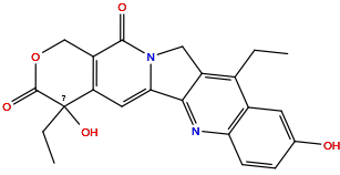
|
|||||
| Formula | C22H20N2O5 |
|||||
| Isosmiles | CCc1c2c(nc3ccc(O)cc13)-c1cc3c(c(=O)n1C2)COC(=O)C3(O)CC |
|||||
| InChI |
InChI=1S/C22H20N2O5/c1-3-12-13-7-11(25)5-6-17(13)23-19-14(12)9-24-18(19)8-16-15(20(24)26)10-29-21(27)22(16,28)4-2/h5-8,25,28H,3-4,9-10H2,1-2H3
|
|||||
| InChIKey |
FJHBVJOVLFPMQE-UHFFFAOYSA-N
|
|||||
| Pharmaceutical Properties | Molecule Weight |
392.411 |
Polar area |
101.65 |
||
Complexity |
29 |
xlogp Value |
2.3476 |
|||
Heavy Count |
29 |
Rot Bonds |
2 |
|||
Hbond acc |
7 |
Hbond Donor |
2 |
|||
The activity data of This Payload
| Standard Type | Value | Units | Cell line | Disease Model | Cell line ID | Reference |
|---|---|---|---|---|---|---|
| Half Maximal Inhibitory Concentration (IC50) | 0.13 | nM |
Daudi cells
|
Burkitt lymphoma
|
[1] | |
| Half Maximal Inhibitory Concentration (IC50) | 0.4 | nM |
Ramos cells
|
Burkitt lymphoma
|
[1] | |
| Half Maximal Inhibitory Concentration (IC50) | 0.47 | nM |
Reh cells
|
B acute lymphoblastic leukemia
|
[1] | |
| Half Maximal Inhibitory Concentration (IC50) | 0.5 | nM |
WSU-FSCCL cells
|
Follicular lymphoma
|
[1] | |
| Half-maximal effective concentration (EC50) | 0.6 | nM |
Ramos cells
|
Burkitt lymphoma
|
[1] | |
| Half Maximal Inhibitory Concentration (IC50) | 1.2±0.9 | nM |
DoHH2 cells
|
Diffuse large B-cell lymphoma germinal center B-cell type
|
[2] | |
| Half Maximal Inhibitory Concentration (IC50) | 1.42 | nM |
Raji cells
|
EBV-related Burkitt lymphoma
|
[1] | |
| Half-maximal effective concentration (EC50) | 1.6 | nM |
Raji cells
|
EBV-related Burkitt lymphoma
|
[1] | |
| Half Maximal Inhibitory Concentration (IC50) | 123 | nM |
MN-60 cells
|
Burkitt lymphoma
|
[1] | |
| Half Maximal Inhibitory Concentration (IC50) | 2.23 | nM |
697 cells
|
Childhood B acute lymphoblastic leukemia
|
[1] | |
| Half Maximal Inhibitory Concentration (IC50) | 2.28 | nM |
RS4
11 cells |
Adult B acute lymphoblastic leukemia
|
[1] | |
| Half Maximal Inhibitory Concentration (IC50) | 5±1 | nM |
Raji cells
|
EBV-related Burkitt lymphoma
|
[2] | |
| Half Maximal Inhibitory Concentration (IC50) | 9±7 | nM |
Daudi cells
|
Burkitt lymphoma
|
[2] | |
| Half-maximal effective concentration (EC50) | 9.1 | nM |
Reh cells
|
B acute lymphoblastic leukemia
|
[1] | |
| Half Maximal Inhibitory Concentration (IC50) | >10 | uM |
JeKo-1 cells
|
Mantle cell lymphoma
|
[1] |
Each Antibody-drug Conjugate Related to This Payload
Full Information of The Activity Data of The ADC(s) Related to This Payload
M603-SN38 DAR6 [Investigative]
Discovered Using Cell Line-derived Xenograft Model
| Experiment 1 Reporting the Activity Date of This ADC | [3] | ||||
| Efficacy Data | Tumor Growth Inhibition value (TGI) | ≈ 0% | High 5T4 expression (5T4+++) | ||
| Method Description |
Treated with m603.
|
||||
| In Vivo Model | Pancreatic cancer CDX model | ||||
| In Vitro Model | Pancreatic cancer | Pancreatic cancer cells | Homo sapiens | ||
| Experiment 2 Reporting the Activity Date of This ADC | [3] | ||||
| Efficacy Data | Tumor Growth Inhibition value (TGI) | ≈ 37.83% | High 5T4 expression (5T4+++) | ||
| Method Description |
Treated with m603-SN38.
|
||||
| In Vivo Model | Pancreatic cancer CDX model | ||||
| In Vitro Model | Pancreatic cancer | Pancreatic cancer cells | Homo sapiens | ||
Revealed Based on the Cell Line Data
| Experiment 1 Reporting the Activity Date of This ADC | [3] | ||||
| Efficacy Data | Half Maximal Inhibitory Concentration (IC50) |
3.9 nM
|
High 5T4 expression (5T4+++) | ||
| Method Description |
Cells in the logarithmic phase of growth were collected and seeded in 96-well plates at a density of 510 3 cells/well and cultured overnight. The culture medium was removed and serial dilution ADCs or naked antibodies were added into the wells of 96-well plates in 200 L medium.
|
||||
| In Vitro Model | Pancreatic ductal adenocarcinoma | BxPC-3 cells | CVCL_0186 | ||
| Experiment 2 Reporting the Activity Date of This ADC | [3] | ||||
| Efficacy Data | Half Maximal Inhibitory Concentration (IC50) |
244.2 nM
|
Moderate 5T4 expression (5T4++) | ||
| Method Description |
Cells in the logarithmic phase of growth were collected and seeded in 96-well plates at a density of 510 3 cells/well and cultured overnight. The culture medium was removed and serial dilution ADCs or naked antibodies were added into the wells of 96-well plates in 200 L medium.
|
||||
| In Vitro Model | Adult hepatocellular carcinoma | Huh-7 cells | CVCL_0336 | ||
N501-SN38 [Investigative]
Discovered Using Cell Line-derived Xenograft Model
| Experiment 1 Reporting the Activity Date of This ADC | [3] | ||||
| Efficacy Data | Tumor Growth Inhibition value (TGI) | ≈ 0% | High 5T4 expression (5T4+++) | ||
| Method Description |
Treated with n501.
|
||||
| In Vivo Model | Pancreatic cancer CDX model | ||||
| In Vitro Model | Pancreatic cancer | Pancreatic cancer cells | Homo sapiens | ||
| Experiment 2 Reporting the Activity Date of This ADC | [3] | ||||
| Efficacy Data | Tumor Growth Inhibition value (TGI) | ≈ 62.93% | High 5T4 expression (5T4+++) | ||
| Method Description |
Treated with n501-SN38.
|
||||
| In Vivo Model | Pancreatic cancer CDX model | ||||
| In Vitro Model | Pancreatic cancer | Pancreatic cancer cells | Homo sapiens | ||
Revealed Based on the Cell Line Data
| Experiment 1 Reporting the Activity Date of This ADC | [3] | ||||
| Efficacy Data | Half Maximal Inhibitory Concentration (IC50) |
11.9 nM
|
High 5T4 expression (5T4+++) | ||
| Method Description |
Cells in the logarithmic phase of growth were collected and seeded in 96-well plates at a density of 510 3 cells/well and cultured overnight. The culture medium was removed and serial dilution ADCs or naked antibodies were added into the wells of 96-well plates in 200 L medium.
|
||||
| In Vitro Model | Pancreatic ductal adenocarcinoma | BxPC-3 cells | CVCL_0186 | ||
| Experiment 2 Reporting the Activity Date of This ADC | [3] | ||||
| Efficacy Data | Half Maximal Inhibitory Concentration (IC50) |
49.8 nM
|
Moderate 5T4 expression (5T4++) | ||
| Method Description |
Cells in the logarithmic phase of growth were collected and seeded in 96-well plates at a density of 510 3 cells/well and cultured overnight. The culture medium was removed and serial dilution ADCs or naked antibodies were added into the wells of 96-well plates in 200 L medium.
|
||||
| In Vitro Model | Adult hepatocellular carcinoma | Huh-7 cells | CVCL_0336 | ||
Cot-APTEDB-SN38 [Investigative]
Discovered Using Cell Line-derived Xenograft Model
| Experiment 1 Reporting the Activity Date of This ADC | [4] | ||||
| Efficacy Data | Tumor Growth Inhibition value (TGI) | ≈ 22.20% | Positive EDB expression (EDB +++/++) | ||
| Method Description |
BALB/c nude mice bearing U87MG tumors (450 mm3) were injected intravenously via the tail vein with Abcot (10 mg/kg) or cot-APTEDB-SN38 (equivalent to 2 mg SN38/kg).
|
||||
| In Vivo Model | U87MG CDX model | ||||
| In Vitro Model | Glioblastoma | U-87MG cells | CVCL_0022 | ||
CAC10-GT [Investigative]
Discovered Using Cell Line-derived Xenograft Model
| Experiment 1 Reporting the Activity Date of This ADC | [5] | ||||
| Efficacy Data | Tumor Growth Inhibition value (TGI) | ≈ 67% | Positive CD30 expression (CD30+++/++) | ||
| Method Description |
Tumor cells, as suspensions, were implanted subcutaneously in SCID or nude mice. Upon tumor engraftment, mice were randomized to study groups (5 mice per group) when the average tumor volume reached about 100 mm3. The ADC or vehicle controls were dosed once via intraperitoneal injection. The dose of cAC10-GT=30mg/kg.
|
||||
| In Vivo Model | HD CDX model | ||||
| In Vitro Model | Hodgkin's disease | L540cy cells | Homo sapiens | ||
| Experiment 2 Reporting the Activity Date of This ADC | [5] | ||||
| Efficacy Data | Tumor Growth Inhibition value (TGI) | ≈ 67% | Positive CD30 expression (CD30+++/++) | ||
| Method Description |
Tumor cells, as suspensions, were implanted subcutaneously in SCID or nude mice. Upon tumor engraftment, mice were randomized to study groups (5 mice per group) when the average tumor volume reached about 100 mm3. The ADC or vehicle controls were dosed once via intraperitoneal injection. The dose of cAC10-GT=10 mg/kg.
|
||||
| In Vivo Model | HD CDX model | ||||
| In Vitro Model | Hodgkin's disease | L540cy cells | Homo sapiens | ||
| Experiment 3 Reporting the Activity Date of This ADC | [5] | ||||
| Efficacy Data | Half Maximal Inhibitory Concentration (IC50) |
20 ng/mL
|
High CD30 expression (CD30+++; 285,000 CD30 molecules/cell) | ||
| Method Description |
Serial dilutions of ADCs in cell culture media were prepared at 4x working concentrations, and 50 uL of each dilution was added to the 96-well plates. Following addition of test articles, cells were incubated for 4 days at 37°C, after which growth inhibition was assessed by the addition of CellTiter-Glo and luminescence was measured on a plate reader.
Click to Show/Hide
|
||||
| In Vitro Model | Precursor T-cell acute lymphoblastic leukemia | ALCL cells | CVCL_A036 | ||
| Experiment 4 Reporting the Activity Date of This ADC | [5] | ||||
| Efficacy Data | Half Maximal Inhibitory Concentration (IC50) |
27 ng/mL
|
High CD30 expression (CD30+++; 180,000 CD30 molecules/cell) | ||
| Method Description |
Serial dilutions of ADCs in cell culture media were prepared at 4x working concentrations, and 50 uL of each dilution was added to the 96-well plates. Following addition of test articles, cells were incubated for 4 days at 37°C, after which growth inhibition was assessed by the addition of CellTiter-Glo and luminescence was measured on a plate reader.
Click to Show/Hide
|
||||
| In Vitro Model | Anaplastic large cell lymphoma | DEL/BVR cells | CVCL_1170 | ||
| Experiment 5 Reporting the Activity Date of This ADC | [5] | ||||
| Efficacy Data | Half Maximal Inhibitory Concentration (IC50) |
79 ng/mL
|
High CD30 expression (CD30+++; 400,000 CD30 molecules/cell) | ||
| Method Description |
Serial dilutions of ADCs in cell culture media were prepared at 4x working concentrations, and 50 uL of each dilution was added to the 96-well plates. Following addition of test articles, cells were incubated for 4 days at 37°C, after which growth inhibition was assessed by the addition of CellTiter-Glo and luminescence was measured on a plate reader.
Click to Show/Hide
|
||||
| In Vitro Model | Hodgkin's disease | L540cy cells | Homo sapiens | ||
| Experiment 6 Reporting the Activity Date of This ADC | [5] | ||||
| Efficacy Data | Half Maximal Inhibitory Concentration (IC50) |
160 ng/mL
|
High CD30 expression (CD30+++; 320,000 CD30 molecules/cell) | ||
| Method Description |
Serial dilutions of ADCs in cell culture media were prepared at 4x working concentrations, and 50 uL of each dilution was added to the 96-well plates. Following addition of test articles, cells were incubated for 4 days at 37°C, after which growth inhibition was assessed by the addition of CellTiter-Glo and luminescence was measured on a plate reader.
Click to Show/Hide
|
||||
| In Vitro Model | ALK-positive anaplastic large cell lymphoma | Karpas-299 cells | CVCL_1324 | ||
| Experiment 7 Reporting the Activity Date of This ADC | [5] | ||||
| Efficacy Data | Half Maximal Inhibitory Concentration (IC50) |
850 ng/mL
|
Low CD30 expression (CD30+; 70,000 CD30 molecules/cell) | ||
| Method Description |
Serial dilutions of ADCs in cell culture media were prepared at 4x working concentrations, and 50 uL of each dilution was added to the 96-well plates. Following addition of test articles, cells were incubated for 4 days at 37°C, after which growth inhibition was assessed by the addition of CellTiter-Glo and luminescence was measured on a plate reader.
Click to Show/Hide
|
||||
| In Vitro Model | Hodgkin lymphoma | L-428 cells | CVCL_1361 | ||
Epratuzumab-CL2E-SN-38 [Investigative]
Discovered Using Cell Line-derived Xenograft Model
| Experiment 1 Reporting the Activity Date of This ADC | [6] | ||||
| Efficacy Data | Median survival time (MST) |
42 Day
|
Low CD22 expression (CD22+) | ||
| Method Description |
The intravenous WSU-FSCCL models were initiated by intravenous injection of 2.5 x 10⁶ cells, in female severe combined immunodeficient (SCID) mice (Taconic). The dose was 0.15 mg/dose (2.4 g SN-38 equivalents).
|
||||
| In Vivo Model | WSU-FSCCL CDX model | ||||
| In Vitro Model | Follicular lymphoma | WSU-FSCCL cells | CVCL_1903 | ||
| Experiment 2 Reporting the Activity Date of This ADC | [6] | ||||
| Efficacy Data | Median survival time (MST) |
63 Day
|
Low CD22 expression (CD22+) | ||
| Method Description |
The intravenous WSU-FSCCL models were initiated by intravenous injection of 2.5 x 10⁶ cells, in female severe combined immunodeficient (SCID) mice (Taconic). The dose was 0.30 mg/dose (4.8 g SN-38 equivalents).
|
||||
| In Vivo Model | WSU-FSCCL CDX model | ||||
| In Vitro Model | Follicular lymphoma | WSU-FSCCL cells | CVCL_1903 | ||
| Experiment 3 Reporting the Activity Date of This ADC | [6] | ||||
| Efficacy Data | Median survival time (MST) |
140 Day
|
Low CD22 expression (CD22+) | ||
| Method Description |
The intravenous WSU-FSCCL models were initiated by intravenous injection of 2.5 x 10⁶ cells, in female severe combined immunodeficient (SCID) mice (Taconic). The dose was 0.15 mg/dose (2.4 g SN-38 equivalents) plus Veltuzumab, 35 ug/dose.
|
||||
| In Vivo Model | WSU-FSCCL CDX model | ||||
| In Vitro Model | Follicular lymphoma | WSU-FSCCL cells | CVCL_1903 | ||
| Experiment 4 Reporting the Activity Date of This ADC | [6] | ||||
| Efficacy Data | Median survival time (MST) | > 161 Day | Low CD22 expression (CD22+) | ||
| Method Description |
The intravenous WSU-FSCCL models were initiated by intravenous injection of 2.5 x 10⁶ cells, in female severe combined immunodeficient (SCID) mice (Taconic). The dose was 0.30 mg/dose (4.8 g SN-38 equivalents) plus Veltuzumab, 35 ug/dose.
|
||||
| In Vivo Model | WSU-FSCCL CDX model | ||||
| In Vitro Model | Follicular lymphoma | WSU-FSCCL cells | CVCL_1903 | ||
Revealed Based on the Cell Line Data
| Experiment 1 Reporting the Activity Date of This ADC | [6] | ||||
| Efficacy Data | Half Maximal Effective Concentration (EC50) |
0.06 nM
|
High CD22 expression (CD22+++) | ||
| Method Description |
Cytotoxicity was determined using the MTS dye reduction assay. Cells were co-incubated with veltuzumab (133 nmol/L) and increasing concentrations of Emab-SN-38.
|
||||
| In Vitro Model | Burkitt lymphoma | Daudi cells | CVCL_0008 | ||
| Experiment 2 Reporting the Activity Date of This ADC | [6] | ||||
| Efficacy Data | Half Maximal Effective Concentration (EC50) |
0.19 nM
|
Moderate CD22 expression (CD22++) | ||
| Method Description |
Cytotoxicity was determined using the MTS dye reduction assay. Cells were co-incubated with veltuzumab (1.33 nmol/L) and increasing concentrations of Emab-SN-38.
|
||||
| In Vitro Model | EBV-related Burkitt lymphoma | Raji cells | CVCL_0511 | ||
| Experiment 3 Reporting the Activity Date of This ADC | [6] | ||||
| Efficacy Data | Half Maximal Effective Concentration (EC50) |
0.2 nM
|
High CD22 expression (CD22+++) | ||
| Method Description |
Cytotoxicity was determined using the MTS dye reduction assay. Cells were co-incubated with veltuzumab (1.33 nmol/L) and increasing concentrations of Emab-SN-38.
|
||||
| In Vitro Model | Burkitt lymphoma | Daudi cells | CVCL_0008 | ||
| Experiment 4 Reporting the Activity Date of This ADC | [6] | ||||
| Efficacy Data | Half Maximal Effective Concentration (EC50) |
0.34 nM
|
Low CD22 expression (CD22+) | ||
| Method Description |
Cytotoxicity was determined using the MTS dye reduction assay. Cells were co-incubated with veltuzumab (1.33 nmol/L) and increasing concentrations of Emab-SN-38.
|
||||
| In Vitro Model | Follicular lymphoma | WSU-FSCCL cells | CVCL_1903 | ||
| Experiment 5 Reporting the Activity Date of This ADC | [6] | ||||
| Efficacy Data | Half Maximal Effective Concentration (EC50) |
0.38 nM
|
Low CD22 expression (CD22+) | ||
| Method Description |
Cytotoxicity was determined using the MTS dye reduction assay. Cells were co-incubated with SN38 and increasing concentrations of Emab-SN-38.
|
||||
| In Vitro Model | Follicular lymphoma | WSU-FSCCL cells | CVCL_1903 | ||
| Experiment 6 Reporting the Activity Date of This ADC | [6] | ||||
| Efficacy Data | Half Maximal Effective Concentration (EC50) |
0.41 nM
|
Low CD22 expression (CD22+) | ||
| Method Description |
Cytotoxicity was determined using the MTS dye reduction assay. Cells were co-incubated with veltuzumab (133 nmol/L) and increasing concentrations of Emab-SN-38.
|
||||
| In Vitro Model | Follicular lymphoma | WSU-FSCCL cells | CVCL_1903 | ||
| Experiment 7 Reporting the Activity Date of This ADC | [6] | ||||
| Efficacy Data | Half Maximal Effective Concentration (EC50) |
0.46 nM
|
Low CD22 expression (CD22+) | ||
| Method Description |
Cytotoxicity was determined using the MTS dye reduction assay. Cells were co-incubated with hRS7 (1.33 nmol/L) and increasing concentrations of Emab-SN-38.
|
||||
| In Vitro Model | Follicular lymphoma | WSU-FSCCL cells | CVCL_1903 | ||
| Experiment 8 Reporting the Activity Date of This ADC | [6] | ||||
| Efficacy Data | Half Maximal Effective Concentration (EC50) |
0.46 nM
|
Low CD22 expression (CD22+) | ||
| Method Description |
Cytotoxicity was determined using the MTS dye reduction assay. Cells were co-incubated with hRS7 (133 nmol/L) and increasing concentrations of Emab-SN-38.
|
||||
| In Vitro Model | Follicular lymphoma | WSU-FSCCL cells | CVCL_1903 | ||
| Experiment 9 Reporting the Activity Date of This ADC | [6] | ||||
| Efficacy Data | Half Maximal Effective Concentration (EC50) |
0.5 nM
|
High CD22 expression (CD22+++) | ||
| Method Description |
Cytotoxicity was determined using the MTS dye reduction assay. Cells were co-incubated with SN38 and increasing concentrations of Emab-SN-38.
|
||||
| In Vitro Model | Burkitt lymphoma | Daudi cells | CVCL_0008 | ||
| Experiment 10 Reporting the Activity Date of This ADC | [6] | ||||
| Efficacy Data | Half Maximal Effective Concentration (EC50) |
0.51 nM
|
Moderate CD22 expression (CD22++) | ||
| Method Description |
Cytotoxicity was determined using the MTS dye reduction assay. Cells were co-incubated with veltuzumab (1.33 nmol/L) and increasing concentrations of Emab-SN-38.
|
||||
| In Vitro Model | Burkitt lymphoma | Ramos cells | CVCL_0597 | ||
| Experiment 11 Reporting the Activity Date of This ADC | [6] | ||||
| Efficacy Data | Half Maximal Effective Concentration (EC50) |
0.52 nM
|
High CD22 expression (CD22+++) | ||
| Method Description |
Cytotoxicity was determined using the MTS dye reduction assay.
|
||||
| In Vitro Model | Burkitt lymphoma | Daudi cells | CVCL_0008 | ||
| Experiment 12 Reporting the Activity Date of This ADC | [6] | ||||
| Efficacy Data | Half Maximal Effective Concentration (EC50) |
0.68 nM
|
Low CD22 expression (CD22+) | ||
| Method Description |
Cytotoxicity was determined using the MTS dye reduction assay.
|
||||
| In Vitro Model | Follicular lymphoma | WSU-FSCCL cells | CVCL_1903 | ||
| Experiment 13 Reporting the Activity Date of This ADC | [6] | ||||
| Efficacy Data | Half Maximal Effective Concentration (EC50) |
0.73 nM
|
Moderate CD22 expression (CD22++) | ||
| Method Description |
Cytotoxicity was determined using the MTS dye reduction assay. Cells were co-incubated with veltuzumab (133 nmol/L) and increasing concentrations of Emab-SN-38.
|
||||
| In Vitro Model | Burkitt lymphoma | Ramos cells | CVCL_0597 | ||
| Experiment 14 Reporting the Activity Date of This ADC | [6] | ||||
| Efficacy Data | Half Maximal Effective Concentration (EC50) |
0.81 nM
|
Low CD22 expression (CD22+) | ||
| Method Description |
Cytotoxicity was determined using the MTS dye reduction assay. Cells were co-incubated with veltuzumab (133 nmol/L) and increasing concentrations of Emab-SN-38.
|
||||
| In Vitro Model | Mantle cell lymphoma | JeKo-1 cells | CVCL_1865 | ||
| Experiment 15 Reporting the Activity Date of This ADC | [6] | ||||
| Efficacy Data | Half Maximal Effective Concentration (EC50) |
0.83 nM
|
Low CD22 expression (CD22+) | ||
| Method Description |
Cytotoxicity was determined using the MTS dye reduction assay. Cells were co-incubated with veltuzumab (1.33 nmol/L) and increasing concentrations of Emab-SN-38.
|
||||
| In Vitro Model | Mantle cell lymphoma | JeKo-1 cells | CVCL_1865 | ||
| Experiment 16 Reporting the Activity Date of This ADC | [6] | ||||
| Efficacy Data | Half Maximal Effective Concentration (EC50) |
0.84 nM
|
High CD22 expression (CD22+++) | ||
| Method Description |
Cytotoxicity was determined using the MTS dye reduction assay. Cells were co-incubated with hRS7 (133 nmol/L) and increasing concentrations of Emab-SN-38.
|
||||
| In Vitro Model | Burkitt lymphoma | Daudi cells | CVCL_0008 | ||
| Experiment 17 Reporting the Activity Date of This ADC | [6] | ||||
| Efficacy Data | Half Maximal Effective Concentration (EC50) |
1.16 nM
|
Low CD22 expression (CD22+) | ||
| Method Description |
Cytotoxicity was determined using the MTS dye reduction assay. Cells were co-incubated with hRS7 (1.33 nmol/L) and increasing concentrations of Emab-SN-38.
|
||||
| In Vitro Model | Mantle cell lymphoma | JeKo-1 cells | CVCL_1865 | ||
| Experiment 18 Reporting the Activity Date of This ADC | [6] | ||||
| Efficacy Data | Half Maximal Effective Concentration (EC50) |
1.16 nM
|
Moderate CD22 expression (CD22++) | ||
| Method Description |
Cytotoxicity was determined using the MTS dye reduction assay. Cells were co-incubated with veltuzumab (133 nmol/L) and increasing concentrations of Emab-SN-38.
|
||||
| In Vitro Model | EBV-related Burkitt lymphoma | Raji cells | CVCL_0511 | ||
| Experiment 19 Reporting the Activity Date of This ADC | [6] | ||||
| Efficacy Data | Half Maximal Effective Concentration (EC50) |
1.22 nM
|
Moderate CD22 expression (CD22++) | ||
| Method Description |
Cytotoxicity was determined using the MTS dye reduction assay.
|
||||
| In Vitro Model | B acute lymphoblastic leukemia | Reh cells | CVCL_1650 | ||
| Experiment 20 Reporting the Activity Date of This ADC | [6] | ||||
| Efficacy Data | Half Maximal Effective Concentration (EC50) |
1.5 nM
|
High CD22 expression (CD22+++) | ||
| Method Description |
Cytotoxicity was determined using the MTS dye reduction assay. Cells were co-incubated with hRS7 (1.33 nmol/L) and increasing concentrations of Emab-SN-38.
|
||||
| In Vitro Model | Burkitt lymphoma | Daudi cells | CVCL_0008 | ||
| Experiment 21 Reporting the Activity Date of This ADC | [6] | ||||
| Efficacy Data | Half Maximal Effective Concentration (EC50) |
1.66 nM
|
Low CD22 expression (CD22+) | ||
| Method Description |
Cytotoxicity was determined using the MTS dye reduction assay. Cells were co-incubated with hRS7 (133 nmol/L) and increasing concentrations of Emab-SN-38.
|
||||
| In Vitro Model | Mantle cell lymphoma | JeKo-1 cells | CVCL_1865 | ||
| Experiment 22 Reporting the Activity Date of This ADC | [6] | ||||
| Efficacy Data | Half Maximal Effective Concentration (EC50) |
1.68 nM
|
Moderate CD22 expression (CD22++) | ||
| Method Description |
Cytotoxicity was determined using the MTS dye reduction assay.
|
||||
| In Vitro Model | Adult B acute lymphoblastic leukemia | RS4;11 cells | CVCL_0093 | ||
| Experiment 23 Reporting the Activity Date of This ADC | [6] | ||||
| Efficacy Data | Half Maximal Effective Concentration (EC50) |
1.72 nM
|
Moderate CD22 expression (CD22++) | ||
| Method Description |
Cytotoxicity was determined using the MTS dye reduction assay. Cells were co-incubated with SN38 and increasing concentrations of Emab-SN-38.
|
||||
| In Vitro Model | EBV-related Burkitt lymphoma | Raji cells | CVCL_0511 | ||
| Experiment 24 Reporting the Activity Date of This ADC | [6] | ||||
| Efficacy Data | Half Maximal Effective Concentration (EC50) |
1.77 nM
|
Low CD22 expression (CD22+) | ||
| Method Description |
Cytotoxicity was determined using the MTS dye reduction assay. Cells were co-incubated with SN38 and increasing concentrations of Emab-SN-38.
|
||||
| In Vitro Model | Mantle cell lymphoma | JeKo-1 cells | CVCL_1865 | ||
| Experiment 25 Reporting the Activity Date of This ADC | [6] | ||||
| Efficacy Data | Half Maximal Effective Concentration (EC50) |
1.84 nM
|
Moderate CD22 expression (CD22++) | ||
| Method Description |
Cytotoxicity was determined using the MTS dye reduction assay. Cells were co-incubated with SN38 and increasing concentrations of Emab-SN-38.
|
||||
| In Vitro Model | Burkitt lymphoma | Ramos cells | CVCL_0597 | ||
| Experiment 26 Reporting the Activity Date of This ADC | [6] | ||||
| Efficacy Data | Half Maximal Effective Concentration (EC50) | . 2.1 nM | Moderate CD22 expression (CD22++) | ||
| Method Description |
Cytotoxicity was determined using the MTS dye reduction assay.
|
||||
| In Vitro Model | EBV-related Burkitt lymphoma | Raji cells | CVCL_0511 | ||
| Experiment 27 Reporting the Activity Date of This ADC | [6] | ||||
| Efficacy Data | Half Maximal Effective Concentration (EC50) |
2.21 nM
|
Moderate CD22 expression (CD22++) | ||
| Method Description |
Cytotoxicity was determined using the MTS dye reduction assay. Cells were co-incubated with hRS7 (133 nmol/L) and increasing concentrations of Emab-SN-38.
|
||||
| In Vitro Model | Burkitt lymphoma | Ramos cells | CVCL_0597 | ||
| Experiment 28 Reporting the Activity Date of This ADC | [6] | ||||
| Efficacy Data | Half Maximal Effective Concentration (EC50) |
2.25 nM
|
Low CD22 expression (CD22+) | ||
| Method Description |
Cytotoxicity was determined using the MTS dye reduction assay.
|
||||
| In Vitro Model | Mantle cell lymphoma | JeKo-1 cells | CVCL_1865 | ||
| Experiment 29 Reporting the Activity Date of This ADC | [6] | ||||
| Efficacy Data | Half Maximal Effective Concentration (EC50) |
2.29 nM
|
Moderate CD22 expression (CD22++) | ||
| Method Description |
Cytotoxicity was determined using the MTS dye reduction assay. Cells were co-incubated with hRS7 (1.33 nmol/L) and increasing concentrations of Emab-SN-38.
|
||||
| In Vitro Model | Burkitt lymphoma | Ramos cells | CVCL_0597 | ||
| Experiment 30 Reporting the Activity Date of This ADC | [6] | ||||
| Efficacy Data | Half Maximal Effective Concentration (EC50) |
2.45 nM
|
Moderate CD22 expression (CD22++) | ||
| Method Description |
Cytotoxicity was determined using the MTS dye reduction assay. Cells were co-incubated with hRS7 (1.33 nmol/L) and increasing concentrations of Emab-SN-38.
|
||||
| In Vitro Model | EBV-related Burkitt lymphoma | Raji cells | CVCL_0511 | ||
| Experiment 31 Reporting the Activity Date of This ADC | [6] | ||||
| Efficacy Data | Half Maximal Effective Concentration (EC50) |
2.67 nM
|
Low CD22 expression (CD22+) | ||
| Method Description |
Cytotoxicity was determined using the MTS dye reduction assay.
|
||||
| In Vitro Model | Childhood B acute lymphoblastic leukemia | 697 cells | CVCL_0079 | ||
| Experiment 32 Reporting the Activity Date of This ADC | [6] | ||||
| Efficacy Data | Half Maximal Effective Concentration (EC50) |
2.88 nM
|
Moderate CD22 expression (CD22++) | ||
| Method Description |
Cytotoxicity was determined using the MTS dye reduction assay. Cells were co-incubated with hRS7 (133 nmol/L) and increasing concentrations of Emab-SN-38.
|
||||
| In Vitro Model | EBV-related Burkitt lymphoma | Raji cells | CVCL_0511 | ||
| Experiment 33 Reporting the Activity Date of This ADC | [6] | ||||
| Efficacy Data | Half Maximal Effective Concentration (EC50) |
2.92 nM
|
Moderate CD22 expression (CD22++) | ||
| Method Description |
Cytotoxicity was determined using the MTS dye reduction assay.
|
||||
| In Vitro Model | Burkitt lymphoma | Ramos cells | CVCL_0597 | ||
| Experiment 34 Reporting the Activity Date of This ADC | [6] | ||||
| Efficacy Data | Half Maximal Effective Concentration (EC50) |
3.65 nM
|
Low CD22 expression (CD22+) | ||
| Method Description |
Cytotoxicity was determined using the MTS dye reduction assay.
|
||||
| In Vitro Model | Burkitt lymphoma | MN-60 cells | CVCL_1421 | ||
| Experiment 35 Reporting the Activity Date of This ADC | [6] | ||||
| Efficacy Data | Half Maximal Effective Concentration (EC50) |
135.8 nM
|
Moderate CD22 expression (CD22++) | ||
| Method Description |
Cytotoxicity was determined using the MTS dye reduction assay.
|
||||
| In Vitro Model | EBV-related Burkitt lymphoma | Raji cells | CVCL_0511 | ||
| Experiment 36 Reporting the Activity Date of This ADC | [6] | ||||
| Efficacy Data | Half Maximal Effective Concentration (EC50) |
152.3 nM
|
Moderate CD22 expression (CD22++) | ||
| Method Description |
Cytotoxicity was determined using the MTS dye reduction assay.
|
||||
| In Vitro Model | Burkitt lymphoma | Ramos cells | CVCL_0597 | ||
| Experiment 37 Reporting the Activity Date of This ADC | [6] | ||||
| Efficacy Data | Half Maximal Effective Concentration (EC50) |
271 nM
|
High CD20 expression (CD20+++) | ||
| Method Description |
Cytotoxicity was determined using the MTS dye reduction assay.
|
||||
| In Vitro Model | Burkitt lymphoma | Ramos cells | CVCL_0597 | ||
Labetuzumab-CL2E-SN-38 [Investigative]
Discovered Using Cell Line-derived Xenograft Model
| Experiment 1 Reporting the Activity Date of This ADC | [6] | ||||
| Efficacy Data | Median survival time (MST) |
63 Day
|
Low CEACAM5 expression (CEACAM5+) | ||
| Method Description |
The intravenous WSU-FSCCL models were initiated by intravenous injection of 2.5 x 10⁶ cells, in female severe combined immunodeficient (SCID) mice (Taconic). The dose was 0.30 mg/dose (4.8 g SN-38 equivalents).
|
||||
| In Vivo Model | WSU-FSCCL CDX model | ||||
| In Vitro Model | Follicular lymphoma | WSU-FSCCL cells | CVCL_1903 | ||
| Experiment 2 Reporting the Activity Date of This ADC | [6] | ||||
| Efficacy Data | Median survival time (MST) |
76 Day
|
Low CEACAM5 expression (CEACAM5+) | ||
| Method Description |
The intravenous WSU-FSCCL models were initiated by intravenous injection of 2.5 x 10⁶ cells, in female severe combined immunodeficient (SCID) mice (Taconic). The dose was 0.15 mg/dose (2.4 g SN-38 equivalents).
|
||||
| In Vivo Model | WSU-FSCCL CDX model | ||||
| In Vitro Model | Follicular lymphoma | WSU-FSCCL cells | CVCL_1903 | ||
| Experiment 3 Reporting the Activity Date of This ADC | [6] | ||||
| Efficacy Data | Median survival time (MST) |
91 Day
|
Low CEACAM5 expression (CEACAM5+) | ||
| Method Description |
The intravenous WSU-FSCCL models were initiated by intravenous injection of 2.5 x 10⁶ cells, in female severe combined immunodeficient (SCID) mice (Taconic). The dose was 0.30 mg/dose (4.8 g SN-38 equivalents) plus Veltuzumab, 35 ug/dose.
|
||||
| In Vivo Model | WSU-FSCCL CDX model | ||||
| In Vitro Model | Follicular lymphoma | WSU-FSCCL cells | CVCL_1903 | ||
| Experiment 4 Reporting the Activity Date of This ADC | [6] | ||||
| Efficacy Data | Median survival time (MST) |
98 Day
|
Low CEACAM5 expression (CEACAM5+) | ||
| Method Description |
The intravenous WSU-FSCCL models were initiated by intravenous injection of 2.5 x 10⁶ cells, in female severe combined immunodeficient (SCID) mice (Taconic). The dose was 0.15 mg/dose (2.4 g SN-38 equivalents) plus Veltuzumab, 35 ug/dose.
|
||||
| In Vivo Model | WSU-FSCCL CDX model | ||||
| In Vitro Model | Follicular lymphoma | WSU-FSCCL cells | CVCL_1903 | ||
Revealed Based on the Cell Line Data
| Experiment 1 Reporting the Activity Date of This ADC | [6] | ||||
| Efficacy Data | Half Maximal Effective Concentration (EC50) |
1.17 nM
|
Low CEACAM5 expression (CEACAM5+) | ||
| Method Description |
Cytotoxicity was determined using the MTS dye reduction assay.
|
||||
| In Vitro Model | Mantle cell lymphoma | JeKo-1 cells | CVCL_1865 | ||
| Experiment 2 Reporting the Activity Date of This ADC | [6] | ||||
| Efficacy Data | Half Maximal Effective Concentration (EC50) |
3.73 nM
|
Low CEACAM5 expression (CEACAM5+) | ||
| Method Description |
Cytotoxicity was determined using the MTS dye reduction assay.
|
||||
| In Vitro Model | EBV-related Burkitt lymphoma | Raji cells | CVCL_0511 | ||
| Experiment 3 Reporting the Activity Date of This ADC | [6] | ||||
| Efficacy Data | Half Maximal Effective Concentration (EC50) |
8.08 nM
|
Low CEACAM5 expression (CEACAM5+) | ||
| Method Description |
Cytotoxicity was determined using the MTS dye reduction assay.
|
||||
| In Vitro Model | Burkitt lymphoma | Daudi cells | CVCL_0008 | ||
| Experiment 4 Reporting the Activity Date of This ADC | [6] | ||||
| Efficacy Data | Half Maximal Effective Concentration (EC50) | > 50 nM | Negative CEACAM5 expression (CEACAM5-) | ||
| Method Description |
Cytotoxicity was determined using the MTS dye reduction assay.
|
||||
| In Vitro Model | Burkitt lymphoma | MN-60 cells | CVCL_1421 | ||
| Experiment 5 Reporting the Activity Date of This ADC | [6] | ||||
| Efficacy Data | Half Maximal Effective Concentration (EC50) | > 50 nM | Negative CEACAM5 expression (CEACAM5-) | ||
| Method Description |
Cytotoxicity was determined using the MTS dye reduction assay.
|
||||
| In Vitro Model | Adult B acute lymphoblastic leukemia | RS4;11 cells | CVCL_0093 | ||
| Experiment 6 Reporting the Activity Date of This ADC | [6] | ||||
| Efficacy Data | Half Maximal Effective Concentration (EC50) | > 50 nM | Negative CEACAM5 expression (CEACAM5-) | ||
| Method Description |
Cytotoxicity was determined using the MTS dye reduction assay.
|
||||
| In Vitro Model | Childhood B acute lymphoblastic leukemia | 697 cells | CVCL_0079 | ||
| Experiment 7 Reporting the Activity Date of This ADC | [6] | ||||
| Efficacy Data | Half Maximal Effective Concentration (EC50) | > 50 nM | Negative CEACAM5 expression (CEACAM5-) | ||
| Method Description |
Cytotoxicity was determined using the MTS dye reduction assay.
|
||||
| In Vitro Model | B acute lymphoblastic leukemia | Reh cells | CVCL_1650 | ||
| Experiment 8 Reporting the Activity Date of This ADC | [6] | ||||
| Efficacy Data | Half Maximal Effective Concentration (EC50) | > 50 nM | Low CEACAM5 expression (CEACAM5+) | ||
| Method Description |
Cytotoxicity was determined using the MTS dye reduction assay.
|
||||
| In Vitro Model | Follicular lymphoma | WSU-FSCCL cells | CVCL_1903 | ||
| Experiment 9 Reporting the Activity Date of This ADC | [6] | ||||
| Efficacy Data | Half Maximal Effective Concentration (EC50) | > 50 nM | Negative CEACAM5 expression (CEACAM5-) | ||
| Method Description |
Cytotoxicity was determined using the MTS dye reduction assay.
|
||||
| In Vitro Model | Burkitt lymphoma | Ramos cells | CVCL_0597 | ||
H00-GT [Investigative]
Discovered Using Cell Line-derived Xenograft Model
| Experiment 1 Reporting the Activity Date of This ADC | [5] | ||||
| Efficacy Data | Half Maximal Inhibitory Concentration (IC50) |
19 ng/mL
|
High CD30 expression (CD30+++; 285,000 CD30 molecules/cell) | ||
| Method Description |
Serial dilutions of ADCs in cell culture media were prepared at 4x working concentrations, and 50 uL of each dilution was added to the 96-well plates. Following addition of test articles, cells were incubated for 4 days at 37°C, after which growth inhibition was assessed by the addition of CellTiter-Glo and luminescence was measured on a plate reader.
Click to Show/Hide
|
||||
| In Vitro Model | Precursor T-cell acute lymphoblastic leukemia | ALCL cells | CVCL_A036 | ||
| Experiment 2 Reporting the Activity Date of This ADC | [5] | ||||
| Efficacy Data | Half Maximal Inhibitory Concentration (IC50) |
25 ng/mL
|
High CD30 expression (CD30+++; 180,000 CD30 molecules/cell) | ||
| Method Description |
Serial dilutions of ADCs in cell culture media were prepared at 4x working concentrations, and 50 uL of each dilution was added to the 96-well plates. Following addition of test articles, cells were incubated for 4 days at 37°C, after which growth inhibition was assessed by the addition of CellTiter-Glo and luminescence was measured on a plate reader.
Click to Show/Hide
|
||||
| In Vitro Model | Anaplastic large cell lymphoma | DEL/BVR cells | CVCL_1170 | ||
| Experiment 3 Reporting the Activity Date of This ADC | [5] | ||||
| Efficacy Data | Half Maximal Inhibitory Concentration (IC50) |
46 ng/mL
|
High CD30 expression (CD30+++; 400,000 CD30 molecules/cell) | ||
| Method Description |
Serial dilutions of ADCs in cell culture media were prepared at 4x working concentrations, and 50 uL of each dilution was added to the 96-well plates. Following addition of test articles, cells were incubated for 4 days at 37°C, after which growth inhibition was assessed by the addition of CellTiter-Glo and luminescence was measured on a plate reader.
Click to Show/Hide
|
||||
| In Vitro Model | Hodgkin's disease | L540cy cells | Homo sapiens | ||
| Experiment 4 Reporting the Activity Date of This ADC | [5] | ||||
| Efficacy Data | Half Maximal Inhibitory Concentration (IC50) |
160 ng/mL
|
High CD30 expression (CD30+++; 320,000 CD30 molecules/cell) | ||
| Method Description |
Serial dilutions of ADCs in cell culture media were prepared at 4x working concentrations, and 50 uL of each dilution was added to the 96-well plates. Following addition of test articles, cells were incubated for 4 days at 37°C, after which growth inhibition was assessed by the addition of CellTiter-Glo and luminescence was measured on a plate reader.
Click to Show/Hide
|
||||
| In Vitro Model | ALK-positive anaplastic large cell lymphoma | Karpas-299 cells | CVCL_1324 | ||
| Experiment 5 Reporting the Activity Date of This ADC | [5] | ||||
| Efficacy Data | Half Maximal Inhibitory Concentration (IC50) |
820 ng/mL
|
Low CD30 expression (CD30+; 70,000 CD30 molecules/cell) | ||
| Method Description |
Serial dilutions of ADCs in cell culture media were prepared at 4x working concentrations, and 50 uL of each dilution was added to the 96-well plates. Following addition of test articles, cells were incubated for 4 days at 37°C, after which growth inhibition was assessed by the addition of CellTiter-Glo and luminescence was measured on a plate reader.
Click to Show/Hide
|
||||
| In Vitro Model | Hodgkin lymphoma | L-428 cells | CVCL_1361 | ||
Mil40-5 [Investigative]
Revealed Based on the Cell Line Data
| Experiment 1 Reporting the Activity Date of This ADC | [7] | ||||
| Efficacy Data | Half Maximal Inhibitory Concentration (IC50) |
14.5 nM
|
High HER2 expression (HER2+++) | ||
| Method Description |
SKOV-3, BT474 HerDR, MDA-MB-231 and MCF-7 cells were cultured under various concentrations of Mil40, SN-38 and ADCs for 10days, 9days, 6days, and 6days, respectively. Cytotoxicity assays were established using the CellTiter-Go assay kit (CTG).
|
||||
| In Vitro Model | Invasive breast carcinoma | BT474 HerDR cells | CVCL_0179 | ||
| Experiment 2 Reporting the Activity Date of This ADC | [7] | ||||
| Efficacy Data | Half Maximal Inhibitory Concentration (IC50) |
157.6 nM
|
High HER2 expression (HER2+++) | ||
| Method Description |
SKOV-3, BT474 HerDR, MDA-MB-231 and MCF-7 cells were cultured under various concentrations of Mil40, SN-38 and ADCs for 10days, 9days, 6days, and 6days, respectively. Cytotoxicity assays were established using the CellTiter-Go assay kit (CTG).
|
||||
| In Vitro Model | Ovarian serous cystadenocarcinoma | SK-OV-3 cells | CVCL_0532 | ||
| Experiment 3 Reporting the Activity Date of This ADC | [7] | ||||
| Efficacy Data | Half Maximal Inhibitory Concentration (IC50) | > 1000 nM | Negative HER2 expression (HER2-) | ||
| Method Description |
SKOV-3, BT474 HerDR, MDA-MB-231 and MCF-7 cells were cultured under various concentrations of Mil40, SN-38 and ADCs for 10days, 9days, 6days, and 6days, respectively. Cytotoxicity assays were established using the CellTiter-Go assay kit (CTG).
|
||||
| In Vitro Model | Invasive breast carcinoma | MCF-7 cells | CVCL_0031 | ||
| Experiment 4 Reporting the Activity Date of This ADC | [7] | ||||
| Efficacy Data | Half Maximal Inhibitory Concentration (IC50) | > 1000 nM | Negative HER2 expression (HER2-) | ||
| Method Description |
SKOV-3, BT474 HerDR, MDA-MB-231 and MCF-7 cells were cultured under various concentrations of Mil40, SN-38 and ADCs for 10days, 9days, 6days, and 6days, respectively. Cytotoxicity assays were established using the CellTiter-Go assay kit (CTG).
|
||||
| In Vitro Model | Breast adenocarcinoma | MDA-MB-231 cells | CVCL_0062 | ||
Mil40-6 [Investigative]
Revealed Based on the Cell Line Data
| Experiment 1 Reporting the Activity Date of This ADC | [7] | ||||
| Efficacy Data | Half Maximal Inhibitory Concentration (IC50) |
15.9 nM
|
High HER2 expression (HER2+++) | ||
| Method Description |
SKOV-3, BT474 HerDR, MDA-MB-231 and MCF-7 cells were cultured under various concentrations of Mil40, SN-38 and ADCs for 10days, 9days, 6days, and 6days, respectively. Cytotoxicity assays were established using the CellTiter-Go assay kit (CTG).
|
||||
| In Vitro Model | Invasive breast carcinoma | BT474 HerDR cells | CVCL_0179 | ||
| Experiment 2 Reporting the Activity Date of This ADC | [7] | ||||
| Efficacy Data | Half Maximal Inhibitory Concentration (IC50) |
320.8 nM
|
High HER2 expression (HER2+++) | ||
| Method Description |
SKOV-3, BT474 HerDR, MDA-MB-231 and MCF-7 cells were cultured under various concentrations of Mil40, SN-38 and ADCs for 10days, 9days, 6days, and 6days, respectively. Cytotoxicity assays were established using the CellTiter-Go assay kit (CTG).
|
||||
| In Vitro Model | Ovarian serous cystadenocarcinoma | SK-OV-3 cells | CVCL_0532 | ||
| Experiment 3 Reporting the Activity Date of This ADC | [7] | ||||
| Efficacy Data | Half Maximal Inhibitory Concentration (IC50) |
484.71 nM
|
Negative HER2 expression (HER2-) | ||
| Method Description |
SKOV-3, BT474 HerDR, MDA-MB-231 and MCF-7 cells were cultured under various concentrations of Mil40, SN-38 and ADCs for 10days, 9days, 6days, and 6days, respectively. Cytotoxicity assays were established using the CellTiter-Go assay kit (CTG).
|
||||
| In Vitro Model | Invasive breast carcinoma | MCF-7 cells | CVCL_0031 | ||
| Experiment 4 Reporting the Activity Date of This ADC | [7] | ||||
| Efficacy Data | Half Maximal Inhibitory Concentration (IC50) | > 1000 nM | Negative HER2 expression (HER2-) | ||
| Method Description |
SKOV-3, BT474 HerDR, MDA-MB-231 and MCF-7 cells were cultured under various concentrations of Mil40, SN-38 and ADCs for 10days, 9days, 6days, and 6days, respectively. Cytotoxicity assays were established using the CellTiter-Go assay kit (CTG).
|
||||
| In Vitro Model | Breast adenocarcinoma | MDA-MB-231 cells | CVCL_0062 | ||
40H3-CL2A-SN38 [Investigative]
Revealed Based on the Cell Line Data
| Experiment 1 Reporting the Activity Date of This ADC | [8] | ||||
| Efficacy Data | Half Maximal Inhibitory Concentration (IC50) |
19.71 nM
|
High EGFR expression (EGFR+++) | ||
| Method Description |
1 x10⁴ cells per well in a volume of 100 ul were plated in 96-well tissue culture plates. After 24 h, ADCs were added at the indicated concentrations. After 72 h, the medium was removed and the viability was determined using the CellTiter-Glo luminescent cell viability assay kit.
|
||||
| In Vitro Model | Skin squamous cell carcinoma | A431 cells | CVCL_0037 | ||
| Experiment 2 Reporting the Activity Date of This ADC | [8] | ||||
| Efficacy Data | Half Maximal Inhibitory Concentration (IC50) |
27.83 nM
|
High EGFR expression (EGFR+++) | ||
| Method Description |
1 x10⁴ cells per well in a volume of 100 ul were plated in 96-well tissue culture plates. After 24 h, ADCs were added at the indicated concentrations. After 72 h, the medium was removed and the viability was determined using the CellTiter-Glo luminescent cell viability assay kit.
|
||||
| In Vitro Model | Breast adenocarcinoma | MDA-MB-468 cells | CVCL_0419 | ||
| Experiment 3 Reporting the Activity Date of This ADC | [8] | ||||
| Efficacy Data | Half Maximal Inhibitory Concentration (IC50) |
98.81 nM
|
Moderate EGFR expression (EGFR++) | ||
| Method Description |
1 x10⁴ cells per well in a volume of 100 ul were plated in 96-well tissue culture plates. After 24 h, ADCs were added at the indicated concentrations. After 72 h, the medium was removed and the viability was determined using the CellTiter-Glo luminescent cell viability assay kit.
|
||||
| In Vitro Model | Invasive breast carcinoma of no special type | BT-20 cells | CVCL_0178 | ||
| Experiment 4 Reporting the Activity Date of This ADC | [8] | ||||
| Efficacy Data | Half Maximal Inhibitory Concentration (IC50) | > 100 nM | Negative EGFR expression (EGFR-) | ||
| Method Description |
1 x10⁴ cells per well in a volume of 100 ul were plated in 96-well tissue culture plates. After 24 h, ADCs were added at the indicated concentrations. After 72 h, the medium was removed and the viability was determined using the CellTiter-Glo luminescent cell viability assay kit.
|
||||
| In Vitro Model | Breast adenocarcinoma | MDA-MB-231 cells | CVCL_0062 | ||
Mil40-7 [Investigative]
Revealed Based on the Cell Line Data
| Experiment 1 Reporting the Activity Date of This ADC | [7] | ||||
| Efficacy Data | Half Maximal Inhibitory Concentration (IC50) |
22 nM
|
High HER2 expression (HER2+++) | ||
| Method Description |
SKOV-3, BT474 HerDR, MDA-MB-231 and MCF-7 cells were cultured under various concentrations of Mil40, SN-38 and ADCs for 10days, 9days, 6days, and 6days, respectively. Cytotoxicity assays were established using the CellTiter-Go assay kit (CTG).
|
||||
| In Vitro Model | Invasive breast carcinoma | BT474 HerDR cells | CVCL_0179 | ||
| Experiment 2 Reporting the Activity Date of This ADC | [7] | ||||
| Efficacy Data | Half Maximal Inhibitory Concentration (IC50) |
159.4 nM
|
High HER2 expression (HER2+++) | ||
| Method Description |
SKOV-3, BT474 HerDR, MDA-MB-231 and MCF-7 cells were cultured under various concentrations of Mil40, SN-38 and ADCs for 10days, 9days, 6days, and 6days, respectively. Cytotoxicity assays were established using the CellTiter-Go assay kit (CTG).
|
||||
| In Vitro Model | Ovarian serous cystadenocarcinoma | SK-OV-3 cells | CVCL_0532 | ||
| Experiment 3 Reporting the Activity Date of This ADC | [7] | ||||
| Efficacy Data | Half Maximal Inhibitory Concentration (IC50) | > 1000 nM | Negative HER2 expression (HER2-) | ||
| Method Description |
SKOV-3, BT474 HerDR, MDA-MB-231 and MCF-7 cells were cultured under various concentrations of Mil40, SN-38 and ADCs for 10days, 9days, 6days, and 6days, respectively. Cytotoxicity assays were established using the CellTiter-Go assay kit (CTG).
|
||||
| In Vitro Model | Invasive breast carcinoma | MCF-7 cells | CVCL_0031 | ||
| Experiment 4 Reporting the Activity Date of This ADC | [7] | ||||
| Efficacy Data | Half Maximal Inhibitory Concentration (IC50) | > 1000 nM | Negative HER2 expression (HER2-) | ||
| Method Description |
SKOV-3, BT474 HerDR, MDA-MB-231 and MCF-7 cells were cultured under various concentrations of Mil40, SN-38 and ADCs for 10days, 9days, 6days, and 6days, respectively. Cytotoxicity assays were established using the CellTiter-Go assay kit (CTG).
|
||||
| In Vitro Model | Breast adenocarcinoma | MDA-MB-231 cells | CVCL_0062 | ||
Mil40-8 [Investigative]
Revealed Based on the Cell Line Data
| Experiment 1 Reporting the Activity Date of This ADC | [7] | ||||
| Efficacy Data | Half Maximal Inhibitory Concentration (IC50) |
235.6 nM
|
High HER2 expression (HER2+++) | ||
| Method Description |
SKOV-3, BT474 HerDR, MDA-MB-231 and MCF-7 cells were cultured under various concentrations of Mil40, SN-38 and ADCs for 10days, 9days, 6days, and 6days, respectively. Cytotoxicity assays were established using the CellTiter-Go assay kit (CTG).
|
||||
| In Vitro Model | Invasive breast carcinoma | BT474 HerDR cells | CVCL_0179 | ||
| Experiment 2 Reporting the Activity Date of This ADC | [7] | ||||
| Efficacy Data | Half Maximal Inhibitory Concentration (IC50) |
283.8 nM
|
High HER2 expression (HER2+++) | ||
| Method Description |
SKOV-3, BT474 HerDR, MDA-MB-231 and MCF-7 cells were cultured under various concentrations of Mil40, SN-38 and ADCs for 10days, 9days, 6days, and 6days, respectively. Cytotoxicity assays were established using the CellTiter-Go assay kit (CTG).
|
||||
| In Vitro Model | Ovarian serous cystadenocarcinoma | SK-OV-3 cells | CVCL_0532 | ||
| Experiment 3 Reporting the Activity Date of This ADC | [7] | ||||
| Efficacy Data | Half Maximal Inhibitory Concentration (IC50) | > 1000 nM | Negative HER2 expression (HER2-) | ||
| Method Description |
SKOV-3, BT474 HerDR, MDA-MB-231 and MCF-7 cells were cultured under various concentrations of Mil40, SN-38 and ADCs for 10days, 9days, 6days, and 6days, respectively. Cytotoxicity assays were established using the CellTiter-Go assay kit (CTG).
|
||||
| In Vitro Model | Invasive breast carcinoma | MCF-7 cells | CVCL_0031 | ||
| Experiment 4 Reporting the Activity Date of This ADC | [7] | ||||
| Efficacy Data | Half Maximal Inhibitory Concentration (IC50) | > 1000 nM | Negative HER2 expression (HER2-) | ||
| Method Description |
SKOV-3, BT474 HerDR, MDA-MB-231 and MCF-7 cells were cultured under various concentrations of Mil40, SN-38 and ADCs for 10days, 9days, 6days, and 6days, respectively. Cytotoxicity assays were established using the CellTiter-Go assay kit (CTG).
|
||||
| In Vitro Model | Breast adenocarcinoma | MDA-MB-231 cells | CVCL_0062 | ||
Veltuzumab-SN-38 [Investigative]
Revealed Based on the Cell Line Data
| Experiment 1 Reporting the Activity Date of This ADC | [6] | ||||
| Efficacy Data | Half Maximal Effective Concentration (EC50) |
0.83 nM
|
Moderate CD20 expression (CD20++) | ||
| Method Description |
Cytotoxicity was determined using the MTS dye reduction assay.
|
||||
| In Vitro Model | Follicular lymphoma | WSU-FSCCL cells | CVCL_1903 | ||
| Experiment 2 Reporting the Activity Date of This ADC | [6] | ||||
| Efficacy Data | Half Maximal Effective Concentration (EC50) |
1.98 nM
|
High CD20 expression (CD20+++) | ||
| Method Description |
Cytotoxicity was determined using the MTS dye reduction assay.
|
||||
| In Vitro Model | Mantle cell lymphoma | JeKo-1 cells | CVCL_1865 | ||
| Experiment 3 Reporting the Activity Date of This ADC | [6] | ||||
| Efficacy Data | Half Maximal Effective Concentration (EC50) |
4.88 nM
|
High CD20 expression (CD20+++) | ||
| Method Description |
Cytotoxicity was determined using the MTS dye reduction assay.
|
||||
| In Vitro Model | EBV-related Burkitt lymphoma | Raji cells | CVCL_0511 | ||
| Experiment 4 Reporting the Activity Date of This ADC | [6] | ||||
| Efficacy Data | Half Maximal Effective Concentration (EC50) |
13.56 nM
|
High CD20 expression (CD20+++) | ||
| Method Description |
Cytotoxicity was determined using the MTS dye reduction assay.
|
||||
| In Vitro Model | Burkitt lymphoma | Daudi cells | CVCL_0008 | ||
| Experiment 5 Reporting the Activity Date of This ADC | [6] | ||||
| Efficacy Data | Half Maximal Effective Concentration (EC50) | > 50 nM | Low CD20 expression (CD20+) | ||
| Method Description |
Cytotoxicity was determined using the MTS dye reduction assay.
|
||||
| In Vitro Model | Burkitt lymphoma | MN-60 cells | CVCL_1421 | ||
| Experiment 6 Reporting the Activity Date of This ADC | [6] | ||||
| Efficacy Data | Half Maximal Effective Concentration (EC50) | > 50 nM | Low CD20 expression (CD20+) | ||
| Method Description |
Cytotoxicity was determined using the MTS dye reduction assay.
|
||||
| In Vitro Model | Adult B acute lymphoblastic leukemia | RS4;11 cells | CVCL_0093 | ||
| Experiment 7 Reporting the Activity Date of This ADC | [6] | ||||
| Efficacy Data | Half Maximal Effective Concentration (EC50) | > 50 nM | Low CD20 expression (CD20+) | ||
| Method Description |
Cytotoxicity was determined using the MTS dye reduction assay.
|
||||
| In Vitro Model | Childhood B acute lymphoblastic leukemia | 697 cells | CVCL_0079 | ||
| Experiment 8 Reporting the Activity Date of This ADC | [6] | ||||
| Efficacy Data | Half Maximal Effective Concentration (EC50) | > 50 nM | Low CD20 expression (CD20+) | ||
| Method Description |
Cytotoxicity was determined using the MTS dye reduction assay.
|
||||
| In Vitro Model | B acute lymphoblastic leukemia | Reh cells | CVCL_1650 | ||
| Experiment 9 Reporting the Activity Date of This ADC | [6] | ||||
| Efficacy Data | Half Maximal Effective Concentration (EC50) | > 50 nM | High CD20 expression (CD20+++) | ||
| Method Description |
Cytotoxicity was determined using the MTS dye reduction assay.
|
||||
| In Vitro Model | Burkitt lymphoma | Ramos cells | CVCL_0597 | ||
| Experiment 10 Reporting the Activity Date of This ADC | [6] | ||||
| Efficacy Data | Half Maximal Effective Concentration (EC50) |
77.9 nM
|
Moderate CD22 expression (CD22++) | ||
| Method Description |
Cytotoxicity was determined using the MTS dye reduction assay.
|
||||
| In Vitro Model | B acute lymphoblastic leukemia | Reh cells | CVCL_1650 | ||
| Experiment 11 Reporting the Activity Date of This ADC | [6] | ||||
| Efficacy Data | Half Maximal Effective Concentration (EC50) |
162 nM
|
Low CD20 expression (CD20+) | ||
| Method Description |
Cytotoxicity was determined using the MTS dye reduction assay.
|
||||
| In Vitro Model | B acute lymphoblastic leukemia | Reh cells | CVCL_1650 | ||
| Experiment 12 Reporting the Activity Date of This ADC | [6] | ||||
| Efficacy Data | Half Maximal Effective Concentration (EC50) |
246.6 nM
|
High CD20 expression (CD20+++) | ||
| Method Description |
Cytotoxicity was determined using the MTS dye reduction assay.
|
||||
| In Vitro Model | EBV-related Burkitt lymphoma | Raji cells | CVCL_0511 | ||
Lmab-CL2A-SN38 [Investigative]
Revealed Based on the Cell Line Data
| Experiment 1 Reporting the Activity Date of This ADC | [1] | ||||
| Efficacy Data | Half Maximal Effective Concentration (EC50) |
1.7 nM
|
|||
| Method Description |
In vitro cytotoxicity by MTS assay of SN-38 and specific ADC conjugates against several hematopoietic tumor cell lines. The effect of linkage stability on the cytotoxicity of antibody conjugates as determined by a 4-day MTS assay.
|
||||
| In Vitro Model | B acute lymphoblastic leukemia | Reh cells | CVCL_1650 | ||
| Experiment 2 Reporting the Activity Date of This ADC | [1] | ||||
| Efficacy Data | Half Maximal Effective Concentration (EC50) |
2.8 nM
|
|||
| Method Description |
In vitro cytotoxicity by MTS assay of SN-38 and specific ADC conjugates against several hematopoietic tumor cell lines. The effect of linkage stability on the cytotoxicity of antibody conjugates as determined by a 4-day MTS assay.
|
||||
| In Vitro Model | EBV-related Burkitt lymphoma | Raji cells | CVCL_0511 | ||
| Experiment 3 Reporting the Activity Date of This ADC | [1] | ||||
| Efficacy Data | Half Maximal Effective Concentration (EC50) |
5.1 nM
|
|||
| Method Description |
In vitro cytotoxicity by MTS assay of SN-38 and specific ADC conjugates against several hematopoietic tumor cell lines. The effect of linkage stability on the cytotoxicity of antibody conjugates as determined by a 4-day MTS assay.
|
||||
| In Vitro Model | Burkitt lymphoma | Ramos cells | CVCL_0597 | ||
Emab-CL2E-SN38 [Investigative]
Revealed Based on the Cell Line Data
| Experiment 1 Reporting the Activity Date of This ADC | [1] | ||||
| Efficacy Data | Half Maximal Effective Concentration (EC50) |
77.9 nM
|
Low CD22 expression (CD22+; Median fluorescence=22.9) | ||
| Method Description |
In vitro cytotoxicity by MTS assay of SN-38 and specific ADC conjugates against several hematopoietic tumor cell lines. The effect of linkage stability on the cytotoxicity of antibody conjugates as determined by a 4-day MTS assay.
|
||||
| In Vitro Model | B acute lymphoblastic leukemia | Reh cells | CVCL_1650 | ||
| Experiment 2 Reporting the Activity Date of This ADC | [1] | ||||
| Efficacy Data | Half Maximal Effective Concentration (EC50) |
135.8 nM
|
Moderate CD22 expression (CD22++; Median fluorescence=45.9) | ||
| Method Description |
In vitro cytotoxicity by MTS assay of SN-38 and specific ADC conjugates against several hematopoietic tumor cell lines. The effect of linkage stability on the cytotoxicity of antibody conjugates as determined by a 4-day MTS assay.
|
||||
| In Vitro Model | EBV-related Burkitt lymphoma | Raji cells | CVCL_0511 | ||
| Experiment 3 Reporting the Activity Date of This ADC | [1] | ||||
| Efficacy Data | Half Maximal Effective Concentration (EC50) |
152.3 nM
|
Moderate CD22 expression (CD22++; Median fluorescence=40.8) | ||
| Method Description |
In vitro cytotoxicity by MTS assay of SN-38 and specific ADC conjugates against several hematopoietic tumor cell lines. The effect of linkage stability on the cytotoxicity of antibody conjugates as determined by a 4-day MTS assay.
|
||||
| In Vitro Model | Burkitt lymphoma | Ramos cells | CVCL_0597 | ||
IN202417078684A+ADC10 [Investigative]
Revealed Based on the Cell Line Data
| Experiment 1 Reporting the Activity Date of This ADC | [9] | ||||
| Efficacy Data | Half Maximal inhibitory Concentration (lC51) |
2330 ng/ml
|
|||
| Method Description |
The cytotoxicity of ADC against cell lines was evaluated by cell viability test with CellTiter-Glo 2.1 luminescence method.
|
||||
| Experiment 2 Reporting the Activity Date of This ADC | [9] | ||||
| Efficacy Data | Half Maximal inhibitory Concentration (lC50) |
244 ng/ml
|
|||
| Method Description |
The cytotoxicity of ADC against cell lines was evaluated by cell viability test with CellTiter-Glo 2.0 luminescence method.
|
||||
WO2023124537A1+ADC1 [Investigative]
Revealed Based on the Cell Line Data
| Experiment 1 Reporting the Activity Date of This ADC | [10] | ||||
| Efficacy Data | Half Maximal inhibitory Concentration (lC50) |
0.112 nM
|
Positive EGFR expression (EGFR+++/++) | ||
| Method Description |
Cells were cultured in a 5% CO2 cell incubator at 37°C for 24 hours. Cytotoxicity test contains about 10000 cells when 100uL is added to each well.
|
||||
| In Vitro Model | Pancreatic ductal adenocarcinoma | BxPC-3 cells | CVCL_0186 | ||
WO2023124537A1+ADC2 [Investigative]
Revealed Based on the Cell Line Data
| Experiment 1 Reporting the Activity Date of This ADC | [10] | ||||
| Efficacy Data | Half Maximal inhibitory Concentration (lC50) |
0.126 nM
|
Positive EGFR expression (EGFR+++/++) | ||
| Method Description |
Cells were cultured in a 5% CO2 cell incubator at 37°C for 24 hours. Cytotoxicity test contains about 10000 cells when 101uL is added to each well.
|
||||
| In Vitro Model | Pancreatic ductal adenocarcinoma | BxPC-3 cells | CVCL_0186 | ||
WO2023124537A1+ADC3 [Investigative]
Revealed Based on the Cell Line Data
| Experiment 1 Reporting the Activity Date of This ADC | [10] | ||||
| Efficacy Data | Half Maximal inhibitory Concentration (lC50) |
0.267 nM
|
Positive EGFR expression (EGFR+++/++) | ||
| Method Description |
Cells were cultured in a 5% CO2 cell incubator at 37°C for 24 hours. Cytotoxicity test contains about 10000 cells when 102uL is added to each well.
|
||||
| In Vitro Model | Pancreatic ductal adenocarcinoma | BxPC-3 cells | CVCL_0186 | ||
Fab-SN38 [Investigative]
Revealed Based on the Cell Line Data
| Experiment 1 Reporting the Activity Date of This ADC | [11] | ||||
| Efficacy Data | Half Maximal inhibitory Concentration (lC50) |
2.23۪.14 nM
|
High HER2 expression (HER2 +++) | ||
| Method Description |
The cytotoxicity of Fab-SN38 was evaluated using the Cell Counting Kit-8 (CCK-8) assay. Cells (MDA-MB-231, Calu-3, NCI-N87 and SK-OV-3 cells) were seeded into clear-bottom 96-well plates (~3000 cells per well). After overnight culture, the medium was removed and the fresh medium containing SN38, Glu (SN38)-OH, Glu-SN38, trastuzumab, the Fab protein or the Fab-SN38 conjugate was added and incubated for 72 h at 37 °C in a CO2 incubator. Cell viability was measured using the CCK-8 reagent according to the protocol. Briefly, 15 uL of CCK-8 solution was added to the culture media. Two hours after incubation at 37 °C, the absorbance at a wavelength of 450 nm was measured by a microplate reader (TECAN, Switzerland) and then the IC50 value was profiled using the Graphpad Prism 8 software.
Click to Show/Hide
|
||||
| In Vitro Model | Ovarian serous cystadenocarcinoma | SK-OV-3 cells | CVCL_0532 | ||
| Experiment 2 Reporting the Activity Date of This ADC | [11] | ||||
| Efficacy Data | Half Maximal inhibitory Concentration (lC50) |
3.5۪.16 nM
|
High HER2 expression (HER2 +++) | ||
| Method Description |
The cytotoxicity of Fab-SN38 was evaluated using the Cell Counting Kit-8 (CCK-8) assay. Cells (MDA-MB-231, Calu-3, NCI-N87 and SK-OV-3 cells) were seeded into clear-bottom 96-well plates (~3000 cells per well). After overnight culture, the medium was removed and the fresh medium containing SN38, Glu (SN38)-OH, Glu-SN38, trastuzumab, the Fab protein or the Fab-SN38 conjugate was added and incubated for 72 h at 37 °C in a CO2 incubator. Cell viability was measured using the CCK-8 reagent according to the protocol. Briefly, 15 uL of CCK-8 solution was added to the culture media. Two hours after incubation at 37 °C, the absorbance at a wavelength of 450 nm was measured by a microplate reader (TECAN, Switzerland) and then the IC50 value was profiled using the Graphpad Prism 8 software.
Click to Show/Hide
|
||||
| In Vitro Model | Lung adenocarcinoma | Calu-3 cells | CVCL_0609 | ||
| Experiment 3 Reporting the Activity Date of This ADC | [11] | ||||
| Efficacy Data | Half Maximal inhibitory Concentration (lC50) |
8.47۪.29 nM
|
Positive HER2 expression (HER2+++/++) | ||
| Method Description |
The cytotoxicity of Fab-SN38 was evaluated using the Cell Counting Kit-8 (CCK-8) assay. Cells (MDA-MB-231, Calu-3, NCI-N87 and SK-OV-3 cells) were seeded into clear-bottom 96-well plates (~3000 cells per well). After overnight culture, the medium was removed and the fresh medium containing SN38, Glu (SN38)-OH, Glu-SN38, trastuzumab, the Fab protein or the Fab-SN38 conjugate was added and incubated for 72 h at 37 °C in a CO2 incubator. Cell viability was measured using the CCK-8 reagent according to the protocol. Briefly, 15 uL of CCK-8 solution was added to the culture media. Two hours after incubation at 37 °C, the absorbance at a wavelength of 450 nm was measured by a microplate reader (TECAN, Switzerland) and then the IC50 value was profiled using the Graphpad Prism 8 software.
Click to Show/Hide
|
||||
| In Vitro Model | Gastric tubular adenocarcinoma | NCI-N87 cells | CVCL_1603 | ||
| Experiment 4 Reporting the Activity Date of This ADC | [11] | ||||
| Efficacy Data | Half Maximal inhibitory Concentration (lC50) | > 1000 nM | Negative HER2 expression (HER2-) | ||
| Method Description |
The cytotoxicity of Fab-SN38 was evaluated using the Cell Counting Kit-8 (CCK-8) assay. Cells (MDA-MB-231, Calu-3, NCI-N87 and SK-OV-3 cells) were seeded into clear-bottom 96-well plates (~3000 cells per well). After overnight culture, the medium was removed and the fresh medium containing SN38, Glu (SN38)-OH, Glu-SN38, trastuzumab, the Fab protein or the Fab-SN38 conjugate was added and incubated for 72 h at 37 °C in a CO2 incubator. Cell viability was measured using the CCK-8 reagent according to the protocol. Briefly, 15 uL of CCK-8 solution was added to the culture media. Two hours after incubation at 37 °C, the absorbance at a wavelength of 450 nm was measured by a microplate reader (TECAN, Switzerland) and then the IC50 value was profiled using the Graphpad Prism 8 software.
Click to Show/Hide
|
||||
| In Vitro Model | Breast adenocarcinoma | MDA-MB-231 cells | CVCL_0062 | ||
YC1663 [Investigative]
Revealed Based on the Cell Line Data
| Experiment 1 Reporting the Activity Date of This ADC | [12] | ||||
| Efficacy Data | Half Maximal inhibitory Concentration (lC50) |
9.269 ng/mL
|
Positive PSMA expression (PSMA+++/++) | ||
| Method Description |
ADCs (10x the final top concentration) was tested in LNCap cells for 3 days.
|
||||
| In Vitro Model | Prostate carcinoma | LNCaP cells | CVCL_0395 | ||
| Experiment 2 Reporting the Activity Date of This ADC | [12] | ||||
| Efficacy Data | Half Maximal inhibitory Concentration (lC50) |
9.269 ng/mL
|
Positive PSMA expression (PSMA+++/++) | ||
| Method Description |
ADCs (10x the final top concentration) was tested in C4-2B cells for 3 days.
|
||||
| In Vitro Model | Prostate carcinoma | LNCaP cells | CVCL_0395 | ||
aPDL1-NPLG-SN38 [Investigative]
Revealed Based on the Cell Line Data
| Experiment 1 Reporting the Activity Date of This ADC | [13] | ||||
| Efficacy Data | Half Maximal inhibitory Concentration (lC50) |
2.27 uM
|
High PD-L1 expression (PD-L1 +++) | ||
| Method Description |
The cytotoxicity of SN38, IgG-NPLG-SN38 and aPDL1-NPLG-SN38 toward MC38 cells was evaluated using a CCK-8 assay. Specifically, the MC38 cells were seeded into 96-well plates (5 × 10³ cells per well) and incubated overnight. SN38, IgG-NPLG-SN38 or aPDL1-NPLG-SN38 was then added at a range of concentrations (0.01-100 uM SN38) to each well and cultured for 72 h. CCK-8 reagent was then added to each well and incubated for 1 h, after which the plate was measured on a microplate reader at a wavelength of 450 nm. Cell viability was calculated from the ratio of optical density (OD) values of sample to control wells.
Click to Show/Hide
|
||||
| In Vitro Model | Mouse colon adenocarcinoma | MC38 cells | CVCL_B288 | ||
IgG-NPLG-SN38 [Investigative]
Revealed Based on the Cell Line Data
| Experiment 1 Reporting the Activity Date of This ADC | [13] | ||||
| Efficacy Data | Half Maximal inhibitory Concentration (lC50) | > 100 uM | High PD-L1 expression (PD-L1 +++) | ||
| Method Description |
The cytotoxicity of SN38, IgG-NPLG-SN38 and aPDL1-NPLG-SN38 toward MC38 cells was evaluated using a CCK-8 assay. Specifically, the MC38 cells were seeded into 96-well plates (5 × 10³ cells per well) and incubated overnight. SN38, IgG-NPLG-SN38 or aPDL1-NPLG-SN38 was then added at a range of concentrations (0.01-100 uM SN38) to each well and cultured for 72 h. CCK-8 reagent was then added to each well and incubated for 1 h, after which the plate was measured on a microplate reader at a wavelength of 450 nm. Cell viability was calculated from the ratio of optical density (OD) values of sample to control wells.
Click to Show/Hide
|
||||
| In Vitro Model | Mouse colon adenocarcinoma | MC38 cells | CVCL_B288 | ||
CN117337196A Compound91 [Investigative]
Revealed Based on the Cell Line Data
| Experiment 1 Reporting the Activity Date of This ADC | [14] | ||||
| Efficacy Data | Cell growth inhibition ratio | ≈ -20% | Positive CD47/PD-L1 expression (CD47/PD-L1+++/++) | ||
| Method Description |
CD47/PD-L1-positive tumor cell line NCI-H661 cells were selected to be inoculated into 96-well plates with 3x10⁵ cells/well and treated with the compound 91 with a dose of 5 ug/mL. Cell viability was measured by Cell Counting Kit-8 (CCK-8), and cell proliferation inhibition rate was calculated.
|
||||
| In Vitro Model | Lung large cell carcinoma | NCI-H661 cells | CVCL_1577 | ||
| Experiment 2 Reporting the Activity Date of This ADC | [14] | ||||
| Efficacy Data | Cell growth inhibition ratio | ≈ -5% | Positive CD47/PD-L1 expression (CD47/PD-L1+++/++) | ||
| Method Description |
CD47/PD-L1-positive tumor cell line HS746T cells were selected to be inoculated into 96-well plates with 3x10⁵ cells/well and treated with the compound 91 with a dose of 5 ug/mL. Cell viability was measured by Cell Counting Kit-8 (CCK-8), and cell proliferation inhibition rate was calculated.
|
||||
| In Vitro Model | Gastric adenocarcinoma | HS746T cells | CVCL_0333 | ||
| Experiment 3 Reporting the Activity Date of This ADC | [14] | ||||
| Efficacy Data | Cell growth inhibition ratio | ≈ 2% | Positive CD47/PD-L1 expression (CD47/PD-L1+++/++) | ||
| Method Description |
CD47/PD-L1-positive tumor cell line NCI-H661 cells were selected to be inoculated into 96-well plates with 3x10⁵ cells/well and treated with the compound 91 with a dose of 10 ug/mL. Cell viability was measured by Cell Counting Kit-8 (CCK-8), and cell proliferation inhibition rate was calculated.
|
||||
| In Vitro Model | Lung large cell carcinoma | NCI-H661 cells | CVCL_1577 | ||
| Experiment 4 Reporting the Activity Date of This ADC | [14] | ||||
| Efficacy Data | Cell growth inhibition ratio | ≈ 5% | Positive CD47/PD-L1 expression (CD47/PD-L1+++/++) | ||
| Method Description |
CD47/PD-L1-positive tumor cell line BxPC3 cells were selected to be inoculated into 96-well plates with 3x10⁵ cells/well and treated with the compound 91 with a dose of 5 ug/mL. Cell viability was measured by Cell Counting Kit-8 (CCK-8), and cell proliferation inhibition rate was calculated.
|
||||
| In Vitro Model | Pancreatic ductal adenocarcinoma | BxPC3 cells | CVCL_0186 | ||
| Experiment 5 Reporting the Activity Date of This ADC | [14] | ||||
| Efficacy Data | Cell growth inhibition ratio | ≈ 10% | Positive CD47/PD-L1 expression (CD47/PD-L1+++/++) | ||
| Method Description |
CD47/PD-L1-positive tumor cell line NCI-H661 cells were selected to be inoculated into 96-well plates with 3x10⁵ cells/well and treated with the compound 91 with a dose of 20 ug/mL. Cell viability was measured by Cell Counting Kit-8 (CCK-8), and cell proliferation inhibition rate was calculated.
|
||||
| In Vitro Model | Lung large cell carcinoma | NCI-H661 cells | CVCL_1577 | ||
| Experiment 6 Reporting the Activity Date of This ADC | [14] | ||||
| Efficacy Data | Cell growth inhibition ratio | ≈ 10% | Positive CD47/PD-L1 expression (CD47/PD-L1+++/++) | ||
| Method Description |
CD47/PD-L1-positive tumor cell line BxPC3 cells were selected to be inoculated into 96-well plates with 3x10⁵ cells/well and treated with the compound 91 with a dose of 10 ug/mL. Cell viability was measured by Cell Counting Kit-8 (CCK-8), and cell proliferation inhibition rate was calculated.
|
||||
| In Vitro Model | Pancreatic ductal adenocarcinoma | BxPC3 cells | CVCL_0186 | ||
| Experiment 7 Reporting the Activity Date of This ADC | [14] | ||||
| Efficacy Data | Cell growth inhibition ratio | ≈ 18% | Positive CD47/PD-L1 expression (CD47/PD-L1+++/++) | ||
| Method Description |
CD47/PD-L1-positive tumor cell line BxPC3 cells were selected to be inoculated into 96-well plates with 3x10⁵ cells/well and treated with the compound 91 with a dose of 20 ug/mL. Cell viability was measured by Cell Counting Kit-8 (CCK-8), and cell proliferation inhibition rate was calculated.
|
||||
| In Vitro Model | Pancreatic ductal adenocarcinoma | BxPC3 cells | CVCL_0186 | ||
| Experiment 8 Reporting the Activity Date of This ADC | [14] | ||||
| Efficacy Data | Cell growth inhibition ratio | ≈ 25% | Positive CD47/PD-L1 expression (CD47/PD-L1+++/++) | ||
| Method Description |
CD47/PD-L1-positive tumor cell line HS746T cells were selected to be inoculated into 96-well plates with 3x10⁵ cells/well and treated with the compound 91 with a dose of 10 ug/mL. Cell viability was measured by Cell Counting Kit-8 (CCK-8), and cell proliferation inhibition rate was calculated.
|
||||
| In Vitro Model | Gastric adenocarcinoma | HS746T cells | CVCL_0333 | ||
| Experiment 9 Reporting the Activity Date of This ADC | [14] | ||||
| Efficacy Data | Cell growth inhibition ratio | ≈ 42% | Positive CD47/PD-L1 expression (CD47/PD-L1+++/++) | ||
| Method Description |
CD47/PD-L1-positive tumor cell line BxPC3 cells were selected to be inoculated into 96-well plates with 3x10⁵ cells/well and treated with the compound 91 with a dose of 40 ug/mL. Cell viability was measured by Cell Counting Kit-8 (CCK-8), and cell proliferation inhibition rate was calculated.
|
||||
| In Vitro Model | Pancreatic ductal adenocarcinoma | BxPC3 cells | CVCL_0186 | ||
| Experiment 10 Reporting the Activity Date of This ADC | [14] | ||||
| Efficacy Data | Cell growth inhibition ratio | ≈ 50% | Positive CD47/PD-L1 expression (CD47/PD-L1+++/++) | ||
| Method Description |
CD47/PD-L1-positive tumor cell line HS746T cells were selected to be inoculated into 96-well plates with 3x10⁵ cells/well and treated with the compound 91 with a dose of 20 ug/mL. Cell viability was measured by Cell Counting Kit-8 (CCK-8), and cell proliferation inhibition rate was calculated.
|
||||
| In Vitro Model | Gastric adenocarcinoma | HS746T cells | CVCL_0333 | ||
| Experiment 11 Reporting the Activity Date of This ADC | [14] | ||||
| Efficacy Data | Cell growth inhibition ratio | ≈ 55% | Positive CD47/PD-L1 expression (CD47/PD-L1+++/++) | ||
| Method Description |
CD47/PD-L1-positive tumor cell line HS746T cells were selected to be inoculated into 96-well plates with 3x10⁵ cells/well and treated with the compound 91 with a dose of 40 ug/mL. Cell viability was measured by Cell Counting Kit-8 (CCK-8), and cell proliferation inhibition rate was calculated.
|
||||
| In Vitro Model | Gastric adenocarcinoma | HS746T cells | CVCL_0333 | ||
| Experiment 12 Reporting the Activity Date of This ADC | [14] | ||||
| Efficacy Data | Cell growth inhibition ratio | ≈ 70% | Positive CD47/PD-L1 expression (CD47/PD-L1+++/++) | ||
| Method Description |
CD47/PD-L1-positive tumor cell line NCI-H661 cells were selected to be inoculated into 96-well plates with 3x10⁵ cells/well and treated with the compound 91 with a dose of 40 ug/mL. Cell viability was measured by Cell Counting Kit-8 (CCK-8), and cell proliferation inhibition rate was calculated.
|
||||
| In Vitro Model | Lung large cell carcinoma | NCI-H661 cells | CVCL_1577 | ||
CN117337196A Compound92 [Investigative]
Revealed Based on the Cell Line Data
| Experiment 1 Reporting the Activity Date of This ADC | [14] | ||||
| Efficacy Data | Cell growth inhibition ratio | ≈ -5% | Positive CD47/PD-L1 expression (CD47/PD-L1+++/++) | ||
| Method Description |
CD47/PD-L1-positive tumor cell line NCI-H661 cells were selected to be inoculated into 96-well plates with 3x10⁵ cells/well and treated with the compound 92 with a dose of 5 ug/mL. Cell viability was measured by Cell Counting Kit-8 (CCK-8), and cell proliferation inhibition rate was calculated.
|
||||
| In Vitro Model | Lung large cell carcinoma | NCI-H661 cells | CVCL_1577 | ||
| Experiment 2 Reporting the Activity Date of This ADC | [14] | ||||
| Efficacy Data | Cell growth inhibition ratio | ≈ 5% | Positive CD47/PD-L1 expression (CD47/PD-L1+++/++) | ||
| Method Description |
CD47/PD-L1-positive tumor cell line NCI-H661 cells were selected to be inoculated into 96-well plates with 3x10⁵ cells/well and treated with the compound 92 with a dose of 10 ug/mL. Cell viability was measured by Cell Counting Kit-8 (CCK-8), and cell proliferation inhibition rate was calculated.
|
||||
| In Vitro Model | Lung large cell carcinoma | NCI-H661 cells | CVCL_1577 | ||
| Experiment 3 Reporting the Activity Date of This ADC | [14] | ||||
| Efficacy Data | Cell growth inhibition ratio | ≈ 16% | Positive CD47/PD-L1 expression (CD47/PD-L1+++/++) | ||
| Method Description |
CD47/PD-L1-positive tumor cell line BxPC3 cells were selected to be inoculated into 96-well plates with 3x10⁵ cells/well and treated with the compound 92 with a dose of 10 ug/mL. Cell viability was measured by Cell Counting Kit-8 (CCK-8), and cell proliferation inhibition rate was calculated.
|
||||
| In Vitro Model | Pancreatic ductal adenocarcinoma | BxPC3 cells | CVCL_0186 | ||
| Experiment 4 Reporting the Activity Date of This ADC | [14] | ||||
| Efficacy Data | Cell growth inhibition ratio | ≈ 17% | Positive CD47/PD-L1 expression (CD47/PD-L1+++/++) | ||
| Method Description |
CD47/PD-L1-positive tumor cell line BxPC3 cells were selected to be inoculated into 96-well plates with 3x10⁵ cells/well and treated with the compound 92 with a dose of 5 ug/mL. Cell viability was measured by Cell Counting Kit-8 (CCK-8), and cell proliferation inhibition rate was calculated.xPC3 cells were selected to be inoculated into 96-well plates with 3x10⁵ cells/well and treated with the compound 92 with a dose of 5 ug/mL. Cell viability was measured by Cell Counting Kit-8 (CCK-8), and cell proliferation inhibition rate was calculated.
Click to Show/Hide
|
||||
| In Vitro Model | Pancreatic ductal adenocarcinoma | BxPC3 cells | CVCL_0186 | ||
| Experiment 5 Reporting the Activity Date of This ADC | [14] | ||||
| Efficacy Data | Cell growth inhibition ratio | ≈ 20% | Positive CD47/PD-L1 expression (CD47/PD-L1+++/++) | ||
| Method Description |
CD47/PD-L1-positive tumor cell line HS746T cells were selected to be inoculated into 96-well plates with 3x10⁵ cells/well and treated with the compound 92 with a dose of 5 ug/mL. Cell viability was measured by Cell Counting Kit-8 (CCK-8), and cell proliferation inhibition rate was calculated.
|
||||
| In Vitro Model | Gastric adenocarcinoma | HS746T cells | CVCL_0333 | ||
| Experiment 6 Reporting the Activity Date of This ADC | [14] | ||||
| Efficacy Data | Cell growth inhibition ratio | ≈ 25% | Positive CD47/PD-L1 expression (CD47/PD-L1+++/++) | ||
| Method Description |
CD47/PD-L1-positive tumor cell line HS746T cells were selected to be inoculated into 96-well plates with 3x10⁵ cells/well and treated with the compound 92 with a dose of 10 ug/mL. Cell viability was measured by Cell Counting Kit-8 (CCK-8), and cell proliferation inhibition rate was calculated.
|
||||
| In Vitro Model | Gastric adenocarcinoma | HS746T cells | CVCL_0333 | ||
| Experiment 7 Reporting the Activity Date of This ADC | [14] | ||||
| Efficacy Data | Cell growth inhibition ratio | ≈ 38% | Positive CD47/PD-L1 expression (CD47/PD-L1+++/++) | ||
| Method Description |
CD47/PD-L1-positive tumor cell line HS746T cells were selected to be inoculated into 96-well plates with 3x10⁵ cells/well and treated with the compound 92 with a dose of 20 ug/mL. Cell viability was measured by Cell Counting Kit-8 (CCK-8), and cell proliferation inhibition rate was calculated.
|
||||
| In Vitro Model | Gastric adenocarcinoma | HS746T cells | CVCL_0333 | ||
| Experiment 8 Reporting the Activity Date of This ADC | [14] | ||||
| Efficacy Data | Cell growth inhibition ratio | ≈ 38% | Positive CD47/PD-L1 expression (CD47/PD-L1+++/++) | ||
| Method Description |
CD47/PD-L1-positive tumor cell line BxPC3 cells were selected to be inoculated into 96-well plates with 3x10⁵ cells/well and treated with the compound 92 with a dose of 20 ug/mL. Cell viability was measured by Cell Counting Kit-8 (CCK-8), and cell proliferation inhibition rate was calculated.
|
||||
| In Vitro Model | Pancreatic ductal adenocarcinoma | BxPC3 cells | CVCL_0186 | ||
| Experiment 9 Reporting the Activity Date of This ADC | [14] | ||||
| Efficacy Data | Cell growth inhibition ratio | ≈ 40% | Positive CD47/PD-L1 expression (CD47/PD-L1+++/++) | ||
| Method Description |
CD47/PD-L1-positive tumor cell line NCI-H661 cells were selected to be inoculated into 96-well plates with 3x10⁵ cells/well and treated with the compound 92 with a dose of 20 ug/mL. Cell viability was measured by Cell Counting Kit-8 (CCK-8), and cell proliferation inhibition rate was calculated.
|
||||
| In Vitro Model | Lung large cell carcinoma | NCI-H661 cells | CVCL_1577 | ||
| Experiment 10 Reporting the Activity Date of This ADC | [14] | ||||
| Efficacy Data | Cell growth inhibition ratio | ≈ 55% | Positive CD47/PD-L1 expression (CD47/PD-L1+++/++) | ||
| Method Description |
CD47/PD-L1-positive tumor cell line BxPC3 cells were selected to be inoculated into 96-well plates with 3x10⁵ cells/well and treated with the compound 92 with a dose of 40 ug/mL. Cell viability was measured by Cell Counting Kit-8 (CCK-8), and cell proliferation inhibition rate was calculated.
|
||||
| In Vitro Model | Pancreatic ductal adenocarcinoma | BxPC3 cells | CVCL_0186 | ||
| Experiment 11 Reporting the Activity Date of This ADC | [14] | ||||
| Efficacy Data | Cell growth inhibition ratio | ≈ 65% | Positive CD47/PD-L1 expression (CD47/PD-L1+++/++) | ||
| Method Description |
CD47/PD-L1-positive tumor cell line HS746T cells were selected to be inoculated into 96-well plates with 3x10⁵ cells/well and treated with the compound 92 with a dose of 40 ug/mL. Cell viability was measured by Cell Counting Kit-8 (CCK-8), and cell proliferation inhibition rate was calculated.
|
||||
| In Vitro Model | Gastric adenocarcinoma | HS746T cells | CVCL_0333 | ||
| Experiment 12 Reporting the Activity Date of This ADC | [14] | ||||
| Efficacy Data | Cell growth inhibition ratio | ≈ 80% | Positive CD47/PD-L1 expression (CD47/PD-L1+++/++) | ||
| Method Description |
CD47/PD-L1-positive tumor cell line NCI-H661 cells were selected to be inoculated into 96-well plates with 3x10⁵ cells/well and treated with the compound 92 with a dose of 40 ug/mL. Cell viability was measured by Cell Counting Kit-8 (CCK-8), and cell proliferation inhibition rate was calculated.
|
||||
| In Vitro Model | Lung large cell carcinoma | NCI-H661 cells | CVCL_1577 | ||
CN117337196A Compound88 [Investigative]
Revealed Based on the Cell Line Data
| Experiment 1 Reporting the Activity Date of This ADC | [14] | ||||
| Efficacy Data | Cell growth inhibition ratio |
10-40 %
|
Positive HER2 expression (HER2+++/++) | ||
| Method Description |
Her2-positive tumor cell line BXPC-3 cells were selected to be inoculated into 96-well plates with 3000 cells/well and treated with the compound 88 with a dose of 10 ug/mL. Cell viability was measured by Cell Counting Kit-8 (CCK-8), and cell proliferation inhibition rate was calculated
|
||||
| In Vitro Model | Pancreatic ductal adenocarcinoma | BxPC-3 cells | CVCL_0186 | ||
CN117337196A Compound87 [Investigative]
Revealed Based on the Cell Line Data
| Experiment 1 Reporting the Activity Date of This ADC | [14] | ||||
| Efficacy Data | Cell growth inhibition ratio | ≈ 10% | Positive CD47/PD-L1 expression (CD47/PD-L1+++/++) | ||
| Method Description |
CD47/PD-L1-positive tumor cell line BXPC-3 cells were selected to be inoculated into 96-well plates with 3x10⁵ cells/well and treated with the compound 87 with a dose of 10 ug/mL. Cell viability was measured by Cell Counting Kit-8 (CCK-8), and cell proliferation inhibition rate was calculated
|
||||
| In Vitro Model | Pancreatic ductal adenocarcinoma | BxPC-3 cells | CVCL_0186 | ||
| Experiment 2 Reporting the Activity Date of This ADC | [14] | ||||
| Efficacy Data | Cell growth inhibition ratio | ≈ 10% | Positive CD47/PD-L1 expression (CD47/PD-L1+++/++) | ||
| Method Description |
CD47/PD-L1-positive tumor cell line NCI-H661 cells were selected to be inoculated into 96-well plates with 3x10⁵ cells/well and treated with the compound 87 with a dose of 10 ug/mL. Cell viability was measured by Cell Counting Kit-8 (CCK-8), and cell proliferation inhibition rate was calculated
|
||||
| In Vitro Model | Lung large cell carcinoma | NCI-H661 cells | CVCL_1577 | ||
| Experiment 3 Reporting the Activity Date of This ADC | [14] | ||||
| Efficacy Data | Cell growth inhibition ratio | ≈ 15% | Positive CD47/PD-L1 expression (CD47/PD-L1+++/++) | ||
| Method Description |
CD47/PD-L1-positive tumor cell line BXPC-3 cells were selected to be inoculated into 96-well plates with 3x10⁵ cells/well and treated with the compound 87 with a dose of 20 ug/mL. Cell viability was measured by Cell Counting Kit-8 (CCK-8), and cell proliferation inhibition rate was calculated
|
||||
| In Vitro Model | Pancreatic ductal adenocarcinoma | BxPC-3 cells | CVCL_0186 | ||
| Experiment 4 Reporting the Activity Date of This ADC | [14] | ||||
| Efficacy Data | Cell growth inhibition ratio | ≈ 15% | Positive CD47/PD-L1 expression (CD47/PD-L1+++/++) | ||
| Method Description |
CD47/PD-L1-positive tumor cell line NCI-H520 cells were selected to be inoculated into 96-well plates with 3x10⁵ cells/well and treated with the compound 87 with a dose of 10 ug/mL. Cell viability was measured by Cell Counting Kit-8 (CCK-8), and cell proliferation inhibition rate was calculated
|
||||
| In Vitro Model | Lung squamous cell carcinoma | NCI-H520 cells | CVCL_1566 | ||
| Experiment 5 Reporting the Activity Date of This ADC | [14] | ||||
| Efficacy Data | Cell growth inhibition ratio | ≈ 20% | Positive CD47/PD-L1 expression (CD47/PD-L1+++/++) | ||
| Method Description |
CD47/PD-L1-positive tumor cell line NCI-H520 cells were selected to be inoculated into 96-well plates with 3x10⁵ cells/well and treated with the compound 87 with a dose of 20 ug/mL. Cell viability was measured by Cell Counting Kit-8 (CCK-8), and cell proliferation inhibition rate was calculated
|
||||
| In Vitro Model | Lung squamous cell carcinoma | NCI-H520 cells | CVCL_1566 | ||
| Experiment 6 Reporting the Activity Date of This ADC | [14] | ||||
| Efficacy Data | Cell growth inhibition ratio | ≈ 30% | Positive CD47/PD-L1 expression (CD47/PD-L1+++/++) | ||
| Method Description |
CD47/PD-L1-positive tumor cell line NCI-H661 cells were selected to be inoculated into 96-well plates with 3x10⁵ cells/well and treated with the compound 87 with a dose of 20 ug/mL. Cell viability was measured by Cell Counting Kit-8 (CCK-8), and cell proliferation inhibition rate was calculated
|
||||
| In Vitro Model | Lung large cell carcinoma | NCI-H661 cells | CVCL_1577 | ||
| Experiment 7 Reporting the Activity Date of This ADC | [14] | ||||
| Efficacy Data | Cell growth inhibition ratio | ≈ 40% | Positive CD47/PD-L1 expression (CD47/PD-L1+++/++) | ||
| Method Description |
CD47/PD-L1-positive tumor cell line BXPC-3 cells were selected to be inoculated into 96-well plates with 3x10⁵ cells/well and treated with the compound 87 with a dose of 40 ug/mL. Cell viability was measured by Cell Counting Kit-8 (CCK-8), and cell proliferation inhibition rate was calculated
|
||||
| In Vitro Model | Pancreatic ductal adenocarcinoma | BxPC-3 cells | CVCL_0186 | ||
| Experiment 8 Reporting the Activity Date of This ADC | [14] | ||||
| Efficacy Data | Cell growth inhibition ratio | ≈ 42% | Positive CD47/PD-L1 expression (CD47/PD-L1+++/++) | ||
| Method Description |
CD47/PD-L1-positive tumor cell line NCI-H520 cells were selected to be inoculated into 96-well plates with 3x10⁵ cells/well and treated with the compound 87 with a dose of 40 ug/mL. Cell viability was measured by Cell Counting Kit-8 (CCK-8), and cell proliferation inhibition rate was calculated
|
||||
| In Vitro Model | Lung squamous cell carcinoma | NCI-H520 cells | CVCL_1566 | ||
| Experiment 9 Reporting the Activity Date of This ADC | [14] | ||||
| Efficacy Data | Cell growth inhibition ratio | ≈ 50% | Positive CD47/PD-L1 expression (CD47/PD-L1+++/++) | ||
| Method Description |
CD47/PD-L1-positive tumor cell line NCI-H661 cells were selected to be inoculated into 96-well plates with 3x10⁵ cells/well and treated with the compound 87 with a dose of 40 ug/mL. Cell viability was measured by Cell Counting Kit-8 (CCK-8), and cell proliferation inhibition rate was calculated
|
||||
| In Vitro Model | Lung large cell carcinoma | NCI-H661 cells | CVCL_1577 | ||
CN117337196A Compound94 [Investigative]
Revealed Based on the Cell Line Data
| Experiment 1 Reporting the Activity Date of This ADC | [14] | ||||
| Efficacy Data | Cell growth inhibition ratio | ≈ 15% | Positive c-Met expression (c-Met+++/++) | ||
| Method Description |
c-Met-positive tumor cell line BxPC-3 cells were selected to be inoculated into 96-well plates with 3x10⁵ cells/well and treated with the compound 93 with a dose of 5 ug/mL. Cell viability was measured by Cell Counting Kit-8 (CCK-8), and cell proliferation inhibition rate was calculated.
|
||||
| In Vitro Model | Pancreatic ductal adenocarcinoma | BxPC3 cells | CVCL_0186 | ||
| Experiment 2 Reporting the Activity Date of This ADC | [14] | ||||
| Efficacy Data | Cell growth inhibition ratio | ≈ 25% | Positive c-Met expression (c-Met+++/++) | ||
| Method Description |
c-Met-positive tumor cell line BxPC-3 cells were selected to be inoculated into 96-well plates with 3x10⁵ cells/well and treated with the compound 93 with a dose of 10 ug/mL. Cell viability was measured by Cell Counting Kit-8 (CCK-8), and cell proliferation inhibition rate was calculated.
|
||||
| In Vitro Model | Pancreatic ductal adenocarcinoma | BxPC3 cells | CVCL_0186 | ||
| Experiment 3 Reporting the Activity Date of This ADC | [14] | ||||
| Efficacy Data | Cell growth inhibition ratio | ≈ 35% | Positive c-Met expression (c-Met+++/++) | ||
| Method Description |
c-Met-positive tumor cell line BxPC-3 cells were selected to be inoculated into 96-well plates with 3x10⁵ cells/well and treated with the compound 93 with a dose of 20 ug/mL. Cell viability was measured by Cell Counting Kit-8 (CCK-8), and cell proliferation inhibition rate was calculated.
|
||||
| In Vitro Model | Pancreatic ductal adenocarcinoma | BxPC3 cells | CVCL_0186 | ||
| Experiment 4 Reporting the Activity Date of This ADC | [14] | ||||
| Efficacy Data | Cell growth inhibition ratio | ≈ 45% | Positive c-Met expression (c-Met+++/++) | ||
| Method Description |
c-Met-positive tumor cell line BxPC-3 cells were selected to be inoculated into 96-well plates with 3x10⁵ cells/well and treated with the compound 93 with a dose of 40 ug/mL. Cell viability was measured by Cell Counting Kit-8 (CCK-8), and cell proliferation inhibition rate was calculated.
|
||||
| In Vitro Model | Pancreatic ductal adenocarcinoma | BxPC3 cells | CVCL_0186 | ||
CN117337196A Compound90 [Investigative]
Revealed Based on the Cell Line Data
| Experiment 1 Reporting the Activity Date of This ADC | [14] | ||||
| Efficacy Data | Cell growth inhibition ratio | ≈ 20% | Positive CD47/PD-L1 expression (CD47/PD-L1+++/++) | ||
| Method Description |
CD47/PD-L1-positive tumor cell line BXPC-3 cells were selected to be inoculated into 96-well plates with 3x10⁵ cells/well and treated with the compound 90 with a dose of 10 ug/mL. Cell viability was measured by Cell Counting Kit-8 (CCK-8), and cell proliferation inhibition rate was calculated
|
||||
| In Vitro Model | Pancreatic ductal adenocarcinoma | BxPC-3 cells | CVCL_0186 | ||
| Experiment 2 Reporting the Activity Date of This ADC | [14] | ||||
| Efficacy Data | Cell growth inhibition ratio | ≈ 45% | Positive CD47/PD-L1 expression (CD47/PD-L1+++/++) | ||
| Method Description |
CD47/PD-L1-positive tumor cell line BXPC-3 cells were selected to be inoculated into 96-well plates with 3x10⁵ cells/well and treated with the compound 90 with a dose of 20 ug/mL. Cell viability was measured by Cell Counting Kit-8 (CCK-8), and cell proliferation inhibition rate was calculated
|
||||
| In Vitro Model | Pancreatic ductal adenocarcinoma | BxPC-3 cells | CVCL_0186 | ||
| Experiment 3 Reporting the Activity Date of This ADC | [14] | ||||
| Efficacy Data | Cell growth inhibition ratio | ≈ 65% | Positive CD47/PD-L1 expression (CD47/PD-L1+++/++) | ||
| Method Description |
CD47/PD-L1-positive tumor cell line BXPC-3 cells were selected to be inoculated into 96-well plates with 3x10⁵ cells/well and treated with the compound 90 with a dose of 40 ug/mL. Cell viability was measured by Cell Counting Kit-8 (CCK-8), and cell proliferation inhibition rate was calculated
|
||||
| In Vitro Model | Pancreatic ductal adenocarcinoma | BxPC-3 cells | CVCL_0186 | ||
CN117337196A Compound89 [Investigative]
Revealed Based on the Cell Line Data
| Experiment 1 Reporting the Activity Date of This ADC | [14] | ||||
| Efficacy Data | Cell growth inhibition ratio | ≈ 20% | Positive CD47/PD-L1 expression (CD47/PD-L1+++/++) | ||
| Method Description |
CD47/PD-L1-positive tumor cell line NCI-H661 cells were selected to be inoculated into 96-well plates with 3x10⁵ cells/well and treated with the compound 89 with a dose of 10 ug/mL. Cell viability was measured by Cell Counting Kit-8 (CCK-8), and cell proliferation inhibition rate was calculated
|
||||
| In Vitro Model | Lung large cell carcinoma | NCI-H661 cells | CVCL_1577 | ||
| Experiment 2 Reporting the Activity Date of This ADC | [14] | ||||
| Efficacy Data | Cell growth inhibition ratio | ≈ 22% | Positive CD47/PD-L1 expression (CD47/PD-L1+++/++) | ||
| Method Description |
CD47/PD-L1-positive tumor cell line NCI-H520 cells were selected to be inoculated into 96-well plates with 3x10⁵ cells/well and treated with the compound 89 with a dose of 10 ug/mL. Cell viability was measured by Cell Counting Kit-8 (CCK-8), and cell proliferation inhibition rate was calculated
|
||||
| In Vitro Model | Lung squamous cell carcinoma | NCI-H520 cells | CVCL_1566 | ||
| Experiment 3 Reporting the Activity Date of This ADC | [14] | ||||
| Efficacy Data | Cell growth inhibition ratio | ≈ 25% | Positive CD47/PD-L1 expression (CD47/PD-L1+++/++) | ||
| Method Description |
CD47/PD-L1-positive tumor cell line BXPC-3 cells were selected to be inoculated into 96-well plates with 3x10⁵ cells/well and treated with the compound 89 with a dose of 10 ug/mL. Cell viability was measured by Cell Counting Kit-8 (CCK-8), and cell proliferation inhibition rate was calculated
|
||||
| In Vitro Model | Pancreatic ductal adenocarcinoma | BxPC-3 cells | CVCL_0186 | ||
| Experiment 4 Reporting the Activity Date of This ADC | [14] | ||||
| Efficacy Data | Cell growth inhibition ratio | ≈ 35% | Positive CD47/PD-L1 expression (CD47/PD-L1+++/++) | ||
| Method Description |
CD47/PD-L1-positive tumor cell line BXPC-3 cells were selected to be inoculated into 96-well plates with 3x10⁵ cells/well and treated with the compound 89 with a dose of 20 ug/mL. Cell viability was measured by Cell Counting Kit-8 (CCK-8), and cell proliferation inhibition rate was calculated
|
||||
| In Vitro Model | Pancreatic ductal adenocarcinoma | BxPC-3 cells | CVCL_0186 | ||
| Experiment 5 Reporting the Activity Date of This ADC | [14] | ||||
| Efficacy Data | Cell growth inhibition ratio | ≈ 40% | Positive CD47/PD-L1 expression (CD47/PD-L1+++/++) | ||
| Method Description |
CD47/PD-L1-positive tumor cell line NCI-H661 cells were selected to be inoculated into 96-well plates with 3x10⁵ cells/well and treated with the compound 89 with a dose of 20 ug/mL. Cell viability was measured by Cell Counting Kit-8 (CCK-8), and cell proliferation inhibition rate was calculated
|
||||
| In Vitro Model | Lung large cell carcinoma | NCI-H661 cells | CVCL_1577 | ||
| Experiment 6 Reporting the Activity Date of This ADC | [14] | ||||
| Efficacy Data | Cell growth inhibition ratio | ≈ 42% | Positive CD47/PD-L1 expression (CD47/PD-L1+++/++) | ||
| Method Description |
CD47/PD-L1-positive tumor cell line NCI-H520 cells were selected to be inoculated into 96-well plates with 3x10⁵ cells/well and treated with the compound 89 with a dose of 20 ug/mL. Cell viability was measured by Cell Counting Kit-8 (CCK-8), and cell proliferation inhibition rate was calculated
|
||||
| In Vitro Model | Lung squamous cell carcinoma | NCI-H520 cells | CVCL_1566 | ||
| Experiment 7 Reporting the Activity Date of This ADC | [14] | ||||
| Efficacy Data | Cell growth inhibition ratio | ≈ 65% | Positive CD47/PD-L1 expression (CD47/PD-L1+++/++) | ||
| Method Description |
CD47/PD-L1-positive tumor cell line NCI-H520 cells were selected to be inoculated into 96-well plates with 3x10⁵ cells/well and treated with the compound 89 with a dose of 40 ug/mL. Cell viability was measured by Cell Counting Kit-8 (CCK-8), and cell proliferation inhibition rate was calculated
|
||||
| In Vitro Model | Lung squamous cell carcinoma | NCI-H520 cells | CVCL_1566 | ||
| Experiment 8 Reporting the Activity Date of This ADC | [14] | ||||
| Efficacy Data | Cell growth inhibition ratio | ≈ 70% | Positive CD47/PD-L1 expression (CD47/PD-L1+++/++) | ||
| Method Description |
CD47/PD-L1-positive tumor cell line NCI-H661 cells were selected to be inoculated into 96-well plates with 3x10⁵ cells/well and treated with the compound 89 with a dose of 40 ug/mL. Cell viability was measured by Cell Counting Kit-8 (CCK-8), and cell proliferation inhibition rate was calculated
|
||||
| In Vitro Model | Lung large cell carcinoma | NCI-H661 cells | CVCL_1577 | ||
| Experiment 9 Reporting the Activity Date of This ADC | [14] | ||||
| Efficacy Data | Cell growth inhibition ratio | ≈ 75% | Positive CD47/PD-L1 expression (CD47/PD-L1+++/++) | ||
| Method Description |
CD47/PD-L1-positive tumor cell line BXPC-3 cells were selected to be inoculated into 96-well plates with 3x10⁵ cells/well and treated with the compound 89 with a dose of 40 ug/mL. Cell viability was measured by Cell Counting Kit-8 (CCK-8), and cell proliferation inhibition rate was calculated
|
||||
| In Vitro Model | Pancreatic ductal adenocarcinoma | BxPC-3 cells | CVCL_0186 | ||
CN117337196A Compound86 [Investigative]
Revealed Based on the Cell Line Data
| Experiment 1 Reporting the Activity Date of This ADC | [14] | ||||
| Efficacy Data | Cell growth inhibition ratio |
60-80 %
|
Positive HER2 expression (HER2+++/++) | ||
| Method Description |
Her2-positive tumor cell line BXPC-3 cells were selected to be inoculated into 96-well plates with 3000 cells/well and treated with the compound 86 with a dose of 10 ug/mL. Cell viability was measured by Cell Counting Kit-8 (CCK-8), and cell proliferation inhibition rate was calculated
|
||||
| In Vitro Model | Pancreatic ductal adenocarcinoma | BxPC-3 cells | CVCL_0186 | ||
Epratuzumab-SN38 [Phase 1]
Revealed Based on the Cell Line Data
| Experiment 1 Reporting the Activity Date of This ADC | [1] | ||||
| Efficacy Data | Half Maximal Inhibitory Concentration (IC50) |
0.06 nM
|
High CD22 expression (CD22+++; Median fluorescence=145.0) | ||
| Method Description |
To assess the prospect for enhanced cytotoxicity when EmabSN-38 is combined with unconjugated anti-CD20 antibody,cells were co-incubated with veltuzumab (anti-CD20 IgG) and increasing concentrations of EmabSN-38. Humanized RS7 (hRS7) anti-Trop-2 is a nonbinding human IgG1. The test drug was Emab-SN-38 + Vmab (133 nmol/L).
|
||||
| In Vitro Model | Burkitt lymphoma | Daudi cells | CVCL_0008 | ||
| Experiment 2 Reporting the Activity Date of This ADC | [1] | ||||
| Efficacy Data | Half Maximal Inhibitory Concentration (IC50) |
0.19 nM
|
Moderate CD22 expression (CD22++; Median fluorescence=45.9) | ||
| Method Description |
To assess the prospect for enhanced cytotoxicity when EmabSN-38 is combined with unconjugated anti-CD20 antibody,cells were co-incubated with veltuzumab (anti-CD20 IgG) and increasing concentrations of EmabSN-38. Humanized RS7 (hRS7) anti-Trop-2 is a nonbinding human IgG1. The test drug was Emab-SN-38 + Vmab (1330 nmol/L).
|
||||
| In Vitro Model | EBV-related Burkitt lymphoma | Raji cells | CVCL_0511 | ||
| Experiment 3 Reporting the Activity Date of This ADC | [1] | ||||
| Efficacy Data | Half Maximal Inhibitory Concentration (IC50) |
0.2 nM
|
High CD22 expression (CD22+++; Median fluorescence=145.0) | ||
| Method Description |
To assess the prospect for enhanced cytotoxicity when EmabSN-38 is combined with unconjugated anti-CD20 antibody,cells were co-incubated with veltuzumab (anti-CD20 IgG) and increasing concentrations of EmabSN-38. Humanized RS7 (hRS7) anti-Trop-2 is a nonbinding human IgG1. The test drug was Emab-SN-38 + Vmab (1330 nmol/L).
|
||||
| In Vitro Model | Burkitt lymphoma | Daudi cells | CVCL_0008 | ||
| Experiment 4 Reporting the Activity Date of This ADC | [1] | ||||
| Efficacy Data | Half Maximal Inhibitory Concentration (IC50) |
0.34 nM
|
Negative CD22 expression (CD22-; Median fluorescence=7.7) | ||
| Method Description |
To assess the prospect for enhanced cytotoxicity when EmabSN-38 is combined with unconjugated anti-CD20 antibody,cells were co-incubated with veltuzumab (anti-CD20 IgG) and increasing concentrations of EmabSN-38. Humanized RS7 (hRS7) anti-Trop-2 is a nonbinding human IgG1. The test drug was Emab-SN-38 + Vmab (1330 nmol/L).
|
||||
| In Vitro Model | Follicular lymphoma | WSU-FSCCL cells | CVCL_1903 | ||
| Experiment 5 Reporting the Activity Date of This ADC | [1] | ||||
| Efficacy Data | Half Maximal Inhibitory Concentration (IC50) |
0.38 nM
|
Negative CD22 expression (CD22-; Median fluorescence=7.7) | ||
| Method Description |
To assess the prospect for enhanced cytotoxicity when EmabSN-38 is combined with unconjugated anti-CD20 antibody,cells were co-incubated with veltuzumab (anti-CD20 IgG) and increasing concentrations of EmabSN-38. Humanized RS7 (hRS7) anti-Trop-2 is a nonbinding human IgG1. The test drug was Emab-SN-38 alone.
|
||||
| In Vitro Model | Follicular lymphoma | WSU-FSCCL cells | CVCL_1903 | ||
| Experiment 6 Reporting the Activity Date of This ADC | [1] | ||||
| Efficacy Data | Half Maximal Inhibitory Concentration (IC50) |
0.41 nM
|
Negative CD22 expression (CD22-; Median fluorescence=7.7) | ||
| Method Description |
To assess the prospect for enhanced cytotoxicity when EmabSN-38 is combined with unconjugated anti-CD20 antibody,cells were co-incubated with veltuzumab (anti-CD20 IgG) and increasing concentrations of EmabSN-38. Humanized RS7 (hRS7) anti-Trop-2 is a nonbinding human IgG1. The test drug was Emab-SN-38 + Vmab (133 nmol/L).
|
||||
| In Vitro Model | Follicular lymphoma | WSU-FSCCL cells | CVCL_1903 | ||
| Experiment 7 Reporting the Activity Date of This ADC | [1] | ||||
| Efficacy Data | Half Maximal Inhibitory Concentration (IC50) |
0.46 nM
|
Negative CD22 expression (CD22-; Median fluorescence=7.7) | ||
| Method Description |
To assess the prospect for enhanced cytotoxicity when EmabSN-38 is combined with unconjugated anti-CD20 antibody,cells were co-incubated with veltuzumab (anti-CD20 IgG) and increasing concentrations of EmabSN-38. Humanized RS7 (hRS7) anti-Trop-2 is a nonbinding human IgG1. The test drug was EmabSN-38 + hRS7 (1330 nmol/L).
|
||||
| In Vitro Model | Follicular lymphoma | WSU-FSCCL cells | CVCL_1903 | ||
| Experiment 8 Reporting the Activity Date of This ADC | [1] | ||||
| Efficacy Data | Half Maximal Inhibitory Concentration (IC50) |
0.46 nM
|
Negative CD22 expression (CD22-; Median fluorescence=7.7) | ||
| Method Description |
To assess the prospect for enhanced cytotoxicity when EmabSN-38 is combined with unconjugated anti-CD20 antibody,cells were co-incubated with veltuzumab (anti-CD20 IgG) and increasing concentrations of EmabSN-38. Humanized RS7 (hRS7) anti-Trop-2 is a nonbinding human IgG1. The test drug was EmabSN-38 + hRS7 (133 nmol/L).
|
||||
| In Vitro Model | Follicular lymphoma | WSU-FSCCL cells | CVCL_1903 | ||
| Experiment 9 Reporting the Activity Date of This ADC | [1] | ||||
| Efficacy Data | Half Maximal Inhibitory Concentration (IC50) |
0.5 nM
|
High CD22 expression (CD22+++; Median fluorescence=145.0) | ||
| Method Description |
To assess the prospect for enhanced cytotoxicity when EmabSN-38 is combined with unconjugated anti-CD20 antibody,cells were co-incubated with veltuzumab (anti-CD20 IgG) and increasing concentrations of EmabSN-38. Humanized RS7 (hRS7) anti-Trop-2 is a nonbinding human IgG1. The test drug was Emab-SN-38 alone.
|
||||
| In Vitro Model | Burkitt lymphoma | Daudi cells | CVCL_0008 | ||
| Experiment 10 Reporting the Activity Date of This ADC | [1] | ||||
| Efficacy Data | Half Maximal Inhibitory Concentration (IC50) |
0.51 nM
|
Moderate CD22 expression (CD22++; Median fluorescence=40.8) | ||
| Method Description |
To assess the prospect for enhanced cytotoxicity when EmabSN-38 is combined with unconjugated anti-CD20 antibody,cells were co-incubated with veltuzumab (anti-CD20 IgG) and increasing concentrations of EmabSN-38. Humanized RS7 (hRS7) anti-Trop-2 is a nonbinding human IgG1. The test drug was Emab-SN-38 + Vmab (1330 nmol/L).
|
||||
| In Vitro Model | Burkitt lymphoma | Ramos cells | CVCL_0597 | ||
| Experiment 11 Reporting the Activity Date of This ADC | [1] | ||||
| Efficacy Data | Half Maximal Inhibitory Concentration (IC50) |
0.73 nM
|
Moderate CD22 expression (CD22++; Median fluorescence=40.8) | ||
| Method Description |
To assess the prospect for enhanced cytotoxicity when EmabSN-38 is combined with unconjugated anti-CD20 antibody,cells were co-incubated with veltuzumab (anti-CD20 IgG) and increasing concentrations of EmabSN-38. Humanized RS7 (hRS7) anti-Trop-2 is a nonbinding human IgG1. The test drug was Emab-SN-38 + Vmab (133 nmol/L).
|
||||
| In Vitro Model | Burkitt lymphoma | Ramos cells | CVCL_0597 | ||
| Experiment 12 Reporting the Activity Date of This ADC | [1] | ||||
| Efficacy Data | Half Maximal Inhibitory Concentration (IC50) |
0.81 nM
|
Low CD22 expression (CD22+; Median fluorescence=11.2) | ||
| Method Description |
To assess the prospect for enhanced cytotoxicity when EmabSN-38 is combined with unconjugated anti-CD20 antibody,cells were co-incubated with veltuzumab (anti-CD20 IgG) and increasing concentrations of EmabSN-38. Humanized RS7 (hRS7) anti-Trop-2 is a nonbinding human IgG1. The test drug was Emab-SN-38 + Vmab (133 nmol/L).
|
||||
| In Vitro Model | Mantle cell lymphoma | JeKo-1 cells | CVCL_1865 | ||
| Experiment 13 Reporting the Activity Date of This ADC | [1] | ||||
| Efficacy Data | Half Maximal Inhibitory Concentration (IC50) |
0.83 nM
|
Low CD22 expression (CD22+; Median fluorescence=11.2) | ||
| Method Description |
To assess the prospect for enhanced cytotoxicity when EmabSN-38 is combined with unconjugated anti-CD20 antibody,cells were co-incubated with veltuzumab (anti-CD20 IgG) and increasing concentrations of EmabSN-38. Humanized RS7 (hRS7) anti-Trop-2 is a nonbinding human IgG1. The test drug was Emab-SN-38 + Vmab (1330 nmol/L).
|
||||
| In Vitro Model | Mantle cell lymphoma | JeKo-1 cells | CVCL_1865 | ||
| Experiment 14 Reporting the Activity Date of This ADC | [1] | ||||
| Efficacy Data | Half Maximal Inhibitory Concentration (IC50) |
0.84 nM
|
High CD22 expression (CD22+++; Median fluorescence=145.0) | ||
| Method Description |
To assess the prospect for enhanced cytotoxicity when EmabSN-38 is combined with unconjugated anti-CD20 antibody,cells were co-incubated with veltuzumab (anti-CD20 IgG) and increasing concentrations of EmabSN-38. Humanized RS7 (hRS7) anti-Trop-2 is a nonbinding human IgG1. The test drug was EmabSN-38 + hRS7 (133 nmol/L).
|
||||
| In Vitro Model | Burkitt lymphoma | Daudi cells | CVCL_0008 | ||
| Experiment 15 Reporting the Activity Date of This ADC | [1] | ||||
| Efficacy Data | Half Maximal Inhibitory Concentration (IC50) |
1.16 nM
|
Low CD22 expression (CD22+; Median fluorescence=11.2) | ||
| Method Description |
To assess the prospect for enhanced cytotoxicity when EmabSN-38 is combined with unconjugated anti-CD20 antibody,cells were co-incubated with veltuzumab (anti-CD20 IgG) and increasing concentrations of EmabSN-38. Humanized RS7 (hRS7) anti-Trop-2 is a nonbinding human IgG1. The test drug was EmabSN-38 + hRS7 (1330 nmol/L).
|
||||
| In Vitro Model | Mantle cell lymphoma | JeKo-1 cells | CVCL_1865 | ||
| Experiment 16 Reporting the Activity Date of This ADC | [1] | ||||
| Efficacy Data | Half Maximal Inhibitory Concentration (IC50) |
1.16 nM
|
Moderate CD22 expression (CD22++; Median fluorescence=45.9) | ||
| Method Description |
To assess the prospect for enhanced cytotoxicity when EmabSN-38 is combined with unconjugated anti-CD20 antibody,cells were co-incubated with veltuzumab (anti-CD20 IgG) and increasing concentrations of EmabSN-38. Humanized RS7 (hRS7) anti-Trop-2 is a nonbinding human IgG1. The test drug was Emab-SN-38 + Vmab (133 nmol/L).
|
||||
| In Vitro Model | EBV-related Burkitt lymphoma | Raji cells | CVCL_0511 | ||
| Experiment 17 Reporting the Activity Date of This ADC | [1] | ||||
| Efficacy Data | Half Maximal Inhibitory Concentration (IC50) |
1.5 nM
|
High CD22 expression (CD22+++; Median fluorescence=145.0) | ||
| Method Description |
To assess the prospect for enhanced cytotoxicity when EmabSN-38 is combined with unconjugated anti-CD20 antibody,cells were co-incubated with veltuzumab (anti-CD20 IgG) and increasing concentrations of EmabSN-38. Humanized RS7 (hRS7) anti-Trop-2 is a nonbinding human IgG1. The test drug was EmabSN-38 + hRS7 (1330 nmol/L).
|
||||
| In Vitro Model | Burkitt lymphoma | Daudi cells | CVCL_0008 | ||
| Experiment 18 Reporting the Activity Date of This ADC | [1] | ||||
| Efficacy Data | Half Maximal Inhibitory Concentration (IC50) |
1.66 nM
|
Low CD22 expression (CD22+; Median fluorescence=11.2) | ||
| Method Description |
To assess the prospect for enhanced cytotoxicity when EmabSN-38 is combined with unconjugated anti-CD20 antibody,cells were co-incubated with veltuzumab (anti-CD20 IgG) and increasing concentrations of EmabSN-38. Humanized RS7 (hRS7) anti-Trop-2 is a nonbinding human IgG1. The test drug was EmabSN-38 + hRS7 (133 nmol/L).
|
||||
| In Vitro Model | Mantle cell lymphoma | JeKo-1 cells | CVCL_1865 | ||
| Experiment 19 Reporting the Activity Date of This ADC | [1] | ||||
| Efficacy Data | Half Maximal Inhibitory Concentration (IC50) |
1.72 nM
|
Moderate CD22 expression (CD22++; Median fluorescence=45.9) | ||
| Method Description |
To assess the prospect for enhanced cytotoxicity when EmabSN-38 is combined with unconjugated anti-CD20 antibody,cells were co-incubated with veltuzumab (anti-CD20 IgG) and increasing concentrations of EmabSN-38. Humanized RS7 (hRS7) anti-Trop-2 is a nonbinding human IgG1. The test drug was Emab-SN-38 alone.
|
||||
| In Vitro Model | EBV-related Burkitt lymphoma | Raji cells | CVCL_0511 | ||
| Experiment 20 Reporting the Activity Date of This ADC | [1] | ||||
| Efficacy Data | Half Maximal Inhibitory Concentration (IC50) |
1.77 nM
|
Low CD22 expression (CD22+; Median fluorescence=11.2) | ||
| Method Description |
To assess the prospect for enhanced cytotoxicity when EmabSN-38 is combined with unconjugated anti-CD20 antibody,cells were co-incubated with veltuzumab (anti-CD20 IgG) and increasing concentrations of EmabSN-38. Humanized RS7 (hRS7) anti-Trop-2 is a nonbinding human IgG1. The test drug was Emab-SN-38 alone.
|
||||
| In Vitro Model | Mantle cell lymphoma | JeKo-1 cells | CVCL_1865 | ||
| Experiment 21 Reporting the Activity Date of This ADC | [1] | ||||
| Efficacy Data | Half Maximal Inhibitory Concentration (IC50) |
1.84 nM
|
Moderate CD22 expression (CD22++; Median fluorescence=40.8) | ||
| Method Description |
To assess the prospect for enhanced cytotoxicity when EmabSN-38 is combined with unconjugated anti-CD20 antibody,cells were co-incubated with veltuzumab (anti-CD20 IgG) and increasing concentrations of EmabSN-38. Humanized RS7 (hRS7) anti-Trop-2 is a nonbinding human IgG1. The test drug was Emab-SN-38 alone.
|
||||
| In Vitro Model | Burkitt lymphoma | Ramos cells | CVCL_0597 | ||
| Experiment 22 Reporting the Activity Date of This ADC | [1] | ||||
| Efficacy Data | Half Maximal Inhibitory Concentration (IC50) |
2.21 nM
|
Moderate CD22 expression (CD22++; Median fluorescence=40.8) | ||
| Method Description |
To assess the prospect for enhanced cytotoxicity when EmabSN-38 is combined with unconjugated anti-CD20 antibody,cells were co-incubated with veltuzumab (anti-CD20 IgG) and increasing concentrations of EmabSN-38. Humanized RS7 (hRS7) anti-Trop-2 is a nonbinding human IgG1. The test drug was EmabSN-38 + hRS7 (133 nmol/L).
|
||||
| In Vitro Model | Burkitt lymphoma | Ramos cells | CVCL_0597 | ||
| Experiment 23 Reporting the Activity Date of This ADC | [1] | ||||
| Efficacy Data | Half Maximal Inhibitory Concentration (IC50) |
2.29 nM
|
Moderate CD22 expression (CD22++; Median fluorescence=40.8) | ||
| Method Description |
To assess the prospect for enhanced cytotoxicity when EmabSN-38 is combined with unconjugated anti-CD20 antibody,cells were co-incubated with veltuzumab (anti-CD20 IgG) and increasing concentrations of EmabSN-38. Humanized RS7 (hRS7) anti-Trop-2 is a nonbinding human IgG1. The test drug was EmabSN-38 + hRS7 (1330 nmol/L).
|
||||
| In Vitro Model | Burkitt lymphoma | Ramos cells | CVCL_0597 | ||
| Experiment 24 Reporting the Activity Date of This ADC | [1] | ||||
| Efficacy Data | Half Maximal Inhibitory Concentration (IC50) |
2.45 nM
|
Moderate CD22 expression (CD22++; Median fluorescence=45.9) | ||
| Method Description |
To assess the prospect for enhanced cytotoxicity when EmabSN-38 is combined with unconjugated anti-CD20 antibody,cells were co-incubated with veltuzumab (anti-CD20 IgG) and increasing concentrations of EmabSN-38. Humanized RS7 (hRS7) anti-Trop-2 is a nonbinding human IgG1. The test drug was EmabSN-38 + hRS7 (1330 nmol/L).
|
||||
| In Vitro Model | EBV-related Burkitt lymphoma | Raji cells | CVCL_0511 | ||
| Experiment 25 Reporting the Activity Date of This ADC | [1] | ||||
| Efficacy Data | Half Maximal Inhibitory Concentration (IC50) |
2.88 nM
|
Moderate CD22 expression (CD22++; Median fluorescence=45.9) | ||
| Method Description |
To assess the prospect for enhanced cytotoxicity when EmabSN-38 is combined with unconjugated anti-CD20 antibody,cells were co-incubated with veltuzumab (anti-CD20 IgG) and increasing concentrations of EmabSN-38. Humanized RS7 (hRS7) anti-Trop-2 is a nonbinding human IgG1. The test drug was EmabSN-38 + hRS7 (133 nmol/L).
|
||||
| In Vitro Model | EBV-related Burkitt lymphoma | Raji cells | CVCL_0511 | ||
| Experiment 26 Reporting the Activity Date of This ADC | [1] | ||||
| Efficacy Data | Half Maximal Effective Concentration (EC50) |
0.52 nM
|
High CD22 expression (CD22+++; Median fluorescence=145.0) | ||
| Method Description |
In vitro cytotoxicity by MTS assay of SN-38 and specific Emab anti-CD22SN-38 conjugates against several hematopoietic tumor cell lines.
|
||||
| In Vitro Model | Burkitt lymphoma | Daudi cells | CVCL_0008 | ||
| Experiment 27 Reporting the Activity Date of This ADC | [1] | ||||
| Efficacy Data | Half Maximal Effective Concentration (EC50) |
0.68 nM
|
Negative CD22 expression (CD22-; Median fluorescence=7.7) | ||
| Method Description |
In vitro cytotoxicity by MTS assay of SN-38 and specific Emab anti-CD22SN-38 conjugates against several hematopoietic tumor cell lines.
|
||||
| In Vitro Model | Follicular lymphoma | WSU-FSCCL cells | CVCL_1903 | ||
| Experiment 28 Reporting the Activity Date of This ADC | [1] | ||||
| Efficacy Data | Half Maximal Effective Concentration (EC50) |
1.22 nM
|
Low CD22 expression (CD22+; Median fluorescence=22.9) | ||
| Method Description |
In vitro cytotoxicity by MTS assay of SN-38 and specific Emab anti-CD22SN-38 conjugates against several hematopoietic tumor cell lines.
|
||||
| In Vitro Model | B acute lymphoblastic leukemia | Reh cells | CVCL_1650 | ||
| Experiment 29 Reporting the Activity Date of This ADC | [1] | ||||
| Efficacy Data | Half Maximal Effective Concentration (EC50) |
1.5 nM
|
Low CD22 expression (CD22+; Median fluorescence=22.9) | ||
| Method Description |
In vitro cytotoxicity by MTS assay of SN-38 and specific ADC conjugates against several hematopoietic tumor cell lines. The effect of linkage stability on the cytotoxicity of antibody conjugates as determined by a 4-day MTS assay.
|
||||
| In Vitro Model | B acute lymphoblastic leukemia | Reh cells | CVCL_1650 | ||
| Experiment 30 Reporting the Activity Date of This ADC | [1] | ||||
| Efficacy Data | Half Maximal Effective Concentration (EC50) |
1.68 nM
|
Low CD22 expression (CD22+; Median fluorescence=22.9) | ||
| Method Description |
In vitro cytotoxicity by MTS assay of SN-38 and specific Emab anti-CD22SN-38 conjugates against several hematopoietic tumor cell lines.
|
||||
| In Vitro Model | Adult B acute lymphoblastic leukemia | RS4;11 cells | CVCL_0093 | ||
| Experiment 31 Reporting the Activity Date of This ADC | [1] | ||||
| Efficacy Data | Half Maximal Effective Concentration (EC50) |
2.1 nM
|
Moderate CD22 expression (CD22++; Median fluorescence=45.9) | ||
| Method Description |
In vitro cytotoxicity by MTS assay of SN-38 and specific Emab anti-CD22SN-38 conjugates against several hematopoietic tumor cell lines.
|
||||
| In Vitro Model | EBV-related Burkitt lymphoma | Raji cells | CVCL_0511 | ||
| Experiment 32 Reporting the Activity Date of This ADC | [1] | ||||
| Efficacy Data | Half Maximal Effective Concentration (EC50) |
2.25 nM
|
Low CD22 expression (CD22+; Median fluorescence=11.2) | ||
| Method Description |
In vitro cytotoxicity by MTS assay of SN-38 and specific Emab anti-CD22SN-38 conjugates against several hematopoietic tumor cell lines.
|
||||
| In Vitro Model | Mantle cell lymphoma | JeKo-1 cells | CVCL_1865 | ||
| Experiment 33 Reporting the Activity Date of This ADC | [1] | ||||
| Efficacy Data | Half Maximal Effective Concentration (EC50) |
2.67 nM
|
Low CD22 expression (CD22+; Median fluorescence=16.0) | ||
| Method Description |
In vitro cytotoxicity by MTS assay of SN-38 and specific Emab anti-CD22SN-38 conjugates against several hematopoietic tumor cell lines.
|
||||
| In Vitro Model | Childhood B acute lymphoblastic leukemia | 697 cells | CVCL_0079 | ||
| Experiment 34 Reporting the Activity Date of This ADC | [1] | ||||
| Efficacy Data | Half Maximal Effective Concentration (EC50) |
2.7 nM
|
Moderate CD22 expression (CD22++; Median fluorescence=40.8) | ||
| Method Description |
In vitro cytotoxicity by MTS assay of SN-38 and specific ADC conjugates against several hematopoietic tumor cell lines. The effect of linkage stability on the cytotoxicity of antibody conjugates as determined by a 4-day MTS assay.
|
||||
| In Vitro Model | Burkitt lymphoma | Ramos cells | CVCL_0597 | ||
| Experiment 35 Reporting the Activity Date of This ADC | [1] | ||||
| Efficacy Data | Half Maximal Effective Concentration (EC50) |
2.92 nM
|
Moderate CD22 expression (CD22++; Median fluorescence=40.8) | ||
| Method Description |
In vitro cytotoxicity by MTS assay of SN-38 and specific Emab anti-CD22SN-38 conjugates against several hematopoietic tumor cell lines.
|
||||
| In Vitro Model | Burkitt lymphoma | Ramos cells | CVCL_0597 | ||
| Experiment 36 Reporting the Activity Date of This ADC | [1] | ||||
| Efficacy Data | Half Maximal Effective Concentration (EC50) |
3.2 nM
|
Moderate CD22 expression (CD22++; Median fluorescence=45.9) | ||
| Method Description |
In vitro cytotoxicity by MTS assay of SN-38 and specific ADC conjugates against several hematopoietic tumor cell lines. The effect of linkage stability on the cytotoxicity of antibody conjugates as determined by a 4-day MTS assay.
|
||||
| In Vitro Model | EBV-related Burkitt lymphoma | Raji cells | CVCL_0511 | ||
| Experiment 37 Reporting the Activity Date of This ADC | [1] | ||||
| Efficacy Data | Half Maximal Effective Concentration (EC50) |
3.65 nM
|
Low CD205 expression (CD205+; IHC 1+) | ||
| Method Description |
In vitro cytotoxicity by MTS assay of SN-38 and specific Emab anti-CD22SN-38 conjugates against several hematopoietic tumor cell lines.
|
||||
| In Vitro Model | Burkitt lymphoma | MN-60 cells | CVCL_1421 | ||
References