
Payload Information
General Information of This Payload (ID: PAY0SOSMQ)
| Name |
Seco-DUBA
|
|||||
|---|---|---|---|---|---|---|
| Synonyms |
Seco-DUBA; Seco-duocarmycin; CYF7MUE6JF; 1227961-59-2; UNII-CYF7MUE6JF; Benzamide, N-(2-(((1S)-1-(chloromethyl)-1,2-dihydro-5-hydroxy-9-methyl-3H-benz(E)indol-3-yl)carbonyl)imidazo(1,2-a)pyridin-6-yl)-4-hydroxy-; N-(2-(((1S)-1-(Chloromethyl)-1,2-dihydro-5-hydroxy-9-methyl-3H-benz(E)indol-3-yl)carbonyl)imidazo(1,2-a)pyridin-6-yl)-4-hydroxybenzamide; SCHEMBL12289192; EX-A6014; AKOS040756558; HY-132180A; MS-29754; CS-0213565; F81356
Click to Show/Hide
|
|||||
| Target | Human deoxyribonucleic acid (hDNA) | |||||
| Structure |
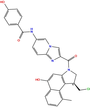
|
|||||
| Formula | C29H23ClN4O4 |
|||||
| Isosmiles | CC1=C2C(=CC=C1)C(=CC3=C2[C@@H](CN3C(=O)C4=CN5C=C(C=CC5=N4)NC(=O)C6=CC=C(C=C6)O)CCl)O |
|||||
| PubChem CID | ||||||
| InChI |
InChI=1S/C29H23ClN4O4/c1-16-3-2-4-21-24(36)11-23-27(26(16)21)18(12-30)13-34(23)29(38)22-15-33-14-19(7-10-25(33)32-22)31-28(37)17-5-8-20(35)9-6-17/h2-11,14-15,18,35-36H,12-13H2,1H3,(H,31,37)/t18-/m1/s1
|
|||||
| InChIKey |
VQAFBYLFRCCWNB-GOSISDBHSA-N
|
|||||
| IUPAC Name |
N-[2-[(1S)-1-(chloromethyl)-5-hydroxy-9-methyl-1,2-dihydrobenzo[e]indole-3-carbonyl]imidazo[1,2-a]pyridin-6-yl]-4-hydroxybenzamide
|
|||||
| Pharmaceutical Properties | Molecule Weight |
527 |
Polar area |
107 |
||
Complexity |
885 |
xlogp Value |
5.3 |
|||
Heavy Count |
38 |
Rot Bonds |
4 |
|||
Hbond acc |
5 |
Hbond Donor |
3 |
|||
The activity data of This Payload
| Standard Type | Value | Units | Cell line | Disease Model | Cell line ID | Reference |
|---|---|---|---|---|---|---|
| Half Maximal Inhibitory Concentration (IC50) | 0.09 | nM |
SK-BR-3 cells
|
Breast adenocarcinoma
|
[1] | |
| Half Maximal Inhibitory Concentration (IC50) | 0.09 | nM |
SW620 cells
|
Colon adenocarcinoma
|
[1] | |
| Half Maximal Inhibitory Concentration (IC50) | 0.43 | nM |
SK-OV-3 cells
|
Ovarian serous cystadenocarcinoma
|
[1] |
Each Antibody-drug Conjugate Related to This Payload
Full Information of The Activity Data of The ADC(s) Related to This Payload
Trastuzumab duocarmazine [New Drug Application (discontinued)]
Identified from the Human Clinical Data
| Experiment 1 Reporting the Activity Date of This ADC | [2] | ||||
| Efficacy Data | Progression Free Survival |
7 months
|
|||
| Patients Enrolled |
Eligible patients were HER2-positive, unresectable/metastatic breast cancer patients progressing after ≥2 HER2-targeted therapies (including ado-trastuzumab emtansine), with ECOG ≤2 and adequate organ function. Exclusions included prohibited medication use, hypersensitivity to HER2 agents, active pneumonitis, uncontrolled cardiovascular disease (LVEF <50% or elevated troponin), untreated/symptomatic brain metastases, or prior significant lung conditions. Measurable/evaluable disease per RECIST 1.1 was required.
Click to Show/Hide
|
||||
| Administration Dosage |
SYD985, every 3 weeks (Q3W)
|
||||
| Related Clinical Trial | |||||
| NCT Number | NCT03262935 | Phase Status | PHASE3 | ||
| Clinical Description |
A Multi-centre, Open-label, Randomized Clinical Trial Comparing the Efficacy and Safety of the Antibody-drug Conjugate SYD985 to Physician's Choice in Patients With HER2-positive Unresectable Locally Advanced or Metastatic Breast Cancer
|
||||
| Primary Endpoint |
Efficacy assessments include progression-free survival (PFS) by central review (RECIST v1.1) and death from randomization through 31March2021, overall survival (OS) monitored until 30June2022, and objective response rate (ORR) via complete/partial responses up to 31March2021. Investigator-assessed PFS and patient-reported quality of life (EORTC QLQ-C30) were also tracked through primary analysis.
Click to Show/Hide
|
||||
| Other Endpoint |
Key efficacy endpoints comprised central and investigator-evaluated PFS (both per RECIST v1.1) from randomization until progression/death (primary data cutoff: 31March2021), OS until death (final cutoff: 30June2022), and ORR. Quality of life was measured via EORTC QLQ-C30's 0-100 scale, with positive/negative changes indicating improvement/decline in health status.
Click to Show/Hide
|
||||
| Experiment 2 Reporting the Activity Date of This ADC | [3] | ||||
| Efficacy Data | Progression Free Survival |
9.4 months
|
|||
| Patients Enrolled |
Phase I (Part I) enrolls any advanced solid tumor patients refractory to standard therapy, while Phase II (Part II) focuses on HER2-positive breast/gastric/urothelial/endometrial cancers. Key requirements: ECOG ≤1, life expectancy >12 weeks, and adequate organ function. Critical exclusions: recent anthracyclines (<3 months) or cardiotoxicity (LVEF <55% /prior trastuzumab-related declines), active brain metastases, or hypersensitivity to HER2-targeted therapies.
Click to Show/Hide
|
||||
| Administration Dosage |
IV (in the vein) infusion every three weeks. Number of Cycles: until cancer progression or unacceptable toxicity develops. Different doses.
|
||||
| Related Clinical Trial | |||||
| NCT Number | NCT02277717 | Phase Status | PHASE1 | ||
| Clinical Description |
A Two Part First-in-human Phase I Study (With Expanded Cohorts) With the Antibody-drug Conjugate SYD985 to Evaluate the Safety, Pharmacokinetics and Efficacy in Patients With Locally Advanced or Metastatic Solid Tumors
|
||||
| Primary Endpoint |
The primary safety endpoint is incidence of dose-limiting toxicities during the first 21-day cycle, establishing the maximum tolerated dose.
|
||||
| Other Endpoint |
Secondary outcomes include long-term safety monitoring (AEs over 2 years), pharmacokinetic profiling (AUC and Cmax of SYD985 at multiple timepoints), hematologic/biochemical changes, immunogenicity (anti-drug antibodies), and antitumor activity (ORR assessed every two cycles).
|
||||
| Experiment 3 Reporting the Activity Date of This ADC | [3] | ||||
| Efficacy Data | Objective Response Rate (ORR) |
33%
|
|||
| Patients Enrolled |
Phase I (Part I) enrolls any advanced solid tumor patients refractory to standard therapy, while Phase II (Part II) focuses on HER2-positive breast/gastric/urothelial/endometrial cancers. Key requirements: ECOG ≤1, life expectancy >12 weeks, and adequate organ function. Critical exclusions: recent anthracyclines (<3 months) or cardiotoxicity (LVEF <55% /prior trastuzumab-related declines), active brain metastases, or hypersensitivity to HER2-targeted therapies.
Click to Show/Hide
|
||||
| Administration Dosage |
IV (in the vein) infusion every three weeks. Number of Cycles: until cancer progression or unacceptable toxicity develops. Different doses.
|
||||
| Related Clinical Trial | |||||
| NCT Number | NCT02277717 | Phase Status | PHASE1 | ||
| Clinical Description |
A Two Part First-in-human Phase I Study (With Expanded Cohorts) With the Antibody-drug Conjugate SYD985 to Evaluate the Safety, Pharmacokinetics and Efficacy in Patients With Locally Advanced or Metastatic Solid Tumors
|
||||
| Primary Endpoint |
The primary safety endpoint is incidence of dose-limiting toxicities during the first 21-day cycle, establishing the maximum tolerated dose.
|
||||
| Other Endpoint |
Secondary outcomes include long-term safety monitoring (AEs over 2 years), pharmacokinetic profiling (AUC and Cmax of SYD985 at multiple timepoints), hematologic/biochemical changes, immunogenicity (anti-drug antibodies), and antitumor activity (ORR assessed every two cycles).
|
||||
| Experiment 4 Reporting the Activity Date of This ADC | [4] | ||||
| Patients Enrolled |
Eligible patients are ≥18 with HER2-positive (IHC 1+ for single-arm; IHC 3+/ISH+ for randomized breast cancer cohort) unresectable/metastatic tumors, ECOG ≤1, and adequate organ function. Key exclusions: prohibited medications, trastuzumab hypersensitivity, keratitis, LVEF <50%, pulmonary/cardiovascular diseases, severe systemic illness, or active brain metastases needing steroids/surgery within 8 weeks. Contact lens use is prohibited during treatment.
Click to Show/Hide
|
||||
| Administration Dosage |
BYON5667 eye drops should be self-administered daily during waking hours. SYD985, every 3 weeks (Q3W)
|
||||
| Related Clinical Trial | |||||
| NCT Number | NCT04983238 | Phase Status | PHASE1|||PHASE2 | ||
| Clinical Description |
A Multicenter, Randomized, Double-blind, Placebo-controlled Trial With a Single Arm run-in Period to Evaluate the Safety and Efficacy of Sodium Thiosulfate (BYON5667) Eye Drops to Reduce Ocular Toxicity in Cancer Patients Treated With SYD985
|
||||
| Primary Endpoint |
The primary efficacy endpoint evaluates the percentage of patients experiencing SYD985-related ocular adverse events (Grade ≥1) by Day 63 after using BYON5667 eye drops.
|
||||
| Other Endpoint |
Secondary assessments include ocular toxicity severity (Day 63/126), tolerability via questionnaire (0-10 discomfort scale), visual function (NEI VFQ-25), time to first ocular AE, discontinuation rates due to toxicity, and SYD985 efficacy/safety endpoints (ORR, PFS, OS, and treatment-emergent AEs) monitored for up to 2 years.
|
||||
| Experiment 5 Reporting the Activity Date of This ADC | [5] | ||||
| Patients Enrolled |
Eligible patients (≥18 years) have HER2-positive/low advanced solid tumors (breast, gastroesophageal, etc.), 1-4 prior therapy lines (dependent on subtype), ECOG 0-2, adequate organ function, and measurable disease (expansion cohort). Exclusions: recent anthracyclines, uncontrolled illness, LVEF <50%, active pneumonitis, corneal disease, CNS metastases, or trastuzumab hypersensitivity. Contraception is mandatory.
Click to Show/Hide
|
||||
| Related Clinical Trial | |||||
| NCT Number | NCT04602117 | Phase Status | PHASE1 | ||
| Clinical Description |
ISPY-P1.01: Evaluating the Safety of Weekly Paclitaxel With Trastuzumab Duocarmazine (SYD985) in Patients With Metastatic Cancer: A Phase I/Ib Trial
|
||||
| Primary Endpoint |
Safety and tolerability of weekly paclitaxel plus tri-weekly trastuzumab duocarmazine (SYD985) will be assessed via CTCAE v5.0 (up to 12 months), focusing on toxicity rates, DLTs, MTD, and RP2D. Preliminary efficacy (RECIST 1.1) includes CBR and ORR at 6 months.
|
||||
| Other Endpoint |
Key efficacy endpoints (RECIST 1.1) are PFS and DOR (up to 12 months) in advanced solid tumor patients treated with paclitaxel-SYD985, alongside safety monitoring of treatment-related AEs.
|
||||
| Experiment 6 Reporting the Activity Date of This ADC | [6] | ||||
| Patients Enrolled |
Eligible patients have untreated, measurable (≥2.5 cm), stage II-III or non-metastatic stage IV breast cancer, ECOG 0-1, adequate organ function, and meet specific biomarker criteria (e.g., MammaPrint High or ER-/HER2+). Exclusions: prior chemotherapy/radiation, uncontrolled cardiac/medical conditions, pregnancy, or recent investigational drug use within 30 days. MRI-incompatible implants are prohibited.
Click to Show/Hide
|
||||
| Related Clinical Trial | |||||
| NCT Number | NCT01042379 | Phase Status | PHASE2 | ||
| Clinical Description |
I-SPY Trial (Investigation of Serial Studies to Predict Your Therapeutic Response With Imaging And moLecular Analysis 2)
|
||||
| Primary Endpoint |
The primary objective is to evaluate if adding experimental agents to standard neoadjuvant chemotherapy improves pathologic complete response (pCR) rates in breast cancer patients, assessed post-surgery after up to 36 weeks of treatment.
|
||||
| Other Endpoint |
Secondary goals include developing biomarker-based predictive models for pCR and residual cancer burden (RCB), tracking 3- and 5-year RFS/OS, monitoring treatment-related AEs/SAEs, and assessing tumor dynamics via MRI at four time points (baseline to pre-surgery).
|
||||
| Experiment 7 Reporting the Activity Date of This ADC | [7] | ||||
| Patients Enrolled |
Eligible patients are females with HER2-expressing (IHC 1+/2+/3+ or ISH+) recurrent/metastatic endometrial carcinoma, progressed after first-line platinum therapy (≤1 prior non-cytotoxic systemic therapy allowed). Key exclusions: LVEF <50%, uncontrolled cardiovascular/pulmonary disease, active brain metastases, or prior trastuzumab hypersensitivity. Measurable disease (RECIST 1.1) and ECOG ≤2 are required.
Click to Show/Hide
|
||||
| Administration Dosage |
SYD985, Intravenous, every 3 weeks (Q3W)
|
||||
| Related Clinical Trial | |||||
| NCT Number | NCT04205630 | Phase Status | PHASE2 | ||
| Clinical Description |
A Single-arm Phase II Trial to Evaluate the Safety and Efficacy of the Antibody-Drug Conjugate (ADC) SYD985 in Patients With Human Epidermal Growth Factor Receptor 2 (HER2)-Expressing Endometrial Carcinoma Who Previously Progressed on or After First Line Platinum-based Chemotherapy
|
||||
| Primary Endpoint |
The primary endpoint evaluates Objective Response Rate (ORR) over 2 years, defined as the proportion of patients achieving complete or partial responses per RECIST v1.1 criteria.The primary endpoint evaluates Objective Response Rate (ORR) over 2 years, defined as the proportion of patients achieving complete or partial responses per RECIST v1.1 criteria.
Click to Show/Hide
|
||||
| Other Endpoint |
Key secondary endpoints include Progression-Free Survival (PFS) and Overall Survival (OS) over 2 years, measuring time to progression/death and overall mortality, respectively. Treatment-emergent AEs will be graded via CTCAE v5.0.
|
||||
| Experiment 8 Reporting the Activity Date of This ADC | [8] | ||||
| Patients Enrolled |
Eligible patients (≥18 years) must have HER2-positive (IHC 1+) advanced/metastatic breast, ovarian, or endometrial cancer (Part 2) or any solid tumor (Part 1) after failing standard therapy, with measurable disease (RECIST 1.1), ECOG ≤1, and adequate organ function. Key exclusions: prior DUBA-ADCs, recent anthracyclines/anticancer therapies, LVEF <50%, significant cardiovascular/pulmonary disease, active brain metastases, or unresolved treatment-related toxicities (>Grade 1). Tumor biopsy (fresh or archival ≤6 months) is mandatory unless unavailable.
Click to Show/Hide
|
||||
| Administration Dosage |
SYD985, Intravenous, every 3 weeks (Q3W) Niraparib taken orally and either 100 mg, 200 mg or 300 mg once daily for either 1, 2 or 3 weeks.
|
||||
| Related Clinical Trial | |||||
| NCT Number | NCT04235101 | Phase Status | PHASE1 | ||
| Clinical Description |
A Two-part Phase I Study With the Antibody-drug Conjugate SYD985 in Combination With Niraparib to Evaluate Safety, Pharmacokinetics and Efficacy in Patients With HER2-expressing Locally Advanced or Metastatic Solid Tumors.
|
||||
| Primary Endpoint |
The primary objective is to evaluate dose-limiting toxicities during the first treatment cycle (21-day timeframe) to establish the safety and tolerability profile of the SYD985 and niraparib combination regimen.
|
||||
| Other Endpoint |
Secondary objectives include safety assessment (AE incidence over 2 years), pharmacokinetic evaluation (AUC and Cmax of SYD985/niraparib up to 6 months), immunogenicity (anti-SYD985 antibodies), hematologic/clinical chemistry changes, and efficacy (ORR assessed every 2 cycles initially, then every 4 cycles).
|
||||
| Experiment 9 Reporting the Activity Date of This ADC | [9] | ||||
| Related Clinical Trial | |||||
| NCT Number | NCT03262935 | Phase Status | Phase 3 | ||
| Clinical Description |
A multi-centre, open-label, randomized clinical trial comparing the efficacy and safety of the antibody-drug conjugate SYD985 to physician's choice in patients with HER2-positive unresectable locally advanced or metastatic breast cancer.
|
||||
| Experiment 10 Reporting the Activity Date of This ADC | [10] | ||||
| Related Clinical Trial | |||||
| NCT Number | NCT01042379 | Phase Status | Phase 2 | ||
| Clinical Description |
I-SPY trial (investigation of serial studies to predict your therapeutic response with imaging and molecular analysis 2).
|
||||
| Experiment 11 Reporting the Activity Date of This ADC | [11] | ||||
| Related Clinical Trial | |||||
| NCT Number | NCT04205630 | Phase Status | Phase 2 | ||
| Clinical Description |
A single-arm phase 2 trial to evaluate the safety and efficacy of the antibody-drug conjugate (ADC) SYD985 in patients with human epidermal growth factor receptor 2 (HER2)-expressing endometrial carcinoma who previously progressed on or after first line platinum-based chemotherapy.
|
||||
| Experiment 12 Reporting the Activity Date of This ADC | [12] | ||||
| Related Clinical Trial | |||||
| NCT Number | NCT02277717 | Phase Status | Phase 1 | ||
| Clinical Description |
A two part first-in-human phase 1 study (with expanded cohorts) with the antibody-drug conjugate SYD985 to evaluate the safety, pharmacokinetics and efficacy in patients with locally advanced or metastatic solid tumors.
|
||||
| Experiment 13 Reporting the Activity Date of This ADC | [13] | ||||
| Related Clinical Trial | |||||
| NCT Number | NCT04235101 | Phase Status | Phase 1 | ||
| Clinical Description |
A two-part phase 1 study with the antibody-drug conjugate SYD985 in combination with niraparib to evaluate safety, pharmacokinetics and efficacy in patients with HER2-expressing locally advanced or metastatic solid tumors.
|
||||
| Experiment 14 Reporting the Activity Date of This ADC | [14] | ||||
| Related Clinical Trial | |||||
| NCT Number | NCT04602117 | Phase Status | Phase 1 | ||
| Clinical Description |
ISPY-P1.01: evaluating the safety of weekly paclitaxel with trastuzumab duocarmazine (SYD985) in patients with metastatic cancer: a phase 1/Ib trial.
|
||||
| Experiment 15 Reporting the Activity Date of This ADC | [15] | ||||
| Related Clinical Trial | |||||
| NCT Number | NCT04983238 | Phase Status | Phase 1 | ||
| Clinical Description |
A multicenter, randomized, double-blind, placebo-controlled trial with a single arm run-in period to evaluate the safety and efficacy of sodium thiosulfate (BYON5667) Eye drops to reduce ocular toxicity in cancer patients treated with SYD985.
|
||||
Discovered Using Patient-derived Xenograft Model
| Experiment 1 Reporting the Activity Date of This ADC | [16] | ||||
| Efficacy Data | Tumor Growth Inhibition value (TGI) | ≈ 33.30% | High HER2 expression (HER2+++; IHC 3+; FISH+) | ||
| Method Description |
All treatments were conducted at day 0 by a single dose, i.v, 3 mg/kg x1 injection into the tail vein, Data, depicted as mean tumour volume, consists of 6-8 animals per experimental group.
|
||||
| In Vivo Model | Breast cancer PDX model (PDX: MAXF-1162) | ||||
| Experiment 2 Reporting the Activity Date of This ADC | [16] | ||||
| Efficacy Data | Tumor Growth Inhibition value (TGI) | ≈ 42.90% | Low HER2 expression (HER2+; IHC +; FISH-) | ||
| Method Description |
All treatments were conducted at day 0 by a single dose, i.v, 3 mg/kg x1 injection into the tail vein, Data, depicted as mean tumour volume, consists of 6-8 animals per experimental group.
|
||||
| In Vivo Model | Breast cancer PDX model (PDX: MAXF 449) | ||||
| Experiment 3 Reporting the Activity Date of This ADC | [16] | ||||
| Efficacy Data | Tumor Growth Inhibition value (TGI) | ≈ 65% | Moderate HER2 expression (HER2++; IHC 2+; FISH-) | ||
| Method Description |
All treatments were conducted at day 0 by a single dose, i.v, 3 mg/kg x1 injection into the tail vein, Data, depicted as mean tumour volume, consists of 6-8 animals per experimental group.
|
||||
| In Vivo Model | Breast cancer PDX model (PDX: HBCx-34) | ||||
| Experiment 4 Reporting the Activity Date of This ADC | [16] | ||||
| Efficacy Data | Tumor Growth Inhibition value (TGI) | ≈ 66.70% | Moderate HER2 expression (HER2++; IHC 2+; FISH-) | ||
| Method Description |
All treatments were conducted at day 0 by a single dose, i.v, 3 mg/kg x1 injection into the tail vein, Data, depicted as mean tumour volume, consists of 6-8 animals per experimental group.
|
||||
| In Vivo Model | Breast cancer PDX model (PDX: ST313) | ||||
| Experiment 5 Reporting the Activity Date of This ADC | [16] | ||||
| Efficacy Data | Tumor Growth Inhibition value (TGI) | ≈ 75% | Low HER2 expression (HER2+; IHC +; FISH-) | ||
| Method Description |
All treatments were conducted at day 0 by a single dose, i.v, 10 mg/kg x1 injection into the tail vein, Data, depicted as mean tumour volume, consists of 6-8 animals per experimental group.
|
||||
| In Vivo Model | Gastric cancer PDX model (PDX: GXA3057) | ||||
| Experiment 6 Reporting the Activity Date of This ADC | [16] | ||||
| Efficacy Data | Tumor Growth Inhibition value (TGI) | ≈ 90% | Moderate HER2 expression (HER2++; IHC 2+; FISH-) | ||
| Method Description |
All treatments were conducted at day 0 by a single dose, i.v, 10 mg/kg x1 injection into the tail vein, Data, depicted as mean tumour volume, consists of 6-8 animals per experimental group.
|
||||
| In Vivo Model | Gastric cancer PDX model (PDX: GXA3038) | ||||
| Experiment 7 Reporting the Activity Date of This ADC | [16] | ||||
| Efficacy Data | Tumor Growth Inhibition value (TGI) | ≈ 100% | High HER2 expression (HER2+++; IHC 3+; FISH+) | ||
| Method Description |
All treatments were conducted at day 0 by a single dose, i.v, 10 mg/kg x1 injection into the tail vein, Data, depicted as mean tumour volume, consists of 6-8 animals per experimental group.
|
||||
| In Vivo Model | Bladder cancer PDX model (PDX: BXF439) | ||||
| Experiment 8 Reporting the Activity Date of This ADC | [16] | ||||
| Efficacy Data | Tumor Growth Inhibition value (TGI) | ≈ 100% | High HER2 expression (HER2+++; IHC 3+; FISH+) | ||
| Method Description |
All treatments were conducted at day 0 by a single dose, i.v, 10 mg/kg x1 injection into the tail vein, Data, depicted as mean tumour volume, consists of 6-8 animals per experimental group.
|
||||
| In Vivo Model | Gastric cancer PDX model (PDX: GXA3054) | ||||
| Experiment 9 Reporting the Activity Date of This ADC | [16] | ||||
| Efficacy Data | Tumor Growth Inhibition value (TGI) | ≈ 100% | Moderate HER2 expression (HER2++; IHC 2+; FISH+) | ||
| Method Description |
All treatments were conducted at day 0 by a single dose, i.v, 10 mg/kg x1 injection into the tail vein, Data, depicted as mean tumour volume, consists of 6-8 animals per experimental group.
|
||||
| In Vivo Model | Gastric cancer PDX model (PDX: GXA3067) | ||||
| Experiment 10 Reporting the Activity Date of This ADC | [16] | ||||
| Efficacy Data | Tumor Growth Inhibition value (TGI) | ≈ 100% | Low HER2 expression (HER2+; IHC +; FISH-) | ||
| Method Description |
All treatments were conducted at day 0 by a single dose, i.v, 3 mg/kg x1 injection into the tail vein, Data, depicted as mean tumour volume, consists of 6-8 animals per experimental group.
|
||||
| In Vivo Model | Breast cancer PDX model (PDX: MAXF-MX1) | ||||
| Experiment 11 Reporting the Activity Date of This ADC | [16] | ||||
| Efficacy Data | Tumor Growth Inhibition value (TGI) | ≈ 100% | Low HER2 expression (HER2+; IHC +; FISH-) | ||
| Method Description |
All treatments were conducted at day 0 by a single dose, i.v, 3 mg/kg x1 injection into the tail vein, Data, depicted as mean tumour volume, consists of 6-8 animals per experimental group.
|
||||
| In Vivo Model | Breast cancer PDX model (PDX: HBCx-10) | ||||
Vobramitamab duocarmazine [Phase 2 (discontinued)]
Identified from the Human Clinical Data
| Experiment 1 Reporting the Activity Date of This ADC | [17] | ||||
| Patients Enrolled |
Eligible participants include those with prostate adenocarcinoma (Part 1; prior ARAT therapy permitted) or select squamous/small cell carcinomas (Part 2; 1-2 prior therapies). Key exclusions involve excessive prior treatments, active CNS metastases, B7-H3 therapy history, or contraindications to corticosteroids. Archival tissue and measurable disease are required. Contraception and baseline organ function criteria apply.
Click to Show/Hide
|
||||
| Administration Dosage |
MGC018 2.0, 2.7 mg/kg every 4 weeks
|
||||
| Related Clinical Trial | |||||
| NCT Number | NCT05551117 | Phase Status | PHASE2 | ||
| Clinical Description |
A Phase 2, Open-label, Study of Vobramitamab Duocarmazine in Participants With Metastatic Castration-resistant Prostate Cancer and Other Solid Tumors
|
||||
| Primary Endpoint |
In Part 1, radiographic progression-free survival (rPFS) will be assessed every 8-12 weeks for up to 2 years using PCWG3 criteria, with a landmark 6-month rPFS analysis. In Part 2, objective response rate (ORR) will be measured by RECIST 1.1 based on confirmed complete (CR) or partial responses (PR).
|
||||
| Other Endpoint |
Secondary endpoints include prostate-specific outcomes (PSA response, time to PSA progression, symptomatic skeletal events), tumor response metrics (ORR, duration of response, tumor size change), and safety (AEs, SAEs, immunogenicity). Assessments will be performed every 4-12 weeks per protocol-defined intervals.
|
||||
| Experiment 2 Reporting the Activity Date of This ADC | [18] | ||||
| Patients Enrolled |
Eligible patients must be ≥18 with platinum-refractory SCLC (including EGFR-mutant NSCLC-transformed SCLC), measurable disease (RECIST v1.1), ECOG 0-2, and adequate organ function. Key exclusions: untreated/symptomatic brain metastases, leptomeningeal disease, concurrent malignancies, or significant effusions. Women/men must adhere to strict contraception requirements. Archived/fresh tumor tissue is mandatory unless biopsy is medically unfeasible. Concurrent investigational agents or major surgery within 4 weeks pre-treatment are prohibited.
Click to Show/Hide
|
||||
| Administration Dosage |
Intravenous (IV) Infusion, 2.7 mg/kg on Day 1 of each 28 day cycle
|
||||
| Related Clinical Trial | |||||
| NCT Number | NCT06227546 | Phase Status | PHASE2 | ||
| Clinical Description |
A Phase II Study of MGC018 in Patients With Relapsed or Refractory Extensive-Stage Small-Cell Lung Cancer (ES-SCLC)
|
||||
| Primary Endpoint |
The primary efficacy endpoint is investigator-assessed Objective Response Rate (ORR) per RECIST v1.1, with tumor evaluations every 2 cycles (each 28 days) over 1 year.
|
||||
| Other Endpoint |
Safety assessments include treatment-emergent adverse events (per CTCAE v5.0) from treatment start until 60 days post-last dose (~1 year). Secondary efficacy measures include Duration of Response (DOR), median PFS, 6-month PFS (both assessed via Kaplan-Meier over 1 year), and Overall Survival (OS) monitored for 3 years.
|
||||
| Experiment 3 Reporting the Activity Date of This ADC | [19] | ||||
| Related Clinical Trial | |||||
| NCT Number | NCT05551117 | Phase Status | Phase 2 | ||
| Clinical Description |
A phase 2, randomized, open-label, study of two dose levels of obramitamab duocarmazine in participants with metastatic castration-resistant prostate cancer.
|
||||
| Experiment 4 Reporting the Activity Date of This ADC | [20] | ||||
| Patients Enrolled |
Patients of multiple tumor types, which included 3 melanoma patients refractory to 2 prior lines of checkpoint therapy.
|
||||
| Administration Dosage |
6 dose cohorts (0.50-4.00 mg/kg) every 3 weeks.
|
||||
| Related Clinical Trial | |||||
| NCT Number | NCT03729596 | Phase Status | Phase 1/2 | ||
| Clinical Description |
A phase 1/2, first-in-human, open-label, dose-escalation study of MGC018 (anti-B7-H3 antibody drug conjugate) alone and in combination with MGA012 (anti-PD-1 antibody) in patients with advanced solid tumors.
|
||||
| Experiment 5 Reporting the Activity Date of This ADC | [21] | ||||
| Related Clinical Trial | |||||
| NCT Number | NCT05293496 | Phase Status | Phase 1 | ||
| Clinical Description |
A phase 1/1b dose escalation and cohort expansion study of MGC018 in combination with checkpoint inhibitor in participants with advanced solid tumors.
|
||||
Discovered Using Patient-derived Xenograft Model
| Experiment 1 Reporting the Activity Date of This ADC | [22] | ||||
| Efficacy Data | Tumor Growth Inhibition value (TGI) | ≈ 96.10% | High MET expression (MET+++; IHC H-score=265) | ||
| Method Description |
Female Athymic Nude-Foxn1nu from Envigo (head and neck,triple-negative breast cancer models),68 weeks of age,were used for the PDX studies. Low passage tumor fragments were implanted into stock animals. When tumors reached 1.01.5 cm3,they were reimplanted into prestudy animals unilaterally on the left flank. When tumors reached an average tumor volume of 150-300 mm3,animals were matched by tumor volume into treatment or vehicle control groups. Three animals were assigned to each group and dosed intravenously by tail vein injection (10 mL/kg). Tumor volumes were measured twice weekly by calipers. Prostate cancer subcutaneous PDX model treated with MGC018 or control ADC at 3 mg/kg (QW3).
Click to Show/Hide
|
||||
| In Vivo Model | Pancreatic cancer PDX model (PDX: PDX-PAX-13565) | ||||
| Experiment 2 Reporting the Activity Date of This ADC | [22] | ||||
| Efficacy Data | Tumor Growth Inhibition value (TGI) | ≈ 97.60% | Moderate MET expression (MET++; IHC H-score=130) | ||
| Method Description |
Male NOG mice from Taconic (prostate cancer model),68 weeks of age,were used for the PDX studies. Low passage tumor fragments were implanted into stock animals. When tumors reached 1.01.5 cm3,they were reimplanted into prestudy animals unilaterally on the left flank. When tumors reached an average tumor volume of 150-300 mm3,animals were matched by tumor volume into treatment or vehicle control groups. Three animals were assigned to each group and dosed intravenously by tail vein injection (10 mL/kg). Tumor volumes were measured twice weekly by calipers. Head and neck cancer subcutaneous PDX model treated with MGC018 or control ADC at 3 mg/kg (Q2W 2).
Click to Show/Hide
|
||||
| In Vivo Model | Head and neck cancer PDX model | ||||
| Experiment 3 Reporting the Activity Date of This ADC | [22] | ||||
| Efficacy Data | Tumor Growth Inhibition value (TGI) | ≈ 98.20% | High MET expression (MET+++; IHC H-score=240) | ||
| Method Description |
Female Athymic Nude-Foxn1nu from Envigo (head and neck,triple-negative breast cancer models),68 weeks of age,were used for the PDX studies. Low passage tumor fragments were implanted into stock animals. When tumors reached 1.01.5 cm3,they were reimplanted into prestudy animals unilaterally on the left flank. When tumors reached an average tumor volume of 150-300 mm3,animals were matched by tumor volume into treatment or vehicle control groups. Three animals were assigned to each group and dosed intravenously by tail vein injection (10 mL/kg). Tumor volumes were measured twice weekly by calipers. Triple-negative breast cancer subcutaneous PDX model treated with MGC018 or control ADC at 3 mg/kg (QW2).
Click to Show/Hide
|
||||
| In Vivo Model | Triple-negative breast cancer PDX model | ||||
Discovered Using Cell Line-derived Xenograft Model
| Experiment 1 Reporting the Activity Date of This ADC | [22] | ||||
| Efficacy Data | Tumor Growth Inhibition value (TGI) | ≈ 22.40% | High MET expression (MET+++; IHC H-score=287) | ||
| Method Description |
Human tumor cells (5x10⁶) were resuspended in 1:1 medium (DMEM/F-12) and Matrigel basement membrane matrix (Corning) and implanted subcutaneously into the flank (A375.S2,Calu-6,PA-1) or mammary fat pad (MDA-MB-468) of mice. Mice were randomized into groups of 5-7 individuals per group. ADCs or vehicle control (PBS) were administered intravenously by tail vein injection (10 mL/kg) following growth of established tumors (100-150 mm3). Calu-6 lung cancer subcutaneous xenografts were treated with MGC018 or control ADC at 0.3 mg/kg QWx4.
Click to Show/Hide
|
||||
| In Vivo Model | Lung cancer CDX model | ||||
| In Vitro Model | Lung adenocarcinoma | Calu-6 cells | CVCL_0236 | ||
| Experiment 2 Reporting the Activity Date of This ADC | [22] | ||||
| Efficacy Data | Tumor Growth Inhibition value (TGI) | ≈ 30.10% | Moderate MET expression (MET++; IHC H-score=150) | ||
| Method Description |
Human tumor cells (5x10⁶) were resuspended in 1:1 medium (DMEM/F-12) and Matrigel basement membrane matrix (Corning) and implanted subcutaneously into the flank (A375.S2,Calu-6,PA-1) or mammary fat pad (MDA-MB-468) of mice. Mice were randomized into groups of 5-7 individuals per group. ADCs or vehicle control (PBS) were administered intravenously by tail vein injection (10 mL/kg) following growth of established tumors (100-150 mm3). PA-1 ovarian cancer subcutaneous xenografts were treated with MGC018 or control ADC at 3 mg/kg as a single dose,QW1.
Click to Show/Hide
|
||||
| In Vivo Model | Ovarian cancer CDX model | ||||
| In Vitro Model | Ovarian mixed germ cell tumor | PA-1 cells | CVCL_0479 | ||
| Experiment 3 Reporting the Activity Date of This ADC | [22] | ||||
| Efficacy Data | Tumor Growth Inhibition value (TGI) | ≈ 42.90% | High MET expression (MET+++; IHC H-score=287) | ||
| Method Description |
Human tumor cells (5x10⁶) were resuspended in 1:1 medium (DMEM/F-12) and Matrigel basement membrane matrix (Corning) and implanted subcutaneously into the flank (A375.S2,Calu-6,PA-1) or mammary fat pad (MDA-MB-468) of mice. Mice were randomized into groups of 5-7 individuals per group. ADCs or vehicle control (PBS) were administered intravenously by tail vein injection (10 mL/kg) following growth of established tumors (100-150 mm3). A-1 ovarian cancer subcutaneous xenografts were treated with MGC018 or control ADC at 3 mg/kg.
Click to Show/Hide
|
||||
| In Vivo Model | Ovarian cancer CDX model | ||||
| In Vitro Model | Ovarian mixed germ cell tumor | PA-1 cells | CVCL_0479 | ||
| Experiment 4 Reporting the Activity Date of This ADC | [22] | ||||
| Efficacy Data | Tumor Growth Inhibition value (TGI) | ≈ 43.90% | NegativeCD276 expression (CD276-) | ||
| Method Description |
Human tumor cells (5x10⁶) were resuspended in 1:1 medium (DMEM/F-12) and Matrigel basement membrane matrix (Corning) and implanted subcutaneously into the flank (A375.S2,Calu-6,PA-1) or mammary fat pad (MDA-MB-468) of mice. Mice were randomized into groups of 5-7 individuals per group. ADCs or vehicle control (PBS) were administered intravenously by tail vein injection (10 mL/kg) following growth of established tumors (100-150 mm3). MDA-MB-468 triple-negative breast cancer orthotopic xenografts were treated with MGC018 or control ADC at 3 mg/kg.
Click to Show/Hide
|
||||
| In Vivo Model | Triple-negative breast cancer CDX model | ||||
| In Vitro Model | Breast adenocarcinoma | MDA-MB-468 Luc cells | CVCL_0419 | ||
| Experiment 5 Reporting the Activity Date of This ADC | [22] | ||||
| Efficacy Data | Tumor Growth Inhibition value (TGI) | ≈ 59.30% | High MET expression (MET+++; IHC H-score=255) | ||
| Method Description |
Human tumor cells (5x10⁶) were resuspended in 1:1 medium (DMEM/F-12) and Matrigel basement membrane matrix (Corning) and implanted subcutaneously into the flank (A375.S2,Calu-6,PA-1) or mammary fat pad (MDA-MB-468) of mice. Mice were randomized into groups of 5-7 individuals per group. ADCs or vehicle control (PBS) were administered intravenously by tail vein injection (10 mL/kg) following growth of established tumors (100-150 mm3). Calu-6 lung cancer subcutaneous xenografts were treated with MGC018 or control ADC at 3 mg/kg.
Click to Show/Hide
|
||||
| In Vivo Model | Lung cancer CDX model | ||||
| In Vitro Model | Lung adenocarcinoma | Calu-6 cells | CVCL_0236 | ||
| Experiment 6 Reporting the Activity Date of This ADC | [22] | ||||
| Efficacy Data | Tumor Growth Inhibition value (TGI) | ≈ 62.70% | Moderate MET expression (MET++; IHC H-score=150) | ||
| Method Description |
Human tumor cells (5x10⁶) were resuspended in 1:1 medium (DMEM/F-12) and Matrigel basement membrane matrix (Corning) and implanted subcutaneously into the flank (A375.S2,Calu-6,PA-1) or mammary fat pad (MDA-MB-468) of mice. Mice were randomized into groups of 5-7 individuals per group. ADCs or vehicle control (PBS) were administered intravenously by tail vein injection (10 mL/kg) following growth of established tumors (100-150 mm3).MDA-MB-468 triple-negative breast cancer orthotopic xenografts were treated with MGC018 or control ADC at 0.3 mg/kg QW4.
Click to Show/Hide
|
||||
| In Vivo Model | Triple-negative breast cancer CDX model | ||||
| In Vitro Model | Breast adenocarcinoma | MDA-MB-468 Luc cells | CVCL_0419 | ||
| Experiment 7 Reporting the Activity Date of This ADC | [22] | ||||
| Efficacy Data | Tumor Growth Inhibition value (TGI) | ≈ 62.80% | High MET expression (MET+++; IHC H-score=287) | ||
| Method Description |
Human tumor cells (5x10⁶) were resuspended in 1:1 medium (DMEM/F-12) and Matrigel basement membrane matrix (Corning) and implanted subcutaneously into the flank (A375.S2,Calu-6,PA-1) or mammary fat pad (MDA-MB-468) of mice. Mice were randomized into groups of 5-7 individuals per group. ADCs or vehicle control (PBS) were administered intravenously by tail vein injection (10 mL/kg) following growth of established tumors (100-150 mm3). PA-1 ovarian cancer subcutaneous xenografts were treated with MGC018 or control ADC at 0.3 mg/kg QW4.
Click to Show/Hide
|
||||
| In Vivo Model | Ovarian cancer CDX model | ||||
| In Vitro Model | Ovarian mixed germ cell tumor | PA-1 cells | CVCL_0479 | ||
| Experiment 8 Reporting the Activity Date of This ADC | [22] | ||||
| Efficacy Data | Tumor Growth Inhibition value (TGI) | ≈ 66.50% | High MET expression (MET+++; IHC H-score=255) | ||
| Method Description |
Human tumor cells (5x10⁶) were resuspended in 1:1 medium (DMEM/F-12) and Matrigel basement membrane matrix (Corning) and implanted subcutaneously into the flank (A375.S2,Calu-6,PA-1) or mammary fat pad (MDA-MB-468) of mice. Mice were randomized into groups of 5-7 individuals per group. ADCs or vehicle control (PBS) were administered intravenously by tail vein injection (10 mL/kg) following growth of established tumors (100-150 mm3). Calu-6 lung cancer subcutaneous xenografts were treated with MGC018 or control ADC at 3 mg/kg.
Click to Show/Hide
|
||||
| In Vivo Model | Lung cancer CDX model | ||||
| In Vitro Model | Lung adenocarcinoma | Calu-6 cells | CVCL_0236 | ||
| Experiment 9 Reporting the Activity Date of This ADC | [22] | ||||
| Efficacy Data | Tumor Growth Inhibition value (TGI) | ≈ 71.90% | High MET expression (MET+++; IHC H-score=255) | ||
| Method Description |
Human tumor cells (5x10⁶) were resuspended in 1:1 medium (DMEM/F-12) and Matrigel basement membrane matrix (Corning) and implanted subcutaneously into the flank (A375.S2,Calu-6,PA-1) or mammary fat pad (MDA-MB-468) of mice. Mice were randomized into groups of 5-7 individuals per group. ADCs or vehicle control (PBS) were administered intravenously by tail vein injection (10 mL/kg) following growth of established tumors (100-150 mm3). Calu-6 lung cancer subcutaneous xenografts were treated with MGC018 or control ADC at 1 mg/kg QWx4.
Click to Show/Hide
|
||||
| In Vivo Model | Lung cancer CDX model | ||||
| In Vitro Model | Lung adenocarcinoma | Calu-6 cells | CVCL_0236 | ||
| Experiment 10 Reporting the Activity Date of This ADC | [22] | ||||
| Efficacy Data | Tumor Growth Inhibition value (TGI) | ≈ 76.30% | High MET expression (MET+++; IHC H-score=255) | ||
| Method Description |
Human tumor cells (5x10⁶) were resuspended in 1:1 medium (DMEM/F-12) and Matrigel basement membrane matrix (Corning) and implanted subcutaneously into the flank (A375.S2,Calu-6,PA-1) or mammary fat pad (MDA-MB-468) of mice. Mice were randomized into groups of 5-7 individuals per group. ADCs or vehicle control (PBS) were administered intravenously by tail vein injection (10 mL/kg) following growth of established tumors (100-150 mm3). Calu-6 lung cancer subcutaneous xenografts were treated with MGC018 or control ADC at 6 mg/kg.
Click to Show/Hide
|
||||
| In Vivo Model | Lung cancer CDX model | ||||
| In Vitro Model | Lung adenocarcinoma | Calu-6 cells | CVCL_0236 | ||
| Experiment 11 Reporting the Activity Date of This ADC | [22] | ||||
| Efficacy Data | Tumor Growth Inhibition value (TGI) | ≈ 76.60% | High MET expression (MET+++; IHC H-score=255) | ||
| Method Description |
Human tumor cells (5x10⁶) were resuspended in 1:1 medium (DMEM/F-12) and Matrigel basement membrane matrix (Corning) and implanted subcutaneously into the flank (A375.S2,Calu-6,PA-1) or mammary fat pad (MDA-MB-468) of mice. Mice were randomized into groups of 5-7 individuals per group. ADCs or vehicle control (PBS) were administered intravenously by tail vein injection (10 mL/kg) following growth of established tumors (100-150 mm3). A375.S2 melanoma subcutaneous xenografts were treated with MGC018 or control ADC at 0.3 mg/kg QW4.
Click to Show/Hide
|
||||
| In Vivo Model | Melanoma CDX model | ||||
| In Vitro Model | Amelanotic melanoma | A375.S2 cells | CVCL_0136 | ||
| Experiment 12 Reporting the Activity Date of This ADC | [22] | ||||
| Efficacy Data | Tumor Growth Inhibition value (TGI) | ≈ 82.80% | High MET expression (MET+++; IHC H-score=255) | ||
| Method Description |
Human tumor cells (5x10⁶) were resuspended in 1:1 medium (DMEM/F-12) and Matrigel basement membrane matrix (Corning) and implanted subcutaneously into the flank (A375.S2,Calu-6,PA-1) or mammary fat pad (MDA-MB-468) of mice. Mice were randomized into groups of 5-7 individuals per group. ADCs or vehicle control (PBS) were administered intravenously by tail vein injection (10 mL/kg) following growth of established tumors (100-150 mm3). Calu-6 lung cancer subcutaneous xenografts were treated with MGC018 or control ADC at 3 mg/kg QWx4.
Click to Show/Hide
|
||||
| In Vivo Model | Lung cancer CDX model | ||||
| In Vitro Model | Lung adenocarcinoma | Calu-6 cells | CVCL_0236 | ||
| Experiment 13 Reporting the Activity Date of This ADC | [22] | ||||
| Efficacy Data | Tumor Growth Inhibition value (TGI) | ≈ 83.90% | High MET expression (MET+++; IHC H-score=265) | ||
| Method Description |
Human tumor cells (5x10⁶) were resuspended in 1:1 medium (DMEM/F-12) and Matrigel basement membrane matrix (Corning) and implanted subcutaneously into the flank (A375.S2,Calu-6,PA-1) or mammary fat pad (MDA-MB-468) of mice. Mice were randomized into groups of 5-7 individuals per group. ADCs or vehicle control (PBS) were administered intravenously by tail vein injection (10 mL/kg) following growth of established tumors (100-150 mm3). A375.S2 melanoma subcutaneous xenografts were treated with MGC018 or control ADC at 1 mg/kg.
Click to Show/Hide
|
||||
| In Vivo Model | Melanoma CDX model | ||||
| In Vitro Model | Amelanotic melanoma | A375.S2 cells | CVCL_0136 | ||
| Experiment 14 Reporting the Activity Date of This ADC | [22] | ||||
| Efficacy Data | Tumor Growth Inhibition value (TGI) | ≈ 87.50% | High MET expression (MET+++; IHC H-score=255) | ||
| Method Description |
Human tumor cells (5x10⁶) were resuspended in 1:1 medium (DMEM/F-12) and Matrigel basement membrane matrix (Corning) and implanted subcutaneously into the flank (A375.S2,Calu-6,PA-1) or mammary fat pad (MDA-MB-468) of mice. Mice were randomized into groups of 5-7 individuals per group. ADCs or vehicle control (PBS) were administered intravenously by tail vein injection (10 mL/kg) following growth of established tumors (100-150 mm3). Calu-6 lung cancer subcutaneous xenografts were treated with MGC018 or control ADC at 10 mg/kg.
Click to Show/Hide
|
||||
| In Vivo Model | Lung cancer CDX model | ||||
| In Vitro Model | Lung adenocarcinoma | Calu-6 cells | CVCL_0236 | ||
| Experiment 15 Reporting the Activity Date of This ADC | [22] | ||||
| Efficacy Data | Tumor Growth Inhibition value (TGI) | ≈ 88.80% | High MET expression (MET+++; IHC H-score=287) | ||
| Method Description |
Human tumor cells (5x10⁶) were resuspended in 1:1 medium (DMEM/F-12) and Matrigel basement membrane matrix (Corning) and implanted subcutaneously into the flank (A375.S2,Calu-6,PA-1) or mammary fat pad (MDA-MB-468) of mice. Mice were randomized into groups of 5-7 individuals per group. ADCs or vehicle control (PBS) were administered intravenously by tail vein injection (10 mL/kg) following growth of established tumors (100-150 mm3). A-1 ovarian cancer subcutaneous xenografts were treated with MGC018 or control ADC at 6 mg/kg.
Click to Show/Hide
|
||||
| In Vivo Model | Ovarian cancer CDX model | ||||
| In Vitro Model | Ovarian mixed germ cell tumor | PA-1 cells | CVCL_0479 | ||
| Experiment 16 Reporting the Activity Date of This ADC | [22] | ||||
| Efficacy Data | Tumor Growth Inhibition value (TGI) | ≈ 90.50% | High MET expression (MET+++; IHC H-score=287) | ||
| Method Description |
Human tumor cells (5x10⁶) were resuspended in 1:1 medium (DMEM/F-12) and Matrigel basement membrane matrix (Corning) and implanted subcutaneously into the flank (A375.S2,Calu-6,PA-1) or mammary fat pad (MDA-MB-468) of mice. Mice were randomized into groups of 5-7 individuals per group. ADCs or vehicle control (PBS) were administered intravenously by tail vein injection (10 mL/kg) following growth of established tumors (100-150 mm3). A-1 ovarian cancer subcutaneous xenografts were treated with MGC018 or control ADC at 10 mg/kg.
Click to Show/Hide
|
||||
| In Vivo Model | Ovarian cancer CDX model | ||||
| In Vitro Model | Ovarian mixed germ cell tumor | PA-1 cells | CVCL_0479 | ||
| Experiment 17 Reporting the Activity Date of This ADC | [22] | ||||
| Efficacy Data | Tumor Growth Inhibition value (TGI) | ≈ 91.40% | High MET expression (MET+++; IHC H-score=287) | ||
| Method Description |
Human tumor cells (5x10⁶) were resuspended in 1:1 medium (DMEM/F-12) and Matrigel basement membrane matrix (Corning) and implanted subcutaneously into the flank (A375.S2,Calu-6,PA-1) or mammary fat pad (MDA-MB-468) of mice. Mice were randomized into groups of 5-7 individuals per group. ADCs or vehicle control (PBS) were administered intravenously by tail vein injection (10 mL/kg) following growth of established tumors (100-150 mm3). PA-1 ovarian cancer subcutaneous xenografts were treated with MGC018 or control ADC at 3 mg/kg as a single dose,QW4.
Click to Show/Hide
|
||||
| In Vivo Model | Ovarian cancer CDX model | ||||
| In Vitro Model | Ovarian mixed germ cell tumor | PA-1 cells | CVCL_0479 | ||
| Experiment 18 Reporting the Activity Date of This ADC | [22] | ||||
| Efficacy Data | Tumor Growth Inhibition value (TGI) | ≈ 94.60% | High MET expression (MET+++; IHC H-score=287) | ||
| Method Description |
Human tumor cells (5x10⁶) were resuspended in 1:1 medium (DMEM/F-12) and Matrigel basement membrane matrix (Corning) and implanted subcutaneously into the flank (A375.S2,Calu-6,PA-1) or mammary fat pad (MDA-MB-468) of mice. Mice were randomized into groups of 5-7 individuals per group. ADCs or vehicle control (PBS) were administered intravenously by tail vein injection (10 mL/kg) following growth of established tumors (100-150 mm3). PA-1 ovarian cancer subcutaneous xenografts were treated with MGC018 or control ADC at 1 mg/kg QW4.
Click to Show/Hide
|
||||
| In Vivo Model | Ovarian cancer CDX model | ||||
| In Vitro Model | Ovarian mixed germ cell tumor | PA-1 cells | CVCL_0479 | ||
| Experiment 19 Reporting the Activity Date of This ADC | [22] | ||||
| Efficacy Data | Tumor Growth Inhibition value (TGI) | ≈ 96.70% | High MET expression (MET+++; IHC H-score=255) | ||
| Method Description |
Human tumor cells (5x10⁶) were resuspended in 1:1 medium (DMEM/F-12) and Matrigel basement membrane matrix (Corning) and implanted subcutaneously into the flank (A375.S2,Calu-6,PA-1) or mammary fat pad (MDA-MB-468) of mice. Mice were randomized into groups of 5-7 individuals per group. ADCs or vehicle control (PBS) were administered intravenously by tail vein injection (10 mL/kg) following growth of established tumors (100-150 mm3). Calu-6 lung cancer subcutaneous xenografts were treated with MGC018 or control ADC at 6 mg/kg.
Click to Show/Hide
|
||||
| In Vivo Model | Lung cancer CDX model | ||||
| In Vitro Model | Lung adenocarcinoma | Calu-6 cells | CVCL_0236 | ||
| Experiment 20 Reporting the Activity Date of This ADC | [22] | ||||
| Efficacy Data | Tumor Growth Inhibition value (TGI) | ≈ 97% | Moderate MET expression (MET++; IHC H-score=150) | ||
| Method Description |
Human tumor cells (5x10⁶) were resuspended in 1:1 medium (DMEM/F-12) and Matrigel basement membrane matrix (Corning) and implanted subcutaneously into the flank (A375.S2,Calu-6,PA-1) or mammary fat pad (MDA-MB-468) of mice. Mice were randomized into groups of 5-7 individuals per group. ADCs or vehicle control (PBS) were administered intravenously by tail vein injection (10 mL/kg) following growth of established tumors (100-150 mm3).MDA-MB-468 triple-negative breast cancer orthotopic xenografts were treated with MGC018 or control ADC at 1 mg/kg QW4.
Click to Show/Hide
|
||||
| In Vivo Model | Triple-negative breast cancer CDX model | ||||
| In Vitro Model | Breast adenocarcinoma | MDA-MB-468 Luc cells | CVCL_0419 | ||
| Experiment 21 Reporting the Activity Date of This ADC | [22] | ||||
| Efficacy Data | Tumor Growth Inhibition value (TGI) | ≈ 97.60% | High MET expression (MET+++; IHC H-score=287) | ||
| Method Description |
Human tumor cells (5x10⁶) were resuspended in 1:1 medium (DMEM/F-12) and Matrigel basement membrane matrix (Corning) and implanted subcutaneously into the flank (A375.S2,Calu-6,PA-1) or mammary fat pad (MDA-MB-468) of mice. Mice were randomized into groups of 5-7 individuals per group. ADCs or vehicle control (PBS) were administered intravenously by tail vein injection (10 mL/kg) following growth of established tumors (100-150 mm3). PA-1 ovarian cancer subcutaneous xenografts were treated with MGC018 or control ADC at 3 mg/kg as a single dose,Q2W4.
Click to Show/Hide
|
||||
| In Vivo Model | Ovarian cancer CDX model | ||||
| In Vitro Model | Ovarian mixed germ cell tumor | PA-1 cells | CVCL_0479 | ||
| Experiment 22 Reporting the Activity Date of This ADC | [22] | ||||
| Efficacy Data | Tumor Growth Inhibition value (TGI) | ≈ 97.90% | Moderate MET expression (MET++; IHC H-score=150) | ||
| Method Description |
Human tumor cells (5x10⁶) were resuspended in 1:1 medium (DMEM/F-12) and Matrigel basement membrane matrix (Corning) and implanted subcutaneously into the flank (A375.S2,Calu-6,PA-1) or mammary fat pad (MDA-MB-468) of mice. Mice were randomized into groups of 5-7 individuals per group. ADCs or vehicle control (PBS) were administered intravenously by tail vein injection (10 mL/kg) following growth of established tumors (100-150 mm3). MDA-MB-468 triple-negative breast cancer orthotopic xenografts were treated with MGC018 or control ADC at 6 mg/kg.
Click to Show/Hide
|
||||
| In Vivo Model | Triple-negative breast cancer CDX model | ||||
| In Vitro Model | Breast adenocarcinoma | MDA-MB-468 Luc cells | CVCL_0419 | ||
| Experiment 23 Reporting the Activity Date of This ADC | [22] | ||||
| Efficacy Data | Tumor Growth Inhibition value (TGI) | ≈ 98.10% | High MET expression (MET+++; IHC H-score=265) | ||
| Method Description |
Human tumor cells (5x10⁶) were resuspended in 1:1 medium (DMEM/F-12) and Matrigel basement membrane matrix (Corning) and implanted subcutaneously into the flank (A375.S2,Calu-6,PA-1) or mammary fat pad (MDA-MB-468) of mice. Mice were randomized into groups of 5-7 individuals per group. ADCs or vehicle control (PBS) were administered intravenously by tail vein injection (10 mL/kg) following growth of established tumors (100-150 mm3). A375.S2 melanoma subcutaneous xenografts were treated with MGC018 or control ADC at 3 mg/kg.
Click to Show/Hide
|
||||
| In Vivo Model | Melanoma CDX model | ||||
| In Vitro Model | Amelanotic melanoma | A375.S2 cells | CVCL_0136 | ||
| Experiment 24 Reporting the Activity Date of This ADC | [22] | ||||
| Efficacy Data | Tumor Growth Inhibition value (TGI) | ≈ 98.60% | High MET expression (MET+++; IHC H-score=255) | ||
| Method Description |
Human tumor cells (5x10⁶) were resuspended in 1:1 medium (DMEM/F-12) and Matrigel basement membrane matrix (Corning) and implanted subcutaneously into the flank (A375.S2,Calu-6,PA-1) or mammary fat pad (MDA-MB-468) of mice. Mice were randomized into groups of 5-7 individuals per group. ADCs or vehicle control (PBS) were administered intravenously by tail vein injection (10 mL/kg) following growth of established tumors (100-150 mm3). Calu-6 lung cancer subcutaneous xenografts were treated with MGC018 or control ADC at 10 mg/kg.
Click to Show/Hide
|
||||
| In Vivo Model | Lung cancer CDX model | ||||
| In Vitro Model | Lung adenocarcinoma | Calu-6 cells | CVCL_0236 | ||
| Experiment 25 Reporting the Activity Date of This ADC | [22] | ||||
| Efficacy Data | Tumor Growth Inhibition value (TGI) | ≈ 99.70% | High MET expression (MET+++; IHC H-score=265) | ||
| Method Description |
Human tumor cells (5x10⁶) were resuspended in 1:1 medium (DMEM/F-12) and Matrigel basement membrane matrix (Corning) and implanted subcutaneously into the flank (A375.S2,Calu-6,PA-1) or mammary fat pad (MDA-MB-468) of mice. Mice were randomized into groups of 5-7 individuals per group. ADCs or vehicle control (PBS) were administered intravenously by tail vein injection (10 mL/kg) following growth of established tumors (100-150 mm3). A375.S2 melanoma subcutaneous xenografts were treated with MGC018 or control ADC at 1 mg/kg QW4.
Click to Show/Hide
|
||||
| In Vivo Model | Melanoma CDX model | ||||
| In Vitro Model | Amelanotic melanoma | A375.S2 cells | CVCL_0136 | ||
Revealed Based on the Cell Line Data
| Experiment 1 Reporting the Activity Date of This ADC | [22] | ||||
| Efficacy Data | Half Maximal Inhibitory Concentration (IC50) |
181 pM
|
High CD276 expression (CD276+++; 138,000 CD276 molecules/cell) | ||
| Method Description |
MGC018-mediated in vitro cytotoxicity was evaluated across a set of tumor cell lines representing multiple cancer types expressing varying levels of B7-H3. MDA-MB-468,A375.S2,PA-1,Calu-6,Hs700T,SW48,and LN-229 tumor cell lines were obtained from ATCC and cultured in DMEM/F-12 media containing 10% FBS. NCI-H1703 and Raji tumor cell lines were obtained from ATCC and cultured in RPMI1640 media containing 10% FBS.
Click to Show/Hide
|
||||
| In Vitro Model | Amelanotic melanoma | A375.S2 cells | CVCL_0136 | ||
| Experiment 2 Reporting the Activity Date of This ADC | [22] | ||||
| Efficacy Data | Half Maximal Inhibitory Concentration (IC50) |
260 pM
|
High CD276 expression (CD276+++; 139,000 CD276 molecules/cell) | ||
| Method Description |
MGC018-mediated in vitro cytotoxicity was evaluated across a set of tumor cell lines representing multiple cancer types expressing varying levels of B7-H3. MDA-MB-468,A375.S2,PA-1,Calu-6,Hs700T,SW48,and LN-229 tumor cell lines were obtained from ATCC and cultured in DMEM/F-12 media containing 10% FBS. NCI-H1703 and Raji tumor cell lines were obtained from ATCC and cultured in RPMI1640 media containing 10% FBS.
Click to Show/Hide
|
||||
| In Vitro Model | Lung adenocarcinoma | Calu-6 cells | CVCL_0236 | ||
| Experiment 3 Reporting the Activity Date of This ADC | [22] | ||||
| Efficacy Data | Half Maximal Inhibitory Concentration (IC50) |
275 pM
|
High CD276 expression (CD276+++; 310,000 CD276 molecules/cell) | ||
| Method Description |
MGC018-mediated in vitro cytotoxicity was evaluated across a set of tumor cell lines representing multiple cancer types expressing varying levels of B7-H3. MDA-MB-468,A375.S2,PA-1,Calu-6,Hs700T,SW48,and LN-229 tumor cell lines were obtained from ATCC and cultured in DMEM/F-12 media containing 10% FBS. NCI-H1703 and Raji tumor cell lines were obtained from ATCC and cultured in RPMI1640 media containing 10% FBS.
Click to Show/Hide
|
||||
| In Vitro Model | Ovarian mixed germ cell tumor | PA-1 cells | CVCL_0479 | ||
| Experiment 4 Reporting the Activity Date of This ADC | [22] | ||||
| Efficacy Data | Half Maximal Inhibitory Concentration (IC50) |
319 pM
|
High CD276 expression (CD276+++; 122,000 CD276 molecules/cell) | ||
| Method Description |
MGC018-mediated in vitro cytotoxicity was evaluated across a set of tumor cell lines representing multiple cancer types expressing varying levels of B7-H3. MDA-MB-468,A375.S2,PA-1,Calu-6,Hs700T,SW48,and LN-229 tumor cell lines were obtained from ATCC and cultured in DMEM/F-12 media containing 10% FBS. NCI-H1703 and Raji tumor cell lines were obtained from ATCC and cultured in RPMI1640 media containing 10% FBS.
Click to Show/Hide
|
||||
| In Vitro Model | Neoplasm | Hs 700T cells | CVCL_0858 | ||
| Experiment 5 Reporting the Activity Date of This ADC | [22] | ||||
| Efficacy Data | Half Maximal Inhibitory Concentration (IC50) |
585 pM
|
Moderate MET expression (MET++; IHC H-score=180) | ||
| Method Description |
MGC018-mediated in vitro cytotoxicity was evaluated across a set of tumor cell lines representing multiple cancer types expressing varying levels of B7-H3. MDA-MB-468,A375.S2,PA-1,Calu-6,Hs700T,SW48,and LN-229 tumor cell lines were obtained from ATCC and cultured in DMEM/F-12 media containing 10% FBS. NCI-H1703 and Raji tumor cell lines were obtained from ATCC and cultured in RPMI1640 media containing 10% FBS.
Click to Show/Hide
|
||||
| In Vitro Model | Lung squamous cell carcinoma | NCI-H1703 cells | CVCL_1490 | ||
| Experiment 6 Reporting the Activity Date of This ADC | [22] | ||||
| Efficacy Data | Half Maximal Inhibitory Concentration (IC50) |
767 pM
|
Moderate CD276 expression (CD276++; 57,000 CD276 molecules/cell) | ||
| Method Description |
MGC018-mediated in vitro cytotoxicity was evaluated across a set of tumor cell lines representing multiple cancer types expressing varying levels of B7-H3. MDA-MB-468,A375.S2,PA-1,Calu-6,Hs700T,SW48,and LN-229 tumor cell lines were obtained from ATCC and cultured in DMEM/F-12 media containing 10% FBS. NCI-H1703 and Raji tumor cell lines were obtained from ATCC and cultured in RPMI1640 media containing 10% FBS.
Click to Show/Hide
|
||||
| In Vitro Model | Breast adenocarcinoma | MDA-MB-468 cells | CVCL_0419 | ||
| Experiment 7 Reporting the Activity Date of This ADC | [22] | ||||
| Efficacy Data | Half Maximal Inhibitory Concentration (IC50) |
910 pM
|
Moderate CD276 expression (CD276++; 73,700 CD276 molecules/cell) | ||
| Method Description |
MGC018-mediated in vitro cytotoxicity was evaluated across a set of tumor cell lines representing multiple cancer types expressing varying levels of B7-H3. MDA-MB-468,A375.S2,PA-1,Calu-6,Hs700T,SW48,and LN-229 tumor cell lines were obtained from ATCC and cultured in DMEM/F-12 media containing 10% FBS. NCI-H1703 and Raji tumor cell lines were obtained from ATCC and cultured in RPMI1640 media containing 10% FBS.
Click to Show/Hide
|
||||
| In Vitro Model | Glioblastoma | LN-229 cells | CVCL_0393 | ||
| Experiment 8 Reporting the Activity Date of This ADC | [22] | ||||
| Efficacy Data | Half Maximal Inhibitory Concentration (IC50) |
1447 pM
|
High CD276 expression (CD276+++; 153,000 CD276 molecules/cell) | ||
| Method Description |
MGC018-mediated in vitro cytotoxicity was evaluated across a set of tumor cell lines representing multiple cancer types expressing varying levels of B7-H3. MDA-MB-468,A375.S2,PA-1,Calu-6,Hs700T,SW48,and LN-229 tumor cell lines were obtained from ATCC and cultured in DMEM/F-12 media containing 10% FBS. NCI-H1703 and Raji tumor cell lines were obtained from ATCC and cultured in RPMI1640 media containing 10% FBS.
Click to Show/Hide
|
||||
| In Vitro Model | Colon adenocarcinoma | SW48 cells | CVCL_1724 | ||
| Experiment 9 Reporting the Activity Date of This ADC | [22] | ||||
| Efficacy Data | Half Maximal Inhibitory Concentration (IC50) | > 10 nM | Moderate CD276 expression (CD276++; 59,800 CD276 molecules/cell) | ||
| Method Description |
MGC018-mediated in vitro cytotoxicity was evaluated across a set of tumor cell lines representing multiple cancer types expressing varying levels of B7-H3. MDA-MB-468,A375.S2,PA-1,Calu-6,Hs700T,SW48,and LN-229 tumor cell lines were obtained from ATCC and cultured in DMEM/F-12 media containing 10% FBS. NCI-H1703 and Raji tumor cell lines were obtained from ATCC and cultured in RPMI1640 media containing 10% FBS.
Click to Show/Hide
|
||||
| In Vitro Model | EBV-related Burkitt lymphoma | Raji cells | CVCL_0511 | ||
BYON3521 [Phase 1]
Identified from the Human Clinical Data
| Experiment 1 Reporting the Activity Date of This ADC | [23] | ||||
| Patients Enrolled |
Eligible patients must have advanced/metastatic c-MET-positive tumors (confirmed by IHC/dISH/mutation) refractory to standard therapy, ECOG ≤1, and adequate organ function. Key exclusions include prior SYD985 treatment, recent anticancer therapy (<4 weeks), active pneumonitis/cardiovascular disease, or untreated/symptomatic brain metastases. Keratitis and glomerulonephritis history also preclude participation.
Click to Show/Hide
|
||||
| Administration Dosage |
BYON3521 (in the vein) infusion every three weeks. Number of cycles: until cancer progression or unacceptable toxicity develops. Different doses.
|
||||
| Related Clinical Trial | |||||
| NCT Number | NCT05323045 | Phase Status | PHASE1 | ||
| Clinical Description |
A First-in-human Dose-escalation and Expansion Trial With the Antibody-drug Conjugate BYON3521 to Evaluate the Safety, Pharmacokinetics and Efficacy in Patients With c-MET Expressing Locally Advanced or Metastatic Solid Tumours
|
||||
| Primary Endpoint |
The study evaluates safety in Part 1 (dose-escalation) by monitoring dose-limiting toxicities (DLTs) during the first 21-day cycle to determine the maximum tolerated dose (MTD).
|
||||
| Other Endpoint |
Efficacy in Part 2 (expansion cohorts) is assessed via objective response rate (ORR) at 21-day intervals across four tumor types: non-squamous NSCLC (Cohort A), gynecological cancers (B), pancreatic adenocarcinoma (C), and uveal melanoma (D), all requiring c-MET overexpression (IHC ≥2+).
|
||||
| Experiment 2 Reporting the Activity Date of This ADC | [25] | ||||
| Patients Enrolled |
Patients with previously treated progressive locally advanced or metastatic solid tumors, MET positive.
|
||||
| Related Clinical Trial | |||||
| NCT Number | NCT05323045 | Phase Status | Phase 1 | ||
| Clinical Description |
A first-in-human dose-escalation and expansion trial with the antibody-drug conjugate BYON3521 to evaluate the safety, pharmacokinetics and efficacy in patients with c-met expressing locally advanced or metastatic solid tumours.
|
||||
BYON-4413 [Phase 1]
Identified from the Human Clinical Data
| Experiment 1 Reporting the Activity Date of This ADC | [24] | ||||
| Patients Enrolled |
Eligible patients have R/R AML or high-blast MDS post-HMA failure (≥3 cycles) with ECOG 0-2; exclusions comprise prior CD123 therapy, recent HSCT (<100 days), unresolved toxicities (>CTCAE Gr1), APL/CNS involvement, active hepatitis, or significant cardiovascular/pulmonary/ocular comorbidities.
|
||||
| Administration Dosage |
BYON4413 will be administered by IV infusion.
|
||||
| Related Clinical Trial | |||||
| NCT Number | NCT06359002 | Phase Status | PHASE1 | ||
| Clinical Description |
A First-in-human Dose Escalation and Expansion Trial With the Antibody-drug Conjugate BYON4413 to Evaluate Safety, Pharmacokinetics, and Preliminary Efficacy in Patients With Relapsed/Refractory Acute Myeloid Leukemia or Myelodysplastic Neoplasms.
|
||||
| Primary Endpoint |
The study evaluates dose-limiting toxicities during initial 21 days and assesses composite complete remission rates (CR+CRh+CRi per ELN 2022) up to 24 months in dose escalation and expansion phases.
|
||||
| Other Endpoint |
Safety parameters include AE monitoring and dose modifications for 24 months, while pharmacokinetics (Cmax, Tmax, AUC) and immunogenicity (anti-drug antibodies) are tracked alongside efficacy measures (ORR, DoR, RFS, EFS, OS) using ELN 2022 criteria throughout the study period.
|
||||
SYD1875 [Phase 1 (discontinued)]
Identified from the Human Clinical Data
| Experiment 1 Reporting the Activity Date of This ADC | [26] | ||||
| Patients Enrolled |
Inclusion: Histologically-confirmed advanced/metastatic cancer (Part 1: any solid tumor; Part 2: 3 predefined cohorts) with 5T4 membrane staining, ECOG ≤1, adequate organ function (Part 2 requires measurable disease). Exclusion: Prior 5T4/SYD985 therapy, recent anticancer/hormone therapy (4w/1w), active keratitis/significant organ dysfunction, symptomatic brain metastases or treatment within 8w.
Click to Show/Hide
|
||||
| Administration Dosage |
SYD1875 IV (in the vein) infusion every three weeks. Number of cycles: until cancer progression or unacceptable toxicity develops. Different doses.
|
||||
| Related Clinical Trial | |||||
| NCT Number | NCT04202705 | Phase Status | PHASE1 | ||
| Clinical Description |
A First-in-human Dose-escalation and Expansion Study With the Antibody-drug Conjugate SYD1875 to Evaluate the Safety, Pharmacokinetics and Efficacy in Patients With 5T4-expressing Locally Advanced or Metastatic Solid Tumours
|
||||
| Primary Endpoint |
Incidence of dose-limiting toxicities
|
||||
| Other Endpoint |
Objective response rate
|
||||
| Experiment 2 Reporting the Activity Date of This ADC | [27] | ||||
| Related Clinical Trial | |||||
| NCT Number | NCT04202705 | Phase Status | Phase 1 | ||
| Clinical Description |
A first-in-human dose-escalation and expansion study with the antibody-drug conjugate SYD1875 to evaluate the safety, pharmacokinetics and efficacy in patients with 5T4-expressing locally advanced or metastatic solid tumours.
|
||||
SYD1035 [Investigative]
Discovered Using Cell Line-derived Xenograft Model
| Experiment 1 Reporting the Activity Date of This ADC | [28] | ||||
| Efficacy Data | Tumor Growth Inhibition value (TGI) | ≈ 28.40% | Positive PSMA expression (PSMA+++/++) | ||
| Method Description |
Tumours were induced subcutaneously by injecting of 10,000,000 LnCap C4.2 cells in 200 uL of RPMI 1640 containing matrigel into the right flank of male CB17.SCID mice. Treatments were started when the tumors reached a mean volume of 100-200 mm3. Mice were randomized according to their individual tumor volume into groups and received a single i.v, 2 mg/kg inection of anti-PSMA ADC.
Click to Show/Hide
|
||||
| In Vivo Model | LNCaP C4-2 CDX model | ||||
| In Vitro Model | Lymph node metastasis of prostate carcinoma | LNCaP C4-2 cells | CVCL_4782 | ||
| Experiment 2 Reporting the Activity Date of This ADC | [28] | ||||
| Efficacy Data | Tumor Growth Inhibition value (TGI) | ≈ 28.96% | Positive PSMA expression (PSMA+++/++) | ||
| Method Description |
Tumours were induced subcutaneously by injecting of 10,000,000 LnCap C4.2 cells in 200 uL of RPMI 1640 containing matrigel into the right flank of male CB17.SCID mice. Treatments were started when the tumors reached a mean volume of 100-200 mm3. Mice were randomized according to their individual tumor volume into groups and received a single i.v, 10 mg/kg inection of anti-PSMA ADC.
Click to Show/Hide
|
||||
| In Vivo Model | LNCaP C4-2 CDX model | ||||
| In Vitro Model | Lymph node metastasis of prostate carcinoma | LNCaP C4-2 cells | CVCL_4782 | ||
Revealed Based on the Cell Line Data
| Experiment 1 Reporting the Activity Date of This ADC | [28] | ||||
| Efficacy Data | Max inhibition rate (MIR) |
82%
|
Positive PSMA expression (PSMA+++/++) | ||
| Method Description |
LNCaP C4-2 cells were incubated with increasing concentrations of each ADCs at 37°C for 6 days in complete culture medium.
|
||||
| In Vitro Model | Lymph node metastasis of prostate carcinoma | LNCaP C4-2 cells | CVCL_4782 | ||
| Experiment 2 Reporting the Activity Date of This ADC | [28] | ||||
| Efficacy Data | Max inhibition rate (MIR) |
96%
|
Negative PSMA expression (PSMA-) | ||
| Method Description |
To assess the sensitivity towards cathepsin B, the ADCs were treated for 2 minutes and 4 hours with activated cathepsin B. To measure release of the respective free toxins DUBA or MMAE, PSMA-negative DU-145 cells (1,000 cells/well) was cultured with the ADCs for 6 days,and the cell viability was measured after 6 days using the CellTiter-GloTM (CTG) assay kit.
Click to Show/Hide
|
||||
| In Vitro Model | Prostate carcinoma | DU145 cells | CVCL_0105 | ||
| Experiment 3 Reporting the Activity Date of This ADC | [28] | ||||
| Efficacy Data | Half Maximal Inhibitory Concentration (IC50) |
0.14 nM
|
Positive PSMA expression (PSMA+++/++) | ||
| Method Description |
LNCaP C4-2 cells were incubated with increasing concentrations of each ADCs at 37°C for 6 days in complete culture medium.
|
||||
| In Vitro Model | Lymph node metastasis of prostate carcinoma | LNCaP C4-2 cells | CVCL_4782 | ||
| Experiment 4 Reporting the Activity Date of This ADC | [28] | ||||
| Efficacy Data | Half Maximal Inhibitory Concentration (IC50) |
0.38 nM
|
Negative PSMA expression (PSMA-) | ||
| Method Description |
To assess the sensitivity towards cathepsin B, the ADCs were treated for 2 minutes and 4 hours with activated cathepsin B. To measure release of the respective free toxins DUBA or MMAE, PSMA-negative DU-145 cells (1,000 cells/well) was cultured with the ADCs for 6 days,and the cell viability was measured after 6 days using the CellTiter-GloTM (CTG) assay kit.
Click to Show/Hide
|
||||
| In Vitro Model | Prostate carcinoma | DU145 cells | CVCL_0105 | ||
| Experiment 5 Reporting the Activity Date of This ADC | [28] | ||||
| Efficacy Data | Half Maximal Inhibitory Concentration (IC50) | > 70 nM | Negative PSMA expression (PSMA-) | ||
| Method Description |
DU-145 cells were incubated with increasing concentrations of each ADCs at 37°C for 6 days in complete culture medium.
|
||||
| In Vitro Model | Prostate carcinoma | DU145 cells | CVCL_0105 | ||
SYD998 [Investigative]
Discovered Using Cell Line-derived Xenograft Model
| Experiment 1 Reporting the Activity Date of This ADC | [28] | ||||
| Efficacy Data | Tumor Growth Inhibition value (TGI) | ≈ 35.67% | Positive PSMA expression (PSMA+++/++) | ||
| Method Description |
Tumours were induced subcutaneously by injecting of 10,000,000 LnCap C4.2 cells in 200 uL of RPMI 1640 containing matrigel into the right flank of male CB17.SCID mice. Treatments were started when the tumors reached a mean volume of 100-200 mm3. Mice were randomized according to their individual tumor volume into groups and received a single i.v, 2 mg/kg inection of anti-PSMA ADC.
Click to Show/Hide
|
||||
| In Vivo Model | LNCaP C4-2 CDX model | ||||
| In Vitro Model | Lymph node metastasis of prostate carcinoma | LNCaP C4-2 cells | CVCL_4782 | ||
| Experiment 2 Reporting the Activity Date of This ADC | [28] | ||||
| Efficacy Data | Tumor Growth Inhibition value (TGI) | ≈ 94.68% | Positive PSMA expression (PSMA+++/++) | ||
| Method Description |
Tumours were induced subcutaneously by injecting of 10,000,000 LnCap C4.2 cells in 200 uL of RPMI 1640 containing matrigel into the right flank of male CB17.SCID mice. Treatments were started when the tumors reached a mean volume of 100-200 mm3. Mice were randomized according to their individual tumor volume into groups and received a single i.v, 10 mg/kg inection of anti-PSMA ADC.
Click to Show/Hide
|
||||
| In Vivo Model | LNCaP C4-2 CDX model | ||||
| In Vitro Model | Lymph node metastasis of prostate carcinoma | LNCaP C4-2 cells | CVCL_4782 | ||
Revealed Based on the Cell Line Data
| Experiment 1 Reporting the Activity Date of This ADC | [28] | ||||
| Efficacy Data | Max inhibition rate (MIR) |
82%
|
Positive PSMA expression (PSMA+++/++) | ||
| Method Description |
LNCaP C4-2 cells were incubated with increasing concentrations of each ADCs at 37°C for 6 days in complete culture medium.
|
||||
| In Vitro Model | Lymph node metastasis of prostate carcinoma | LNCaP C4-2 cells | CVCL_4782 | ||
| Experiment 2 Reporting the Activity Date of This ADC | [28] | ||||
| Efficacy Data | Max inhibition rate (MIR) |
97%
|
Negative PSMA expression (PSMA-) | ||
| Method Description |
To assess the sensitivity towards cathepsin B, the ADCs were treated for 2 minutes and 4 hours with activated cathepsin B. To measure release of the respective free toxins DUBA or MMAE, PSMA-negative DU-145 cells (1,000 cells/well) was cultured with the ADCs for 6 days,and the cell viability was measured after 6 days using the CellTiter-GloTM (CTG) assay kit.
Click to Show/Hide
|
||||
| In Vitro Model | Prostate carcinoma | DU145 cells | CVCL_0105 | ||
| Experiment 3 Reporting the Activity Date of This ADC | [28] | ||||
| Efficacy Data | Half Maximal Inhibitory Concentration (IC50) |
0.23 nM
|
Positive PSMA expression (PSMA+++/++) | ||
| Method Description |
LNCaP C4-2 cells were incubated with increasing concentrations of each ADCs at 37°C for 6 days in complete culture medium.
|
||||
| In Vitro Model | Lymph node metastasis of prostate carcinoma | LNCaP C4-2 cells | CVCL_4782 | ||
| Experiment 4 Reporting the Activity Date of This ADC | [28] | ||||
| Efficacy Data | Half Maximal Inhibitory Concentration (IC50) |
0.7 nM
|
Negative PSMA expression (PSMA-) | ||
| Method Description |
To assess the sensitivity towards cathepsin B, the ADCs were treated for 2 minutes and 4 hours with activated cathepsin B. To measure release of the respective free toxins DUBA or MMAE, PSMA-negative DU-145 cells (1,000 cells/well) was cultured with the ADCs for 6 days,and the cell viability was measured after 6 days using the CellTiter-GloTM (CTG) assay kit.
Click to Show/Hide
|
||||
| In Vitro Model | Prostate carcinoma | DU145 cells | CVCL_0105 | ||
| Experiment 5 Reporting the Activity Date of This ADC | [28] | ||||
| Efficacy Data | Half Maximal Inhibitory Concentration (IC50) | > 70 nM | Negative PSMA expression (PSMA-) | ||
| Method Description |
DU-145 cells were incubated with increasing concentrations of each ADCs at 37°C for 6 days in complete culture medium.
|
||||
| In Vitro Model | Prostate carcinoma | DU145 cells | CVCL_0105 | ||
HmAb-C-DUBA [Investigative]
Discovered Using Cell Line-derived Xenograft Model
| Experiment 1 Reporting the Activity Date of This ADC | [29] | ||||
| Efficacy Data | Tumor Growth Inhibition value (TGI) | ≈ 42.39% | Moderate CD276 expression (CD276 ++) | ||
| Method Description |
PA-1 cells were subcutaneously implanted into groups of mice(n=7), which then received a single dose of hmAb-C-DUBA or Ctrl-DUBA (3 mg/kg) at Day 20.
|
||||
| In Vivo Model | PA-1 CDX model | ||||
| In Vitro Model | Ovarian mixed germ cell tumor | PA-1 cells | CVCL_0479 | ||
| Experiment 2 Reporting the Activity Date of This ADC | [29] | ||||
| Efficacy Data | Tumor Growth Inhibition value (TGI) | ≈ 43.85% | Moderate CD276 expression (CD276 ++) | ||
| Method Description |
PA-1 cells were subcutaneously implanted into groups of mice(n=7), which then received a single dose of hmAb-C-DUBA or Ctrl-DUBA (3 mg/kg) at Day 20.
|
||||
| In Vivo Model | PA-1 CDX model | ||||
| In Vitro Model | Ovarian mixed germ cell tumor | PA-1 cells | CVCL_0479 | ||
| Experiment 3 Reporting the Activity Date of This ADC | [29] | ||||
| Efficacy Data | Tumor Growth Inhibition value (TGI) | ≈ 49.48% | Moderate CD276 expression (CD276 ++) | ||
| Method Description |
MDA-MB-468 cells were subcutaneously implanted into groups of mice(n=7), which then received a single dose of hmAb-C-DUBA or Ctrl-DUBA (3 mg/kg) at Day 20.
|
||||
| In Vivo Model | MDA-MB-468 CDX model | ||||
| In Vitro Model | Breast adenocarcinoma | MDA-MB-468 cells | CVCL_0419 | ||
| Experiment 4 Reporting the Activity Date of This ADC | [29] | ||||
| Efficacy Data | Tumor Growth Inhibition value (TGI) | ≈ 59.20% | Moderate CD276 expression (CD276 ++) | ||
| Method Description |
Calu-6 non-small cell lung carcinoma cells were subcutaneously implanted into groups of mice(n=7), which then received a single dose of hmAb-C-DUBA or Ctrl-DUBA (3 mg/kg) at Day 20.
|
||||
| In Vivo Model | Calu-6 CDX model | ||||
| In Vitro Model | Lung adenocarcinoma | Calu-6 cells | CVCL_0236 | ||
| Experiment 5 Reporting the Activity Date of This ADC | [29] | ||||
| Efficacy Data | Tumor Growth Inhibition value (TGI) | ≈ 76.07% | Moderate CD276 expression (CD276 ++) | ||
| Method Description |
PA-1 cells were subcutaneously implanted into groups of mice(n=7), which then received a single dose of hmAb-C-DUBA or Ctrl-DUBA (10 mg/kg) at Day 20.
|
||||
| In Vivo Model | PA-1 CDX model | ||||
| In Vitro Model | Ovarian mixed germ cell tumor | PA-1 cells | CVCL_0479 | ||
| Experiment 6 Reporting the Activity Date of This ADC | [29] | ||||
| Efficacy Data | Tumor Growth Inhibition value (TGI) | ≈ 83.49% | Moderate CD276 expression (CD276 ++) | ||
| Method Description |
PA-1 cells were subcutaneously implanted into groups of mice(n=7), which then received a single dose of hmAb-C-DUBA or Ctrl-DUBA (10 mg/kg x 2) at Day 20.
|
||||
| In Vivo Model | PA-1 CDX model | ||||
| In Vitro Model | Ovarian mixed germ cell tumor | PA-1 cells | CVCL_0479 | ||
| Experiment 7 Reporting the Activity Date of This ADC | [29] | ||||
| Efficacy Data | Tumor Growth Inhibition value (TGI) | ≈ 84.42% | Moderate CD276 expression (CD276 ++) | ||
| Method Description |
Calu-6 non-small cell lung carcinoma cells were subcutaneously implanted intogroups of mice (n=5) essentially, which then received doses of hmAb-C-DUBA (1 mg/kg x 3) at Day 24, 31, 38 and 45 post inoculation, and the animals were evaluated for tumor volume for up to 62 days.
|
||||
| In Vivo Model | Calu-6 CDX model | ||||
| In Vitro Model | Lung adenocarcinoma | Calu-6 cells | CVCL_0236 | ||
| Experiment 8 Reporting the Activity Date of This ADC | [29] | ||||
| Efficacy Data | Tumor Growth Inhibition value (TGI) | ≈ 85.17% | Moderate CD276 expression (CD276 ++) | ||
| Method Description |
A375.52 cells were subcutaneously implanted into groups of mice(n=7), which then received a single dose of hmAb-C-DUBA or Ctrl-DUBA (3 mg/kg) at Day 20.
|
||||
| In Vivo Model | A375.52 CDX model | ||||
| In Vitro Model | Amelanotic melanoma | A375.S2 cells | CVCL_0136 | ||
| Experiment 9 Reporting the Activity Date of This ADC | [29] | ||||
| Efficacy Data | Tumor Growth Inhibition value (TGI) | ≈ 88.65% | Moderate CD276 expression (CD276 ++) | ||
| Method Description |
PA-1 cells were subcutaneously implanted into groups of mice(n=7), which then received a single dose of hmAb-C-DUBA or Ctrl-DUBA (10 mg/kg) at Day 20.
|
||||
| In Vivo Model | PA-1 CDX model | ||||
| In Vitro Model | Ovarian mixed germ cell tumor | PA-1 cells | CVCL_0479 | ||
| Experiment 10 Reporting the Activity Date of This ADC | [29] | ||||
| Efficacy Data | Tumor Growth Inhibition value (TGI) | ≈ 90.47% | Moderate CD276 expression (CD276 ++) | ||
| Method Description |
Calu-6 non-small cell lung carcinoma cells were subcutaneously implanted intogroups of mice (n=5) essentially, which then received doses of hmAb-C-DUBA (3 mg/kg x 3) at Day 24, 31, 38 and 45 post inoculation, and the animals were evaluated for tumor volume for up to 62 days.
|
||||
| In Vivo Model | Calu-6 CDX model | ||||
| In Vitro Model | Lung adenocarcinoma | Calu-6 cells | CVCL_0236 | ||
| Experiment 11 Reporting the Activity Date of This ADC | [29] | ||||
| Efficacy Data | Tumor Growth Inhibition value (TGI) | ≈ 91.81% | Moderate CD276 expression (CD276 ++) | ||
| Method Description |
Calu-6 non-small cell lung carcinoma cells were subcutaneously implanted into groups of mice(n=7), which then received a single dose of hmAb-C-DUBA or Ctrl-DUBA (10 mg/kg) at Day 20.
|
||||
| In Vivo Model | Calu-6 CDX model | ||||
| In Vitro Model | Lung adenocarcinoma | Calu-6 cells | CVCL_0236 | ||
| Experiment 12 Reporting the Activity Date of This ADC | [29] | ||||
| Efficacy Data | Tumor Growth Inhibition value (TGI) | ≈ 92.95% | Moderate CD276 expression (CD276 ++) | ||
| Method Description |
PA-1 cells were subcutaneously implanted into groups of mice(n=7), which then received a single dose of hmAb-C-DUBA or Ctrl-DUBA (6 mg/kg) at Day 20.
|
||||
| In Vivo Model | PA-1 CDX model | ||||
| In Vitro Model | Ovarian mixed germ cell tumor | PA-1 cells | CVCL_0479 | ||
| Experiment 13 Reporting the Activity Date of This ADC | [29] | ||||
| Efficacy Data | Tumor Growth Inhibition value (TGI) | ≈ 97.20% | Moderate CD276 expression (CD276 ++) | ||
| Method Description |
Calu-6 non-small cell lung carcinoma cells were subcutaneously implanted intogroups of mice (n=5) essentially, which then received doses of hmAb-C-DUBA (6 mg/kg x 3) at Day 24, 31, 38 and 45 post inoculation, and the animals were evaluated for tumor volume for up to 62 days.
|
||||
| In Vivo Model | Calu-6 CDX model | ||||
| In Vitro Model | Lung adenocarcinoma | Calu-6 cells | CVCL_0236 | ||
| Experiment 14 Reporting the Activity Date of This ADC | [29] | ||||
| Efficacy Data | Tumor Growth Inhibition value (TGI) | ≈ 98.30% | Moderate CD276 expression (CD276 ++) | ||
| Method Description |
MDA-MB-468 cells were subcutaneously implanted intogroups of mice (n=5) essentially, which then received doses of hmAb-C-DUBA (3 mg/kg x 3), and the animals were evaluated for tumor volume for up to 110 days.
|
||||
| In Vivo Model | MDA-MB-468 CDX model | ||||
| In Vitro Model | Breast adenocarcinoma | MDA-MB-468 cells | CVCL_0419 | ||
| Experiment 15 Reporting the Activity Date of This ADC | [29] | ||||
| Efficacy Data | Tumor Growth Inhibition value (TGI) | ≈ 98.92% | Moderate CD276 expression (CD276 ++) | ||
| Method Description |
A375.52 cells were subcutaneously implanted into groups of mice(n=7), which then received a single dose of hmAb-C-DUBA or Ctrl-DUBA (6 mg/kg) at Day 20.
|
||||
| In Vivo Model | A375.52 CDX model | ||||
| In Vitro Model | Amelanotic melanoma | A375.S2 cells | CVCL_0136 | ||
| Experiment 16 Reporting the Activity Date of This ADC | [29] | ||||
| Efficacy Data | Tumor Growth Inhibition value (TGI) | ≈ 98.97% | Moderate CD276 expression (CD276 ++) | ||
| Method Description |
MDA-MB-468 cells were subcutaneously implanted into groups of mice(n=7), which then received a single dose of hmAb-C-DUBA or Ctrl-DUBA (6 mg/kg) at Day 20.
|
||||
| In Vivo Model | MDA-MB-468 CDX model | ||||
| In Vitro Model | Breast adenocarcinoma | MDA-MB-468 cells | CVCL_0419 | ||
| Experiment 17 Reporting the Activity Date of This ADC | [29] | ||||
| Efficacy Data | Tumor Growth Inhibition value (TGI) | ≈ 99.24% | Moderate CD276 expression (CD276 ++) | ||
| Method Description |
PA-1 cells were subcutaneously implanted into groups of mice(n=7), which then received a single dose of hmAb-C-DUBA or Ctrl-DUBA (10 mg/kg x 4) at Day 20.
|
||||
| In Vivo Model | PA-1 CDX model | ||||
| In Vitro Model | Ovarian mixed germ cell tumor | PA-1 cells | CVCL_0479 | ||
WO2015177360A1 ADC-LC41 [Investigative]
Discovered Using Cell Line-derived Xenograft Model
| Experiment 1 Reporting the Activity Date of This ADC | [28] | ||||
| Efficacy Data | Tumor Growth Inhibition value (TGI) | ≈ 43.85% | Positive 5T4 expression (5T4+++/++) | ||
| Method Description |
Tumours were induced subcutaneously by injecting of 10,000,000 PA-1 cells in 100 uL of RPMI 1640 containing matrigel into the right flank of male Balb/c nude mice. Treatments were started when the tumors reached a mean volume of 200-300 mm3. Mice were randomized according to their individual tumor volume into groups and received a single i.v, 3 mg/kg inection of anti-5T4 ADC.
Click to Show/Hide
|
||||
| In Vivo Model | PA-1 CDX model | ||||
| In Vitro Model | Ovarian mixed germ cell tumor | PA-1 cells | CVCL_0479 | ||
| Experiment 2 Reporting the Activity Date of This ADC | [28] | ||||
| Efficacy Data | Tumor Growth Inhibition value (TGI) | ≈ 85.04% | Positive 5T4 expression (5T4+++/++) | ||
| Method Description |
Tumours were induced subcutaneously by injecting of 10,000,000 PA-1 cells in 100 uL of RPMI 1640 containing matrigel into the right flank of male Balb/c nude mice. Treatments were started when the tumors reached a mean volume of 200-300 mm3. Mice were randomized according to their individual tumor volume into groups and received a single i.v, 10 mg/kg inection of anti-5T4 ADC.
Click to Show/Hide
|
||||
| In Vivo Model | PA-1 CDX model | ||||
| In Vitro Model | Ovarian mixed germ cell tumor | PA-1 cells | CVCL_0479 | ||
Revealed Based on the Cell Line Data
| Experiment 1 Reporting the Activity Date of This ADC | [28] | ||||
| Efficacy Data | Max inhibition rate (MIR) |
50%
|
Negative PSMA expression (PSMA-) | ||
| Method Description |
To assess the sensitivity towards cathepsin B, the ADCs were treated for 2 minutes and 4 hours with activated cathepsin B. To measure release of the respective free toxins DUBA or MMAE, PSMA-negative DU-145 cells (1,000 cells/well) was cultured with the ADCs for 6 days,and the cell viability was measured after 6 days using the CellTiter-GloTM (CTG) assay kit.
Click to Show/Hide
|
||||
| In Vitro Model | Prostate carcinoma | DU145 cells | CVCL_0105 | ||
| Experiment 2 Reporting the Activity Date of This ADC | [28] | ||||
| Efficacy Data | Max inhibition rate (MIR) |
80%
|
Positive PSMA expression (PSMA+++/++) | ||
| Method Description |
LNCaP C4-2 cells were incubated with increasing concentrations of each ADCs at 37°C for 6 days in complete culture medium.
|
||||
| In Vitro Model | Lymph node metastasis of prostate carcinoma | LNCaP C4-2 cells | CVCL_4782 | ||
| Experiment 3 Reporting the Activity Date of This ADC | [28] | ||||
| Efficacy Data | Max inhibition rate (MIR) |
91%
|
Positive 5T4 expression (5T4+++/++) | ||
| Method Description |
MDA-MB-468 cells were incubated with increasing concentrations of each ADCs at 37°C for 6 days in complete culture medium.
|
||||
| In Vitro Model | Breast adenocarcinoma | MDA-MB-468 cells | CVCL_0419 | ||
| Experiment 4 Reporting the Activity Date of This ADC | [28] | ||||
| Efficacy Data | Max inhibition rate (MIR) |
98%
|
Negative 5T4 expression (5T4-) | ||
| Method Description |
To assess the sensitivity towards cathepsin B, the ADCs were treated for 2 minutes and 4 hours with activated cathepsin B. To measure release of the respective free toxins DUBA or MMAE, 5T4-negative SK-MEL-30 cells (2,000 cells/well) was cultured with the ADCs for 6 days, and the cell viability was measured after 6 days using the CellTiter-GloTM (CTG) assay kit.
Click to Show/Hide
|
||||
| In Vitro Model | Cutaneous melanoma | SK-MEL-30 cells | CVCL_0039 | ||
| Experiment 5 Reporting the Activity Date of This ADC | [28] | ||||
| Efficacy Data | Half Maximal Inhibitory Concentration (IC50) |
0.09 nM
|
Positive 5T4 expression (5T4+++/++) | ||
| Method Description |
MDA-MB-468 cells were incubated with increasing concentrations of each ADCs at 37°C for 6 days in complete culture medium.
|
||||
| In Vitro Model | Breast adenocarcinoma | MDA-MB-468 cells | CVCL_0419 | ||
| Experiment 6 Reporting the Activity Date of This ADC | [28] | ||||
| Efficacy Data | Half Maximal Inhibitory Concentration (IC50) |
0.31 nM
|
Positive PSMA expression (PSMA+++/++) | ||
| Method Description |
LNCaP C4-2 cells were incubated with increasing concentrations of each ADCs at 37°C for 6 days in complete culture medium.
|
||||
| In Vitro Model | Lymph node metastasis of prostate carcinoma | LNCaP C4-2 cells | CVCL_4782 | ||
| Experiment 7 Reporting the Activity Date of This ADC | [28] | ||||
| Efficacy Data | Half Maximal Inhibitory Concentration (IC50) |
0.35 nM
|
Negative 5T4 expression (5T4-) | ||
| Method Description |
To assess the sensitivity towards cathepsin B, the ADCs were treated for 2 minutes and 4 hours with activated cathepsin B. To measure release of the respective free toxins DUBA or MMAE, 5T4-negative SK-MEL-30 cells (2,000 cells/well) was cultured with the ADCs for 6 days, and the cell viability was measured after 6 days using the CellTiter-GloTM (CTG) assay kit.
Click to Show/Hide
|
||||
| In Vitro Model | Cutaneous melanoma | SK-MEL-30 cells | CVCL_0039 | ||
| Experiment 8 Reporting the Activity Date of This ADC | [28] | ||||
| Efficacy Data | Half Maximal Inhibitory Concentration (IC50) | > 10 nM | Negative PSMA expression (PSMA-) | ||
| Method Description |
To assess the sensitivity towards cathepsin B, the ADCs were treated for 2 minutes and 4 hours with activated cathepsin B. To measure release of the respective free toxins DUBA or MMAE, PSMA-negative DU-145 cells (1,000 cells/well) was cultured with the ADCs for 6 days,and the cell viability was measured after 6 days using the CellTiter-GloTM (CTG) assay kit.
Click to Show/Hide
|
||||
| In Vitro Model | Prostate carcinoma | DU145 cells | CVCL_0105 | ||
| Experiment 9 Reporting the Activity Date of This ADC | [28] | ||||
| Efficacy Data | Half Maximal Inhibitory Concentration (IC50) | > 70 nM | Negative PSMA expression (PSMA-) | ||
| Method Description |
DU-145 cells were incubated with increasing concentrations of each ADCs at 37°C for 6 days in complete culture medium.
|
||||
| In Vitro Model | Prostate carcinoma | DU145 cells | CVCL_0105 | ||
| Experiment 10 Reporting the Activity Date of This ADC | [28] | ||||
| Efficacy Data | Half Maximal Inhibitory Concentration (IC50) | > 90 nM | Negative 5T4 expression (5T4-) | ||
| Method Description |
SK-MEL-30 cells were incubated with increasing concentrations of each ADCs at 37°C for 6 days in complete culture medium.
|
||||
| In Vitro Model | Cutaneous melanoma | SK-MEL-30 cells | CVCL_0039 | ||
SYD1091 [Investigative]
Discovered Using Cell Line-derived Xenograft Model
| Experiment 1 Reporting the Activity Date of This ADC | [28] | ||||
| Efficacy Data | Tumor Growth Inhibition value (TGI) | ≈ 67.53% | Positive PSMA expression (PSMA+++/++) | ||
| Method Description |
Tumours were induced subcutaneously by injecting of 10,000,000 LnCap C4.2 cells in 200 uL of RPMI 1640 containing matrigel into the right flank of male CB17.SCID mice. Treatments were started when the tumors reached a mean volume of 100-200 mm3. Mice were randomized according to their individual tumor volume into groups and received a single i.v, 2 mg/kg inection of anti-PSMA ADC.
Click to Show/Hide
|
||||
| In Vivo Model | LNCaP C4-2 CDX model | ||||
| In Vitro Model | Lymph node metastasis of prostate carcinoma | LNCaP C4-2 cells | CVCL_4782 | ||
| Experiment 2 Reporting the Activity Date of This ADC | [28] | ||||
| Efficacy Data | Tumor Growth Inhibition value (TGI) | ≈ 93.44% | Positive PSMA expression (PSMA+++/++) | ||
| Method Description |
Tumours were induced subcutaneously by injecting of 10,000,000 LnCap C4.2 cells in 200 uL of RPMI 1640 containing matrigel into the right flank of male CB17.SCID mice. Treatments were started when the tumors reached a mean volume of 100-200 mm3. Mice were randomized according to their individual tumor volume into groups and received a single i.v, 10 mg/kg inection of anti-PSMA ADC.
Click to Show/Hide
|
||||
| In Vivo Model | LNCaP C4-2 CDX model | ||||
| In Vitro Model | Lymph node metastasis of prostate carcinoma | LNCaP C4-2 cells | CVCL_4782 | ||
Revealed Based on the Cell Line Data
| Experiment 1 Reporting the Activity Date of This ADC | [28] | ||||
| Efficacy Data | Max inhibition rate (MIR) |
59%
|
Negative PSMA expression (PSMA-) | ||
| Method Description |
To assess the sensitivity towards cathepsin B, the ADCs were treated for 2 minutes and 4 hours with activated cathepsin B. To measure release of the respective free toxins DUBA or MMAE, PSMA-negative DU-145 cells (1,000 cells/well) was cultured with the ADCs for 6 days,and the cell viability was measured after 6 days using the CellTiter-GloTM (CTG) assay kit.
Click to Show/Hide
|
||||
| In Vitro Model | Prostate carcinoma | DU145 cells | CVCL_0105 | ||
| Experiment 2 Reporting the Activity Date of This ADC | [28] | ||||
| Efficacy Data | Max inhibition rate (MIR) |
78%
|
Positive PSMA expression (PSMA+++/++) | ||
| Method Description |
LNCaP C4-2 cells were incubated with increasing concentrations of each ADCs at 37°C for 6 days in complete culture medium.
|
||||
| In Vitro Model | Lymph node metastasis of prostate carcinoma | LNCaP C4-2 cells | CVCL_4782 | ||
| Experiment 3 Reporting the Activity Date of This ADC | [28] | ||||
| Efficacy Data | Half Maximal Inhibitory Concentration (IC50) |
0.25 nM
|
Positive PSMA expression (PSMA+++/++) | ||
| Method Description |
LNCaP C4-2 cells were incubated with increasing concentrations of each ADCs at 37°C for 6 days in complete culture medium.
|
||||
| In Vitro Model | Lymph node metastasis of prostate carcinoma | LNCaP C4-2 cells | CVCL_4782 | ||
| Experiment 4 Reporting the Activity Date of This ADC | [28] | ||||
| Efficacy Data | Half Maximal Inhibitory Concentration (IC50) |
5 nM
|
Negative PSMA expression (PSMA-) | ||
| Method Description |
To assess the sensitivity towards cathepsin B, the ADCs were treated for 2 minutes and 4 hours with activated cathepsin B. To measure release of the respective free toxins DUBA or MMAE, PSMA-negative DU-145 cells (1,000 cells/well) was cultured with the ADCs for 6 days,and the cell viability was measured after 6 days using the CellTiter-GloTM (CTG) assay kit.
Click to Show/Hide
|
||||
| In Vitro Model | Prostate carcinoma | DU145 cells | CVCL_0105 | ||
| Experiment 5 Reporting the Activity Date of This ADC | [28] | ||||
| Efficacy Data | Half Maximal Inhibitory Concentration (IC50) | > 70 nM | Negative PSMA expression (PSMA-) | ||
| Method Description |
DU-145 cells were incubated with increasing concentrations of each ADCs at 37°C for 6 days in complete culture medium.
|
||||
| In Vitro Model | Prostate carcinoma | DU145 cells | CVCL_0105 | ||
WO2015177360A1 ADC-H8-HC41 [Investigative]
Discovered Using Cell Line-derived Xenograft Model
| Experiment 1 Reporting the Activity Date of This ADC | [28] | ||||
| Efficacy Data | Tumor Growth Inhibition value (TGI) | ≈ 72.01% | Positive 5T4 expression (5T4+++/++) | ||
| Method Description |
Tumours were induced subcutaneously by injecting of 10,000,000 PA-1 cells in 100 uL of RPMI 1640 containing matrigel into the right flank of male Balb/c nude mice. Treatments were started when the tumors reached a mean volume of 200-300 mm3. Mice were randomized according to their individual tumor volume into groups and received a single i.v, 3 mg/kg inection of anti-5T4 ADC.
Click to Show/Hide
|
||||
| In Vivo Model | PA-1 CDX model | ||||
| In Vitro Model | Ovarian mixed germ cell tumor | PA-1 cells | CVCL_0479 | ||
| Experiment 2 Reporting the Activity Date of This ADC | [28] | ||||
| Efficacy Data | Tumor Growth Inhibition value (TGI) | ≈ 86.18% | Positive 5T4 expression (5T4+++/++) | ||
| Method Description |
Tumours were induced subcutaneously by injecting of 10,000,000 PA-1 cells in 100 uL of RPMI 1640 containing matrigel into the right flank of male Balb/c nude mice. Treatments were started when the tumors reached a mean volume of 200-300 mm3. Mice were randomized according to their individual tumor volume into groups and received a single i.v, 10 mg/kg inection of anti-5T4 ADC.
Click to Show/Hide
|
||||
| In Vivo Model | PA-1 CDX model | ||||
| In Vitro Model | Ovarian mixed germ cell tumor | PA-1 cells | CVCL_0479 | ||
Revealed Based on the Cell Line Data
| Experiment 1 Reporting the Activity Date of This ADC | [28] | ||||
| Efficacy Data | Max inhibition rate (MIR) |
88%
|
Positive 5T4 expression (5T4+++/++) | ||
| Method Description |
MDA-MB-468 cells were incubated with increasing concentrations of each ADCs at 37°C for 6 days in complete culture medium.
|
||||
| In Vitro Model | Breast adenocarcinoma | MDA-MB-468 cells | CVCL_0419 | ||
| Experiment 2 Reporting the Activity Date of This ADC | [28] | ||||
| Efficacy Data | Max inhibition rate (MIR) |
98%
|
Negative 5T4 expression (5T4-) | ||
| Method Description |
To assess the sensitivity towards cathepsin B, the ADCs were treated for 2 minutes and 4 hours with activated cathepsin B. To measure release of the respective free toxins DUBA or MMAE, 5T4-negative SK-MEL-30 cells (2,000 cells/well) was cultured with the ADCs for 6 days, and the cell viability was measured after 6 days using the CellTiter-GloTM (CTG) assay kit.
Click to Show/Hide
|
||||
| In Vitro Model | Cutaneous melanoma | SK-MEL-30 cells | CVCL_0039 | ||
| Experiment 3 Reporting the Activity Date of This ADC | [28] | ||||
| Efficacy Data | Half Maximal Inhibitory Concentration (IC50) |
0.07 nM
|
Positive 5T4 expression (5T4+++/++) | ||
| Method Description |
MDA-MB-468 cells were incubated with increasing concentrations of each ADCs at 37°C for 6 days in complete culture medium.
|
||||
| In Vitro Model | Breast adenocarcinoma | MDA-MB-468 cells | CVCL_0419 | ||
| Experiment 4 Reporting the Activity Date of This ADC | [28] | ||||
| Efficacy Data | Half Maximal Inhibitory Concentration (IC50) |
1.27 nM
|
Negative 5T4 expression (5T4-) | ||
| Method Description |
To assess the sensitivity towards cathepsin B, the ADCs were treated for 2 minutes and 4 hours with activated cathepsin B. To measure release of the respective free toxins DUBA or MMAE, 5T4-negative SK-MEL-30 cells (2,000 cells/well) was cultured with the ADCs for 6 days, and the cell viability was measured after 6 days using the CellTiter-GloTM (CTG) assay kit.
Click to Show/Hide
|
||||
| In Vitro Model | Cutaneous melanoma | SK-MEL-30 cells | CVCL_0039 | ||
| Experiment 5 Reporting the Activity Date of This ADC | [28] | ||||
| Efficacy Data | Half Maximal Inhibitory Concentration (IC50) | > 90 nM | Negative 5T4 expression (5T4-) | ||
| Method Description |
SK-MEL-30 cells were incubated with increasing concentrations of each ADCs at 37°C for 6 days in complete culture medium.
|
||||
| In Vitro Model | Cutaneous melanoma | SK-MEL-30 cells | CVCL_0039 | ||
WO2015177360A1 ADC-HC375 [Investigative]
Revealed Based on the Cell Line Data
| Experiment 1 Reporting the Activity Date of This ADC | [28] | ||||
| Efficacy Data | Max inhibition rate (MIR) |
45%
|
Negative PSMA expression (PSMA-) | ||
| Method Description |
To assess the sensitivity towards cathepsin B, the ADCs were treated for 2 minutes and 4 hours with activated cathepsin B. To measure release of the respective free toxins DUBA or MMAE, PSMA-negative DU-145 cells (1,000 cells/well) was cultured with the ADCs for 6 days,and the cell viability was measured after 6 days using the CellTiter-GloTM (CTG) assay kit.
Click to Show/Hide
|
||||
| In Vitro Model | Prostate carcinoma | DU145 cells | CVCL_0105 | ||
| Experiment 2 Reporting the Activity Date of This ADC | [28] | ||||
| Efficacy Data | Max inhibition rate (MIR) |
81%
|
Positive PSMA expression (PSMA+++/++) | ||
| Method Description |
LNCaP C4-2 cells were incubated with increasing concentrations of each ADCs at 37°C for 6 days in complete culture medium.
|
||||
| In Vitro Model | Lymph node metastasis of prostate carcinoma | LNCaP C4-2 cells | CVCL_4782 | ||
| Experiment 3 Reporting the Activity Date of This ADC | [28] | ||||
| Efficacy Data | Half Maximal Inhibitory Concentration (IC50) |
0.25 nM
|
Positive PSMA expression (PSMA+++/++) | ||
| Method Description |
LNCaP C4-2 cells were incubated with increasing concentrations of each ADCs at 37°C for 6 days in complete culture medium.
|
||||
| In Vitro Model | Lymph node metastasis of prostate carcinoma | LNCaP C4-2 cells | CVCL_4782 | ||
| Experiment 4 Reporting the Activity Date of This ADC | [28] | ||||
| Efficacy Data | Half Maximal Inhibitory Concentration (IC50) | > 10 nM | Negative PSMA expression (PSMA-) | ||
| Method Description |
To assess the sensitivity towards cathepsin B, the ADCs were treated for 2 minutes and 4 hours with activated cathepsin B. To measure release of the respective free toxins DUBA or MMAE, PSMA-negative DU-145 cells (1,000 cells/well) was cultured with the ADCs for 6 days,and the cell viability was measured after 6 days using the CellTiter-GloTM (CTG) assay kit.
Click to Show/Hide
|
||||
| In Vitro Model | Prostate carcinoma | DU145 cells | CVCL_0105 | ||
| Experiment 5 Reporting the Activity Date of This ADC | [28] | ||||
| Efficacy Data | Half Maximal Inhibitory Concentration (IC50) | > 70 nM | Negative PSMA expression (PSMA-) | ||
| Method Description |
DU-145 cells were incubated with increasing concentrations of each ADCs at 37°C for 6 days in complete culture medium.
|
||||
| In Vitro Model | Prostate carcinoma | DU145 cells | CVCL_0105 | ||
WO2015177360A1 ADC-LC40 [Investigative]
Revealed Based on the Cell Line Data
| Experiment 1 Reporting the Activity Date of This ADC | [28] | ||||
| Efficacy Data | Max inhibition rate (MIR) |
50%
|
Negative PSMA expression (PSMA-) | ||
| Method Description |
To assess the sensitivity towards cathepsin B, the ADCs were treated for 2 minutes and 4 hours with activated cathepsin B. To measure release of the respective free toxins DUBA or MMAE, PSMA-negative DU-145 cells (1,000 cells/well) was cultured with the ADCs for 6 days,and the cell viability was measured after 6 days using the CellTiter-GloTM (CTG) assay kit.
Click to Show/Hide
|
||||
| In Vitro Model | Prostate carcinoma | DU145 cells | CVCL_0105 | ||
| Experiment 2 Reporting the Activity Date of This ADC | [28] | ||||
| Efficacy Data | Max inhibition rate (MIR) |
79%
|
Positive PSMA expression (PSMA+++/++) | ||
| Method Description |
LNCaP C4-2 cells were incubated with increasing concentrations of each ADCs at 37°C for 6 days in complete culture medium.
|
||||
| In Vitro Model | Lymph node metastasis of prostate carcinoma | LNCaP C4-2 cells | CVCL_4782 | ||
| Experiment 3 Reporting the Activity Date of This ADC | [28] | ||||
| Efficacy Data | Max inhibition rate (MIR) |
80%
|
Positive PSMA expression (PSMA+++/++) | ||
| Method Description |
LNCaP C4-2 cells were incubated with increasing concentrations of each ADCs at 37°C for 6 days in complete culture medium.
|
||||
| In Vitro Model | Lymph node metastasis of prostate carcinoma | LNCaP C4-2 cells | CVCL_4782 | ||
| Experiment 4 Reporting the Activity Date of This ADC | [28] | ||||
| Efficacy Data | Max inhibition rate (MIR) |
82%
|
Positive PSMA expression (PSMA+++/++) | ||
| Method Description |
LNCaP C4-2 cells were incubated with increasing concentrations of each ADCs at 37°C for 6 days in complete culture medium.
|
||||
| In Vitro Model | Lymph node metastasis of prostate carcinoma | LNCaP C4-2 cells | CVCL_4782 | ||
| Experiment 5 Reporting the Activity Date of This ADC | [28] | ||||
| Efficacy Data | Max inhibition rate (MIR) |
83%
|
Positive PSMA expression (PSMA+++/++) | ||
| Method Description |
LNCaP C4-2 cells were incubated with increasing concentrations of each ADCs at 37°C for 6 days in complete culture medium.
|
||||
| In Vitro Model | Lymph node metastasis of prostate carcinoma | LNCaP C4-2 cells | CVCL_4782 | ||
| Experiment 6 Reporting the Activity Date of This ADC | [28] | ||||
| Efficacy Data | Max inhibition rate (MIR) |
96%
|
Negative PSMA expression (PSMA-) | ||
| Method Description |
To assess the sensitivity towards cathepsin B, the ADCs were treated for 2 minutes and 4 hours with activated cathepsin B. To measure release of the respective free toxins DUBA or MMAE, PSMA-negative DU-145 cells (1,000 cells/well) was cultured with the ADCs for 6 days,and the cell viability was measured after 6 days using the CellTiter-GloTM (CTG) assay kit.
Click to Show/Hide
|
||||
| In Vitro Model | Prostate carcinoma | DU145 cells | CVCL_0105 | ||
| Experiment 7 Reporting the Activity Date of This ADC | [28] | ||||
| Efficacy Data | Max inhibition rate (MIR) |
97%
|
Negative PSMA expression (PSMA-) | ||
| Method Description |
To assess the sensitivity towards cathepsin B, the ADCs were treated for 2 minutes and 4 hours with activated cathepsin B. To measure release of the respective free toxins DUBA or MMAE, PSMA-negative DU-145 cells (1,000 cells/well) was cultured with the ADCs for 6 days,and the cell viability was measured after 6 days using the CellTiter-GloTM (CTG) assay kit.
Click to Show/Hide
|
||||
| In Vitro Model | Prostate carcinoma | DU145 cells | CVCL_0105 | ||
| Experiment 8 Reporting the Activity Date of This ADC | [28] | ||||
| Efficacy Data | Max inhibition rate (MIR) |
98%
|
Negative PSMA expression (PSMA-) | ||
| Method Description |
To assess the sensitivity towards cathepsin B, the ADCs were treated for 2 minutes and 4 hours with activated cathepsin B. To measure release of the respective free toxins DUBA or MMAE, PSMA-negative DU-145 cells (1,000 cells/well) was cultured with the ADCs for 6 days,and the cell viability was measured after 6 days using the CellTiter-GloTM (CTG) assay kit.
Click to Show/Hide
|
||||
| In Vitro Model | Prostate carcinoma | DU145 cells | CVCL_0105 | ||
| Experiment 9 Reporting the Activity Date of This ADC | [28] | ||||
| Efficacy Data | Half Maximal Inhibitory Concentration (IC50) |
0.17 nM
|
Positive PSMA expression (PSMA+++/++) | ||
| Method Description |
LNCaP C4-2 cells were incubated with increasing concentrations of each ADCs at 37°C for 6 days in complete culture medium.
|
||||
| In Vitro Model | Lymph node metastasis of prostate carcinoma | LNCaP C4-2 cells | CVCL_4782 | ||
| Experiment 10 Reporting the Activity Date of This ADC | [28] | ||||
| Efficacy Data | Half Maximal Inhibitory Concentration (IC50) |
0.24 nM
|
Positive PSMA expression (PSMA+++/++) | ||
| Method Description |
LNCaP C4-2 cells were incubated with increasing concentrations of each ADCs at 37°C for 6 days in complete culture medium.
|
||||
| In Vitro Model | Lymph node metastasis of prostate carcinoma | LNCaP C4-2 cells | CVCL_4782 | ||
| Experiment 11 Reporting the Activity Date of This ADC | [28] | ||||
| Efficacy Data | Half Maximal Inhibitory Concentration (IC50) |
0.26 nM
|
Negative PSMA expression (PSMA-) | ||
| Method Description |
To assess the sensitivity towards cathepsin B, the ADCs were treated for 2 minutes and 4 hours with activated cathepsin B. To measure release of the respective free toxins DUBA or MMAE, PSMA-negative DU-145 cells (1,000 cells/well) was cultured with the ADCs for 6 days,and the cell viability was measured after 6 days using the CellTiter-GloTM (CTG) assay kit.
Click to Show/Hide
|
||||
| In Vitro Model | Prostate carcinoma | DU145 cells | CVCL_0105 | ||
| Experiment 12 Reporting the Activity Date of This ADC | [28] | ||||
| Efficacy Data | Half Maximal Inhibitory Concentration (IC50) |
0.3 nM
|
Positive PSMA expression (PSMA+++/++) | ||
| Method Description |
LNCaP C4-2 cells were incubated with increasing concentrations of each ADCs at 37°C for 6 days in complete culture medium.
|
||||
| In Vitro Model | Lymph node metastasis of prostate carcinoma | LNCaP C4-2 cells | CVCL_4782 | ||
| Experiment 13 Reporting the Activity Date of This ADC | [28] | ||||
| Efficacy Data | Half Maximal Inhibitory Concentration (IC50) |
0.48 nM
|
Negative PSMA expression (PSMA-) | ||
| Method Description |
To assess the sensitivity towards cathepsin B, the ADCs were treated for 2 minutes and 4 hours with activated cathepsin B. To measure release of the respective free toxins DUBA or MMAE, PSMA-negative DU-145 cells (1,000 cells/well) was cultured with the ADCs for 6 days,and the cell viability was measured after 6 days using the CellTiter-GloTM (CTG) assay kit.
Click to Show/Hide
|
||||
| In Vitro Model | Prostate carcinoma | DU145 cells | CVCL_0105 | ||
| Experiment 14 Reporting the Activity Date of This ADC | [28] | ||||
| Efficacy Data | Half Maximal Inhibitory Concentration (IC50) |
0.51 nM
|
Positive PSMA expression (PSMA+++/++) | ||
| Method Description |
LNCaP C4-2 cells were incubated with increasing concentrations of each ADCs at 37°C for 6 days in complete culture medium.
|
||||
| In Vitro Model | Lymph node metastasis of prostate carcinoma | LNCaP C4-2 cells | CVCL_4782 | ||
| Experiment 15 Reporting the Activity Date of This ADC | [28] | ||||
| Efficacy Data | Half Maximal Inhibitory Concentration (IC50) |
2.11 nM
|
Negative PSMA expression (PSMA-) | ||
| Method Description |
To assess the sensitivity towards cathepsin B, the ADCs were treated for 2 minutes and 4 hours with activated cathepsin B. To measure release of the respective free toxins DUBA or MMAE, PSMA-negative DU-145 cells (1,000 cells/well) was cultured with the ADCs for 6 days,and the cell viability was measured after 6 days using the CellTiter-GloTM (CTG) assay kit.
Click to Show/Hide
|
||||
| In Vitro Model | Prostate carcinoma | DU145 cells | CVCL_0105 | ||
| Experiment 16 Reporting the Activity Date of This ADC | [28] | ||||
| Efficacy Data | Half Maximal Inhibitory Concentration (IC50) | > 10 nM | Negative PSMA expression (PSMA-) | ||
| Method Description |
To assess the sensitivity towards cathepsin B, the ADCs were treated for 2 minutes and 4 hours with activated cathepsin B. To measure release of the respective free toxins DUBA or MMAE, PSMA-negative DU-145 cells (1,000 cells/well) was cultured with the ADCs for 6 days,and the cell viability was measured after 6 days using the CellTiter-GloTM (CTG) assay kit.
Click to Show/Hide
|
||||
| In Vitro Model | Prostate carcinoma | DU145 cells | CVCL_0105 | ||
| Experiment 17 Reporting the Activity Date of This ADC | [28] | ||||
| Efficacy Data | Half Maximal Inhibitory Concentration (IC50) | > 70 nM | Negative PSMA expression (PSMA-) | ||
| Method Description |
DU-145 cells were incubated with increasing concentrations of each ADCs at 37°C for 6 days in complete culture medium.
|
||||
| In Vitro Model | Prostate carcinoma | DU145 cells | CVCL_0105 | ||
| Experiment 18 Reporting the Activity Date of This ADC | [28] | ||||
| Efficacy Data | Half Maximal Inhibitory Concentration (IC50) | > 70 nM | Negative PSMA expression (PSMA-) | ||
| Method Description |
DU-145 cells were incubated with increasing concentrations of each ADCs at 37°C for 6 days in complete culture medium.
|
||||
| In Vitro Model | Prostate carcinoma | DU145 cells | CVCL_0105 | ||
| Experiment 19 Reporting the Activity Date of This ADC | [28] | ||||
| Efficacy Data | Half Maximal Inhibitory Concentration (IC50) | > 70 nM | Negative PSMA expression (PSMA-) | ||
| Method Description |
DU-145 cells were incubated with increasing concentrations of each ADCs at 37°C for 6 days in complete culture medium.
|
||||
| In Vitro Model | Prostate carcinoma | DU145 cells | CVCL_0105 | ||
| Experiment 20 Reporting the Activity Date of This ADC | [28] | ||||
| Efficacy Data | Half Maximal Inhibitory Concentration (IC50) | > 70 nM | Negative PSMA expression (PSMA-) | ||
| Method Description |
DU-145 cells were incubated with increasing concentrations of each ADCs at 37°C for 6 days in complete culture medium.
|
||||
| In Vitro Model | Prostate carcinoma | DU145 cells | CVCL_0105 | ||
WO2015177360A1 ADC-HC152 [Investigative]
Revealed Based on the Cell Line Data
| Experiment 1 Reporting the Activity Date of This ADC | [28] | ||||
| Efficacy Data | Max inhibition rate (MIR) |
50%
|
Negative PSMA expression (PSMA-) | ||
| Method Description |
To assess the sensitivity towards cathepsin B, the ADCs were treated for 2 minutes and 4 hours with activated cathepsin B. To measure release of the respective free toxins DUBA or MMAE, PSMA-negative DU-145 cells (1,000 cells/well) was cultured with the ADCs for 6 days,and the cell viability was measured after 6 days using the CellTiter-GloTM (CTG) assay kit.
Click to Show/Hide
|
||||
| In Vitro Model | Prostate carcinoma | DU145 cells | CVCL_0105 | ||
| Experiment 2 Reporting the Activity Date of This ADC | [28] | ||||
| Efficacy Data | Max inhibition rate (MIR) |
78%
|
Positive PSMA expression (PSMA+++/++) | ||
| Method Description |
LNCaP C4-2 cells were incubated with increasing concentrations of each ADCs at 37°C for 6 days in complete culture medium.
|
||||
| In Vitro Model | Lymph node metastasis of prostate carcinoma | LNCaP C4-2 cells | CVCL_4782 | ||
| Experiment 3 Reporting the Activity Date of This ADC | [28] | ||||
| Efficacy Data | Half Maximal Inhibitory Concentration (IC50) |
0.44 nM
|
Positive PSMA expression (PSMA+++/++) | ||
| Method Description |
LNCaP C4-2 cells were incubated with increasing concentrations of each ADCs at 37°C for 6 days in complete culture medium.
|
||||
| In Vitro Model | Lymph node metastasis of prostate carcinoma | LNCaP C4-2 cells | CVCL_4782 | ||
| Experiment 4 Reporting the Activity Date of This ADC | [28] | ||||
| Efficacy Data | Half Maximal Inhibitory Concentration (IC50) | > 10 nM | Negative PSMA expression (PSMA-) | ||
| Method Description |
To assess the sensitivity towards cathepsin B, the ADCs were treated for 2 minutes and 4 hours with activated cathepsin B. To measure release of the respective free toxins DUBA or MMAE, PSMA-negative DU-145 cells (1,000 cells/well) was cultured with the ADCs for 6 days,and the cell viability was measured after 6 days using the CellTiter-GloTM (CTG) assay kit.
Click to Show/Hide
|
||||
| In Vitro Model | Prostate carcinoma | DU145 cells | CVCL_0105 | ||
| Experiment 5 Reporting the Activity Date of This ADC | [28] | ||||
| Efficacy Data | Half Maximal Inhibitory Concentration (IC50) | > 70 nM | Negative PSMA expression (PSMA-) | ||
| Method Description |
DU-145 cells were incubated with increasing concentrations of each ADCs at 37°C for 6 days in complete culture medium.
|
||||
| In Vitro Model | Prostate carcinoma | DU145 cells | CVCL_0105 | ||
WO2015177360A1 ADC-HC236 [Investigative]
Revealed Based on the Cell Line Data
| Experiment 1 Reporting the Activity Date of This ADC | [28] | ||||
| Efficacy Data | Max inhibition rate (MIR) |
76%
|
Positive PSMA expression (PSMA+++/++) | ||
| Method Description |
LNCaP C4-2 cells were incubated with increasing concentrations of each ADCs at 37°C for 6 days in complete culture medium.
|
||||
| In Vitro Model | Lymph node metastasis of prostate carcinoma | LNCaP C4-2 cells | CVCL_4782 | ||
| Experiment 2 Reporting the Activity Date of This ADC | [28] | ||||
| Efficacy Data | Max inhibition rate (MIR) |
100%
|
Negative PSMA expression (PSMA-) | ||
| Method Description |
To assess the sensitivity towards cathepsin B, the ADCs were treated for 2 minutes and 4 hours with activated cathepsin B. To measure release of the respective free toxins DUBA or MMAE, PSMA-negative DU-145 cells (1,000 cells/well) was cultured with the ADCs for 6 days,and the cell viability was measured after 6 days using the CellTiter-GloTM (CTG) assay kit.
Click to Show/Hide
|
||||
| In Vitro Model | Prostate carcinoma | DU145 cells | CVCL_0105 | ||
| Experiment 3 Reporting the Activity Date of This ADC | [28] | ||||
| Efficacy Data | Half Maximal Inhibitory Concentration (IC50) |
0.22 nM
|
Positive PSMA expression (PSMA+++/++) | ||
| Method Description |
LNCaP C4-2 cells were incubated with increasing concentrations of each ADCs at 37°C for 6 days in complete culture medium.
|
||||
| In Vitro Model | Lymph node metastasis of prostate carcinoma | LNCaP C4-2 cells | CVCL_4782 | ||
| Experiment 4 Reporting the Activity Date of This ADC | [28] | ||||
| Efficacy Data | Half Maximal Inhibitory Concentration (IC50) |
2.08 nM
|
Negative PSMA expression (PSMA-) | ||
| Method Description |
To assess the sensitivity towards cathepsin B, the ADCs were treated for 2 minutes and 4 hours with activated cathepsin B. To measure release of the respective free toxins DUBA or MMAE, PSMA-negative DU-145 cells (1,000 cells/well) was cultured with the ADCs for 6 days,and the cell viability was measured after 6 days using the CellTiter-GloTM (CTG) assay kit.
Click to Show/Hide
|
||||
| In Vitro Model | Prostate carcinoma | DU145 cells | CVCL_0105 | ||
| Experiment 5 Reporting the Activity Date of This ADC | [28] | ||||
| Efficacy Data | Half Maximal Inhibitory Concentration (IC50) | > 70 nM | Negative PSMA expression (PSMA-) | ||
| Method Description |
DU-145 cells were incubated with increasing concentrations of each ADCs at 37°C for 6 days in complete culture medium.
|
||||
| In Vitro Model | Prostate carcinoma | DU145 cells | CVCL_0105 | ||
WO2015177360A1 ADC-HC153 [Investigative]
Revealed Based on the Cell Line Data
| Experiment 1 Reporting the Activity Date of This ADC | [28] | ||||
| Efficacy Data | Max inhibition rate (MIR) |
79%
|
Positive PSMA expression (PSMA+++/++) | ||
| Method Description |
LNCaP C4-2 cells were incubated with increasing concentrations of each ADCs at 37°C for 6 days in complete culture medium.
|
||||
| In Vitro Model | Lymph node metastasis of prostate carcinoma | LNCaP C4-2 cells | CVCL_4782 | ||
| Experiment 2 Reporting the Activity Date of This ADC | [28] | ||||
| Efficacy Data | Max inhibition rate (MIR) |
98%
|
Negative PSMA expression (PSMA-) | ||
| Method Description |
To assess the sensitivity towards cathepsin B, the ADCs were treated for 2 minutes and 4 hours with activated cathepsin B. To measure release of the respective free toxins DUBA or MMAE, PSMA-negative DU-145 cells (1,000 cells/well) was cultured with the ADCs for 6 days,and the cell viability was measured after 6 days using the CellTiter-GloTM (CTG) assay kit.
Click to Show/Hide
|
||||
| In Vitro Model | Prostate carcinoma | DU145 cells | CVCL_0105 | ||
| Experiment 3 Reporting the Activity Date of This ADC | [28] | ||||
| Efficacy Data | Half Maximal Inhibitory Concentration (IC50) |
0.34 nM
|
Positive PSMA expression (PSMA+++/++) | ||
| Method Description |
LNCaP C4-2 cells were incubated with increasing concentrations of each ADCs at 37°C for 6 days in complete culture medium.
|
||||
| In Vitro Model | Lymph node metastasis of prostate carcinoma | LNCaP C4-2 cells | CVCL_4782 | ||
| Experiment 4 Reporting the Activity Date of This ADC | [28] | ||||
| Efficacy Data | Half Maximal Inhibitory Concentration (IC50) |
0.76 nM
|
Negative PSMA expression (PSMA-) | ||
| Method Description |
To assess the sensitivity towards cathepsin B, the ADCs were treated for 2 minutes and 4 hours with activated cathepsin B. To measure release of the respective free toxins DUBA or MMAE, PSMA-negative DU-145 cells (1,000 cells/well) was cultured with the ADCs for 6 days,and the cell viability was measured after 6 days using the CellTiter-GloTM (CTG) assay kit.
Click to Show/Hide
|
||||
| In Vitro Model | Prostate carcinoma | DU145 cells | CVCL_0105 | ||
| Experiment 5 Reporting the Activity Date of This ADC | [28] | ||||
| Efficacy Data | Half Maximal Inhibitory Concentration (IC50) | > 70 nM | Negative PSMA expression (PSMA-) | ||
| Method Description |
DU-145 cells were incubated with increasing concentrations of each ADCs at 37°C for 6 days in complete culture medium.
|
||||
| In Vitro Model | Prostate carcinoma | DU145 cells | CVCL_0105 | ||
WO2015177360A1 ADC-HC376 [Investigative]
Revealed Based on the Cell Line Data
| Experiment 1 Reporting the Activity Date of This ADC | [28] | ||||
| Efficacy Data | Max inhibition rate (MIR) |
82%
|
Positive PSMA expression (PSMA+++/++) | ||
| Method Description |
LNCaP C4-2 cells were incubated with increasing concentrations of each ADCs at 37°C for 6 days in complete culture medium.
|
||||
| In Vitro Model | Lymph node metastasis of prostate carcinoma | LNCaP C4-2 cells | CVCL_4782 | ||
| Experiment 2 Reporting the Activity Date of This ADC | [28] | ||||
| Efficacy Data | Max inhibition rate (MIR) |
98%
|
Negative PSMA expression (PSMA-) | ||
| Method Description |
To assess the sensitivity towards cathepsin B, the ADCs were treated for 2 minutes and 4 hours with activated cathepsin B. To measure release of the respective free toxins DUBA or MMAE, PSMA-negative DU-145 cells (1,000 cells/well) was cultured with the ADCs for 6 days,and the cell viability was measured after 6 days using the CellTiter-GloTM (CTG) assay kit.
Click to Show/Hide
|
||||
| In Vitro Model | Prostate carcinoma | DU145 cells | CVCL_0105 | ||
| Experiment 3 Reporting the Activity Date of This ADC | [28] | ||||
| Efficacy Data | Half Maximal Inhibitory Concentration (IC50) |
0.2 nM
|
Positive PSMA expression (PSMA+++/++) | ||
| Method Description |
LNCaP C4-2 cells were incubated with increasing concentrations of each ADCs at 37°C for 6 days in complete culture medium.
|
||||
| In Vitro Model | Lymph node metastasis of prostate carcinoma | LNCaP C4-2 cells | CVCL_4782 | ||
| Experiment 4 Reporting the Activity Date of This ADC | [28] | ||||
| Efficacy Data | Half Maximal Inhibitory Concentration (IC50) |
0.6 nM
|
Negative PSMA expression (PSMA-) | ||
| Method Description |
To assess the sensitivity towards cathepsin B, the ADCs were treated for 2 minutes and 4 hours with activated cathepsin B. To measure release of the respective free toxins DUBA or MMAE, PSMA-negative DU-145 cells (1,000 cells/well) was cultured with the ADCs for 6 days,and the cell viability was measured after 6 days using the CellTiter-GloTM (CTG) assay kit.
Click to Show/Hide
|
||||
| In Vitro Model | Prostate carcinoma | DU145 cells | CVCL_0105 | ||
| Experiment 5 Reporting the Activity Date of This ADC | [28] | ||||
| Efficacy Data | Half Maximal Inhibitory Concentration (IC50) | > 70 nM | Negative PSMA expression (PSMA-) | ||
| Method Description |
DU-145 cells were incubated with increasing concentrations of each ADCs at 37°C for 6 days in complete culture medium.
|
||||
| In Vitro Model | Prostate carcinoma | DU145 cells | CVCL_0105 | ||
WO2015177360A1 ADC-HC247 [Investigative]
Revealed Based on the Cell Line Data
| Experiment 1 Reporting the Activity Date of This ADC | [28] | ||||
| Efficacy Data | Max inhibition rate (MIR) |
82%
|
Positive PSMA expression (PSMA+++/++) | ||
| Method Description |
LNCaP C4-2 cells were incubated with increasing concentrations of each ADCs at 37°C for 6 days in complete culture medium.
|
||||
| In Vitro Model | Lymph node metastasis of prostate carcinoma | LNCaP C4-2 cells | CVCL_4782 | ||
| Experiment 2 Reporting the Activity Date of This ADC | [28] | ||||
| Efficacy Data | Max inhibition rate (MIR) |
99%
|
Negative PSMA expression (PSMA-) | ||
| Method Description |
To assess the sensitivity towards cathepsin B, the ADCs were treated for 2 minutes and 4 hours with activated cathepsin B. To measure release of the respective free toxins DUBA or MMAE, PSMA-negative DU-145 cells (1,000 cells/well) was cultured with the ADCs for 6 days,and the cell viability was measured after 6 days using the CellTiter-GloTM (CTG) assay kit.
Click to Show/Hide
|
||||
| In Vitro Model | Prostate carcinoma | DU145 cells | CVCL_0105 | ||
| Experiment 3 Reporting the Activity Date of This ADC | [28] | ||||
| Efficacy Data | Half Maximal Inhibitory Concentration (IC50) |
0.1 nM
|
Positive PSMA expression (PSMA+++/++) | ||
| Method Description |
LNCaP C4-2 cells were incubated with increasing concentrations of each ADCs at 37°C for 6 days in complete culture medium.
|
||||
| In Vitro Model | Lymph node metastasis of prostate carcinoma | LNCaP C4-2 cells | CVCL_4782 | ||
| Experiment 4 Reporting the Activity Date of This ADC | [28] | ||||
| Efficacy Data | Half Maximal Inhibitory Concentration (IC50) |
2.01 nM
|
Negative PSMA expression (PSMA-) | ||
| Method Description |
To assess the sensitivity towards cathepsin B, the ADCs were treated for 2 minutes and 4 hours with activated cathepsin B. To measure release of the respective free toxins DUBA or MMAE, PSMA-negative DU-145 cells (1,000 cells/well) was cultured with the ADCs for 6 days,and the cell viability was measured after 6 days using the CellTiter-GloTM (CTG) assay kit.
Click to Show/Hide
|
||||
| In Vitro Model | Prostate carcinoma | DU145 cells | CVCL_0105 | ||
| Experiment 5 Reporting the Activity Date of This ADC | [28] | ||||
| Efficacy Data | Half Maximal Inhibitory Concentration (IC50) | > 70 nM | Negative PSMA expression (PSMA-) | ||
| Method Description |
DU-145 cells were incubated with increasing concentrations of each ADCs at 37°C for 6 days in complete culture medium.
|
||||
| In Vitro Model | Prostate carcinoma | DU145 cells | CVCL_0105 | ||
WO2015177360A1 ADC-HC339 [Investigative]
Revealed Based on the Cell Line Data
| Experiment 1 Reporting the Activity Date of This ADC | [28] | ||||
| Efficacy Data | Max inhibition rate (MIR) |
83%
|
Positive PSMA expression (PSMA+++/++) | ||
| Method Description |
LNCaP C4-2 cells were incubated with increasing concentrations of each ADCs at 37°C for 6 days in complete culture medium.
|
||||
| In Vitro Model | Lymph node metastasis of prostate carcinoma | LNCaP C4-2 cells | CVCL_4782 | ||
| Experiment 2 Reporting the Activity Date of This ADC | [28] | ||||
| Efficacy Data | Max inhibition rate (MIR) |
99%
|
Negative PSMA expression (PSMA-) | ||
| Method Description |
To assess the sensitivity towards cathepsin B, the ADCs were treated for 2 minutes and 4 hours with activated cathepsin B. To measure release of the respective free toxins DUBA or MMAE, PSMA-negative DU-145 cells (1,000 cells/well) was cultured with the ADCs for 6 days,and the cell viability was measured after 6 days using the CellTiter-GloTM (CTG) assay kit.
Click to Show/Hide
|
||||
| In Vitro Model | Prostate carcinoma | DU145 cells | CVCL_0105 | ||
| Experiment 3 Reporting the Activity Date of This ADC | [28] | ||||
| Efficacy Data | Half Maximal Inhibitory Concentration (IC50) |
0.12 nM
|
Positive PSMA expression (PSMA+++/++) | ||
| Method Description |
LNCaP C4-2 cells were incubated with increasing concentrations of each ADCs at 37°C for 6 days in complete culture medium.
|
||||
| In Vitro Model | Lymph node metastasis of prostate carcinoma | LNCaP C4-2 cells | CVCL_4782 | ||
| Experiment 4 Reporting the Activity Date of This ADC | [28] | ||||
| Efficacy Data | Half Maximal Inhibitory Concentration (IC50) |
5 nM
|
Negative PSMA expression (PSMA-) | ||
| Method Description |
To assess the sensitivity towards cathepsin B, the ADCs were treated for 2 minutes and 4 hours with activated cathepsin B. To measure release of the respective free toxins DUBA or MMAE, PSMA-negative DU-145 cells (1,000 cells/well) was cultured with the ADCs for 6 days,and the cell viability was measured after 6 days using the CellTiter-GloTM (CTG) assay kit.
Click to Show/Hide
|
||||
| In Vitro Model | Prostate carcinoma | DU145 cells | CVCL_0105 | ||
| Experiment 5 Reporting the Activity Date of This ADC | [28] | ||||
| Efficacy Data | Half Maximal Inhibitory Concentration (IC50) | > 70 nM | Negative PSMA expression (PSMA-) | ||
| Method Description |
DU-145 cells were incubated with increasing concentrations of each ADCs at 37°C for 6 days in complete culture medium.
|
||||
| In Vitro Model | Prostate carcinoma | DU145 cells | CVCL_0105 | ||
WO2015177360A1 ADC-H8-HC40 [Investigative]
Revealed Based on the Cell Line Data
| Experiment 1 Reporting the Activity Date of This ADC | [28] | ||||
| Efficacy Data | Max inhibition rate (MIR) |
88%
|
Positive 5T4 expression (5T4+++/++) | ||
| Method Description |
MDA-MB-468 cells were incubated with increasing concentrations of each ADCs at 37°C for 6 days in complete culture medium.
|
||||
| In Vitro Model | Breast adenocarcinoma | MDA-MB-468 cells | CVCL_0419 | ||
| Experiment 2 Reporting the Activity Date of This ADC | [28] | ||||
| Efficacy Data | Max inhibition rate (MIR) |
93%
|
Negative 5T4 expression (5T4-) | ||
| Method Description |
To assess the sensitivity towards cathepsin B, the ADCs were treated for 2 minutes and 4 hours with activated cathepsin B. To measure release of the respective free toxins DUBA or MMAE, 5T4-negative SK-MEL-30 cells (2,000 cells/well) was cultured with the ADCs for 6 days, and the cell viability was measured after 6 days using the CellTiter-GloTM (CTG) assay kit.
Click to Show/Hide
|
||||
| In Vitro Model | Cutaneous melanoma | SK-MEL-30 cells | CVCL_0039 | ||
| Experiment 3 Reporting the Activity Date of This ADC | [28] | ||||
| Efficacy Data | Half Maximal Inhibitory Concentration (IC50) |
0.07 nM
|
Positive 5T4 expression (5T4+++/++) | ||
| Method Description |
MDA-MB-468 cells were incubated with increasing concentrations of each ADCs at 37°C for 6 days in complete culture medium.
|
||||
| In Vitro Model | Breast adenocarcinoma | MDA-MB-468 cells | CVCL_0419 | ||
| Experiment 4 Reporting the Activity Date of This ADC | [28] | ||||
| Efficacy Data | Half Maximal Inhibitory Concentration (IC50) |
0.98 nM
|
Negative 5T4 expression (5T4-) | ||
| Method Description |
To assess the sensitivity towards cathepsin B, the ADCs were treated for 2 minutes and 4 hours with activated cathepsin B. To measure release of the respective free toxins DUBA or MMAE, 5T4-negative SK-MEL-30 cells (2,000 cells/well) was cultured with the ADCs for 6 days, and the cell viability was measured after 6 days using the CellTiter-GloTM (CTG) assay kit.
Click to Show/Hide
|
||||
| In Vitro Model | Cutaneous melanoma | SK-MEL-30 cells | CVCL_0039 | ||
| Experiment 5 Reporting the Activity Date of This ADC | [28] | ||||
| Efficacy Data | Half Maximal Inhibitory Concentration (IC50) | > 90 nM | Negative 5T4 expression (5T4-) | ||
| Method Description |
SK-MEL-30 cells were incubated with increasing concentrations of each ADCs at 37°C for 6 days in complete culture medium.
|
||||
| In Vitro Model | Cutaneous melanoma | SK-MEL-30 cells | CVCL_0039 | ||
PSMA-HC41-LD36 [Investigative]
Revealed Based on the Cell Line Data
| Experiment 1 Reporting the Activity Date of This ADC | [30] | ||||
| Efficacy Data | Half Maximal inhibitory Concentration (lC50) |
100 nM
|
Positive PSMA expression (PSMA +++/++) | ||
| Method Description |
In vitro cytotoxicity of anti-PSMA ADCs in human tumour cells expressing PSMAPSMA-positive cell line LNCaP-C4.2
|
||||
| In Vitro Model | Prostate carcinoma | LNCaP-C4.2 cells | CVCL_4782 | ||
References