
Payload Information
General Information of This Payload (ID: PAY0EHKEY)
| Name |
Belotecan
|
|||||
|---|---|---|---|---|---|---|
| Synonyms |
Belotecan
Click to Show/Hide
|
|||||
| Target | DNA topoisomerase 1 (TOP1) | |||||
| Structure |
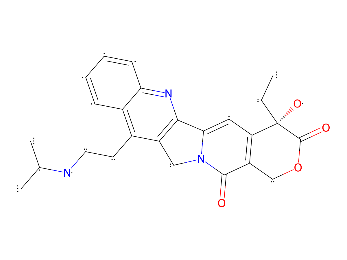
|
|||||
| Formula | C25H27N3O4 |
|||||
| Isosmiles | CC(NCCC(C1=CC=CC=C1N=C2C3=CC([C@@]4(O)CC)=C5COC4=O)=C2CN3C5=O)C |
|||||
| PubChem CID | ||||||
| InChI |
InChI=1S/C25H27N3O4/c1-4-25(31)19-11-21-22-17(12-28(21)23(29)18(19)13-32-24(25)30)15(9-10-26-14(2)3)16-7-5-6-8-20(16)27-22/h5-8,11,14,26,31H,4,9-10,12-13H2,1-3H3/t25-/m0/s1
|
|||||
| InChIKey |
LNHWXBUNXOXMRL-VWLOTQADSA-N
|
|||||
| Pharmaceutical Properties | Molecule Weight |
433.508 |
Polar area |
93.45 |
||
Complexity |
1279.616657 |
xlogp Value |
2.6201 |
|||
Heavy Count |
32 |
Rot Bonds |
5 |
|||
Hbond acc |
7 |
Hbond Donor |
2 |
|||
The activity data of This Payload
Each Antibody-drug Conjugate Related to This Payload
Full Information of The Activity Data of The ADC(s) Related to This Payload
WO2025019776A2ADC 33-8 [Investigative]
Discovered Using Cell Line-derived Xenograft Model
| Experiment 1 Reporting the Activity Date of This ADC | [1] | ||||
| Efficacy Data | Tumor Growth lnhibition value (TGl) |
12.40%
|
Positive IL13Ra2 expression (IL13Ra2+++/++) | ||
| Method Description |
Each female BALB/c Nude mouse was inoculated subcutaneously in the right upper flank region with 0.5x10 6of SK-MES-1 tumor cells (a non-small cell lung cancer (NSCLC)cell line having an IL13Ra2 copy number of about 4x10⁴)in 0.1 mL of PBS for tumor development.When the mean tumor size reached about 80-150 mm 3,mice were randomized into respective treatment groups (8 mice per group)and received intravenous injections of PBS vehicle or a single dose of the tested ADC 33-8 at 1 mg/kg on Day 1. The end of the study was day 44.
Click to Show/Hide
|
||||
| In Vivo Model | CDX Model-SK-MES-1 | ||||
| Experiment 2 Reporting the Activity Date of This ADC | [1] | ||||
| Efficacy Data | Tumor Growth lnhibition value (TGl) |
51.40%
|
Positive IL13Ra2 expression (IL13Ra2+++/++) | ||
| Method Description |
Each female BALB/c Nude mouse was inoculated subcutaneously in the right upper flank region with 0.5x10 6of SK-MES-1 tumor cells (a non-small cell lung cancer (NSCLC)cell line having an IL13Ra2 copy number of about 4x10⁴)in 0.1 mL of PBS for tumor development.When the mean tumor size reached about 80-150 mm 3,mice were randomized into respective treatment groups (8 mice per group)and received intravenous injections of PBS vehicle or a single dose of the tested ADC 33-8 at 3 mg/kg on Day 1. The end of the study was day 44.
Click to Show/Hide
|
||||
| In Vivo Model | CDX Model-SK-MES-1 | ||||
| Experiment 3 Reporting the Activity Date of This ADC | [1] | ||||
| Efficacy Data | Tumor Growth lnhibition value (TGl) |
61.10%
|
Positive IL13Ra2 expression (IL13Ra2+++/++) | ||
| Method Description |
Each female BALB/c Nude mouse was inoculated subcutaneously in the right upper flank region with 0.5x10 6of SK-MES-1 tumor cells (a non-small cell lung cancer (NSCLC)cell line having an IL13Ra2 copy number of about 4x10⁴)in 0.1 mL of PBS for tumor development.When the mean tumor size reached about 80-150 mm 3,mice were randomized into respective treatment groups (8 mice per group)and received intravenous injections of PBS vehicle or a single dose of the tested ADC 33-8 at 6 mg/kg on Day 1. The end of the study was day 44.
Click to Show/Hide
|
||||
| In Vivo Model | CDX Model-SK-MES-1 | ||||
| Experiment 4 Reporting the Activity Date of This ADC | [1] | ||||
| Efficacy Data | Tumor Growth lnhibition value (TGl) |
62%
|
Positive IL13Ra2 expression (IL13Ra2+++/++) | ||
| Method Description |
Each female BALB/c Nude mouse was inoculated subcutaneously in the right upper flank region with 0.5x10 6of SK-MES-1 tumor cells (a non-small cell lung cancer (NSCLC)cell line having an IL13Ra2 copy number of about 4x10⁴)in 0.1 mL of PBS for tumor development.When the mean tumor size reached about 80-150 mm 3,mice were randomized into respective treatment groups (8 mice per group)and received intravenous injections of PBS vehicle or a single dose of the tested ADC 33-8 at 10 mg/kg on Day 1. The end of the study was day 44.
Click to Show/Hide
|
||||
| In Vivo Model | CDX Model-SK-MES-1 | ||||
| Experiment 5 Reporting the Activity Date of This ADC | [1] | ||||
| Efficacy Data | Tumor Growth lnhibition value (TGl) |
80.50%
|
Positive IL13Ra2 expression (IL13Ra2+++/++) | ||
| Method Description |
Each female BALB/c Nude mouse was inoculated subcutaneously in the right upper/lower flank region with 1x10 7of H2228 tumor cells (an NSCLC cell line having an IL13Ra2 copy number of about 2,000)in 0.1 mL of PBS mixed with MATRIGELR (1:1)for tumor development.When the mean tumor size reached about 80-150 mm 3,mice were randomized into respective treatment groups (10 mice per group)and received intravenous injections of vehicle or a single dose of the tested ADCs at 10 mg/kg on Day 1. Body weights and tumor volumes were measured twice per week until the end of the study (Day 48).
Click to Show/Hide
|
||||
| In Vivo Model | CDX Model-H2228 | ||||
| Experiment 6 Reporting the Activity Date of This ADC | [1] | ||||
| Efficacy Data | Tumor Growth lnhibition value (TGl) |
88.90%
|
Positive IL13Ra2 expression (IL13Ra2+++/++) | ||
| Method Description |
Each female BALB/c Nude mouse was inoculated subcutaneously in the right upper flank region with 0.5x10 6of SK-MES-1 tumor cells (a non-small cell lung cancer (NSCLC)cell line having an IL13Ra2 copy number of about 4x10⁴)in 0.1 mL of PBS for tumor development.When the mean tumor size reached about 80-150 mm 3,mice were randomized into respective treatment groups (10 mice per group)and received intravenous injections of PBS vehicle or a single dose of the tested ADCs at 10 mg/kg on Day 1. The end of the study was day 49.
Click to Show/Hide
|
||||
| In Vivo Model | CDX Model-SK-MES-1 | ||||
| Experiment 7 Reporting the Activity Date of This ADC | [1] | ||||
| Efficacy Data | Tumor Growth lnhibition value (TGl) |
105.50%
|
Positive IL13Ra2 expression (IL13Ra2+++/++) | ||
| Method Description |
Each female NOD.Cg-Prkdcscid II2rgmIVst/Vst (NPG)mouse was inoculated subcutaneously in the right upper flank region with 1x10 7of H1792 tumor cells (an NSCLC cell line having an IL13Ra2 copy number of about 47000)in 0.1 mL of PBS mixed with MATRIGELR (1:1)for tumor development.When the mean tumor size reached about 80-150 mm 3,mice were randomized into respective treatment groups (10 mice per group)and received intravenous injections of vehicle or a single dose of the tested ADCs at 10 mg/kg on Day 1. Body weights and tumor volumes were measured twice per week until the end of the study (Day 57).
Click to Show/Hide
|
||||
| In Vivo Model | CDX Model-H1792 | ||||
| Experiment 8 Reporting the Activity Date of This ADC | [1] | ||||
| Efficacy Data | Tumor Growth lnhibition value (TGl) |
125%
|
High IL13Ra2 expression (IL13Ra2 +++) | ||
| Method Description |
0.5x10⁶of A375 tumor cells (a melanoma cancer cell line having an IL13Ra2 copy number of 3.5x10⁵measured as described in Example 11)mixed in 1:10 dilution of MATRIGELR (Corning Life Sciences)with serum free media was implanted subcutaneously at the hind right flank in female BALB/c Nude mice on Day-10.When the average tumor volume reached about 140 mm3,mice were randomized into respective treatment groups (10 mice per group)and received intravenous injections of PBS vehicle or the tested ADC 33-8 just on Day 1 with a dose of 3 mg/kg. Body weights and tumor volumes were measured twice per week until the end of the study. Days post-dosing 18, the TGI (vs isotype ADC) was calculated.
Click to Show/Hide
|
||||
| In Vivo Model | A375 BALB/c Nude mice model | ||||
Discovered Using Patient-derived Xenograft Model
| Experiment 1 Reporting the Activity Date of This ADC | [1] | ||||
| Efficacy Data | Tumor Growth lnhibition value (TGl) |
86%
|
Positive IL13Ra2 expression (IL13Ra2+++/++) | ||
| Method Description |
Female athymic nude-Foxnlnu mice were implanted (left flank)with patient tumor cells. H score: 225. Tumor cells from one NSCLC patient were tested. When sufficient stock animals reached 1000-1500 mm 3,tumors were harvested for re-implantation into pre-study animals.Pre-study animals were implanted unilaterally on the left flank with tumor fragments harvested from stock animals.When the average tumor volume reached 150-300 mm 3on Day 0,mice were randomized into respective treatment groups and received intravenous injections of vehicle or the tested ADC 33-8 (5mg/kg).Body weights and tumor volumes were measured twice per week until the end of the study.Any animal exhibiting>20%net weight loss for a period lasting 7 days or mice displaying>30% net weight loss when compared to Day O was considered moribund and euthanized. Beginning on Day 0,tumor dimensions were measured twice weekly by digital caliper,and data including individual and mean estimated tumor volumes (Mean TV± SEM)was recorded for each group;tumor volume was calculated using the formula (1): TV=width2xlengthx0.52.
Click to Show/Hide
|
||||
| In Vivo Model | Patient-Derived Xenograft (PDX)Models- NSCLC | ||||
Revealed Based on the Cell Line Data
| Experiment 1 Reporting the Activity Date of This ADC | [1] | ||||
| Efficacy Data | the MFI (geometric mean) signal | ≈ 40000-50000 . | Positive hIL13Ra2 expression (hIL13Ra2+++/++) | ||
| Method Description |
FACS was used to evaluate the binding profiles of A375 cells expressing IL13Ra2. The samples were then read by flow cytometry and the MFI (geometric mean) signal was calculated.
|
||||
| In Vitro Model | Amelanotic melanoma | A375 cells | CVCL_0132 | ||
| Experiment 2 Reporting the Activity Date of This ADC | [1] | ||||
| Efficacy Data | the MFI (geometric mean) signal | ≈ 40000-50000 . | Positive hIL13Ra2 expression (hIL13Ra2+++/++) | ||
| Method Description |
FACS was used to evaluate the binding profiles of HEK293 cells engineered to express IL13Ra2 (HEK293-IL13Ra2) cells expressing IL13Ra2. The samples were then read by flow cytometry and the MFI (geometric mean) signal was calculated.
|
||||
| In Vitro Model | Normal | HEK293 cells | CVCL_0045 | ||
| Experiment 3 Reporting the Activity Date of This ADC | [1] | ||||
| Efficacy Data | Half Maximal inhibitory Concentration (lC50) |
2 nM
|
High hIL13Ra2 expression (hIL13Ra2 +++) | ||
| Method Description |
The cytotoxic activity of ADC 33-8 was determined against cancer cell lines A375.FITC and anti-Hen egg-white lysozyme isotype control antibody (HEWL)conjugated to the same linker payloads.
|
||||
| In Vitro Model | Amelanotic melanoma | A375 cells | CVCL_0132 | ||
| Experiment 4 Reporting the Activity Date of This ADC | [1] | ||||
| Efficacy Data | Half Maximal inhibitory Concentration (lC50) | > 100 nM | Positive hIL13Ra2 expression (hIL13Ra2+++/++) | ||
| Method Description |
The cytotoxic activity of ADC 33-8 was determined against cancer cell lines H1792.FITC and anti-Hen egg-white lysozyme isotype control antibody (HEWL)conjugated to the same linker payloads.
|
||||
| In Vitro Model | Lung adenocarcinoma | H1792 cells | CVCL_1495 | ||
| Experiment 5 Reporting the Activity Date of This ADC | [1] | ||||
| Efficacy Data | Half Maximal inhibitory Concentration (lC50) | > 100 nM | Positive hIL13Ra2 expression (hIL13Ra2+++/++) | ||
| Method Description |
The cytotoxic activity of ADC 33-8 was determined against cancer cell lines H2228.FITC and anti-Hen egg-white lysozyme isotype control antibody (HEWL)conjugated to the same linker payloads.
|
||||
| In Vitro Model | Lung adenocarcinoma | H2228 cells | CVCL_1543 | ||
| Experiment 6 Reporting the Activity Date of This ADC | [1] | ||||
| Efficacy Data | Half Maximal inhibitory Concentration (lC50) | > 100 nM | Positive hIL13Ra2 expression (hIL13Ra2+++/++) | ||
| Method Description |
The cytotoxic activity of ADC 33-8 was determined against cancer cell lines SK-MES-1.FITC and anti-Hen egg-white lysozyme isotype control antibody (HEWL)conjugated to the same linker payloads.
|
||||
| In Vitro Model | Lung squamous cell carcinoma | SK-MES-1 cells | CVCL_0630 | ||
WO2025019776A2ADC-00 [Investigative]
Discovered Using Cell Line-derived Xenograft Model
| Experiment 1 Reporting the Activity Date of This ADC | [2] | ||||
| Efficacy Data | Tumor Growth lnhibition value (TGl) | > 30% | Positive ROR1 expression (ROR1+++/++) | ||
| Method Description |
CB.17 female SCID mice were inoculated subcutaneously in the right up flank region with Jeko-1 tumor cells (5×10 6)in 0.1 mL of PBS with Matrigel (1:1)for tumor development.When the tumor volumes reached about 150~250 mm 3on Day 1,mice were randomized into respective treatment groups (8 mice per group).The mice then received intravenous injections of vehicle or the tested ADC-00 at 7.5mg/kg on Day 1 and Day 8.PBS vehicle was tested in parallel,serving as a control.
Click to Show/Hide
|
||||
| In Vivo Model | JEKO-1 MCL Xenograft Model | ||||
| Experiment 2 Reporting the Activity Date of This ADC | [2] | ||||
| Efficacy Data | Tumor Growth lnhibition value (TGl) | > 50% | Positive ROR1 expression (ROR1+++/++) | ||
| Method Description |
CB.17 female SCID mice were implanted (flank)with 1.0x10 7of MDA-MB-231 tumor cells in 50%MATRIGELR (Corning Life Sciences)matrix (1:1 cell to Matrigel) on Day -13.When the average tumor volume reached about 200 mm 3on Day 1,mice were randomized into respective treatment groups (10 mice per group).The mice then received intravenous injections of the tested ADC-00 at 7.5 mg/kg on Day 1 and Day 8.PBS vehicle was tested in parallel,serving as a control.Body weight and tumor volume were measured twice per week.The study was ended on Day 45.
Click to Show/Hide
|
||||
| In Vivo Model | MDA-MB-231 TNBC Xenograft Model | ||||
Revealed Based on the Cell Line Data
| Experiment 1 Reporting the Activity Date of This ADC | [2] | ||||
| Efficacy Data | Half Maximal inhibitory Concentration (lC50) |
0.1-1 nM
|
Positive ROR1 expression (ROR1+++/++) | ||
| Method Description |
In vitro cytotoxicity of the ADC-00 was assessed on hROR1 expressing HEK cells with a top concentration of 10 nM, and ADC-00 was serially diluted 1:4, The treated cells were incubated for 5 days.The cell viability reading was taken. In order to activate the masked ADCs,an enzyme digestion system MMP9 was added.
|
||||
| In Vitro Model | Endocervical adenocarcinoma | HEK cells | CVCL_M624 | ||
WO2025019776A2ADC-11 [Investigative]
Discovered Using Cell Line-derived Xenograft Model
| Experiment 1 Reporting the Activity Date of This ADC | [2] | ||||
| Efficacy Data | Tumor Growth lnhibition value (TGl) | > 50% | Positive ROR1 expression (ROR1+++/++) | ||
| Method Description |
CB.17 female SCID mice were implanted (flank)with 1.0x10 7of MDA-MB-231 tumor cells in 50%MATRIGELR (Corning Life Sciences)matrix (1:1 cell to Matrigel) on Day -13.When the average tumor volume reached about 200 mm 3on Day 1,mice were randomized into respective treatment groups (10 mice per group).The mice then received intravenous injections of the tested ADC-11 at 7.5 mg/kg on Day 1 and Day 8.PBS vehicle was tested in parallel,serving as a control.Body weight and tumor volume were measured twice per week.The study was ended on Day 45.
Click to Show/Hide
|
||||
| In Vivo Model | MDA-MB-231 TNBC Xenograft Model | ||||
| Experiment 2 Reporting the Activity Date of This ADC | [2] | ||||
| Efficacy Data | Tumor Growth lnhibition value (TGl) | > 60% | Positive ROR1 expression (ROR1+++/++) | ||
| Method Description |
CB.17 female SCID mice were inoculated subcutaneously in the right up flank region with Jeko-1 tumor cells (5×10 6)in 0.1 mL of PBS with Matrigel (1:1)for tumor development.When the tumor volumes reached about 150~250 mm 3on Day 1,mice were randomized into respective treatment groups (8 mice per group).The mice then received intravenous injections of vehicle or the tested ADC-11 at 7.5mg/kg on Day 1 and Day 8.PBS vehicle was tested in parallel,serving as a control.
Click to Show/Hide
|
||||
| In Vivo Model | JEKO-1 MCL Xenograft Model | ||||
Revealed Based on the Cell Line Data
| Experiment 1 Reporting the Activity Date of This ADC | [2] | ||||
| Efficacy Data | Half Maximal inhibitory Concentration (lC50) |
0.1-1 nM
|
Positive ROR1 expression (ROR1+++/++) | ||
| Method Description |
In vitro cytotoxicity of the ADC-00 was assessed on hROR1 expressing HEK cells with a top concentration of 10 nM, and ADC-00 was serially diluted 1:4, The treated cells were incubated for 5 days.The cell viability reading was taken. In order to activate the masked ADCs,an enzyme digestion system MMP9 was added.
|
||||
| In Vitro Model | Endocervical adenocarcinoma | HEK cells | CVCL_M624 | ||
WO2025019776A2ADC-13 [Investigative]
Discovered Using Cell Line-derived Xenograft Model
| Experiment 1 Reporting the Activity Date of This ADC | [2] | ||||
| Efficacy Data | Tumor Growth lnhibition value (TGl) | > 50% | Positive ROR1 expression (ROR1+++/++) | ||
| Method Description |
CB.17 female SCID mice were inoculated subcutaneously in the right up flank region with Jeko-1 tumor cells (5×10 6)in 0.1 mL of PBS with Matrigel (1:1)for tumor development.When the tumor volumes reached about 150~250 mm 3on Day 1,mice were randomized into respective treatment groups (8 mice per group).The mice then received intravenous injections of vehicle or the tested ADC-13 at 7.5mg/kg on Day 1 and Day 8.PBS vehicle was tested in parallel,serving as a control.
Click to Show/Hide
|
||||
| In Vivo Model | JEKO-1 MCL Xenograft Model | ||||
| Experiment 2 Reporting the Activity Date of This ADC | [2] | ||||
| Efficacy Data | Tumor Growth lnhibition value (TGl) | > 60% | Positive ROR1 expression (ROR1+++/++) | ||
| Method Description |
CB.17 female SCID mice were implanted (flank)with 1.0x10 7of MDA-MB-231 tumor cells in 50%MATRIGELR (Corning Life Sciences)matrix (1:1 cell to Matrigel) on Day -13.When the average tumor volume reached about 200 mm 3on Day 1,mice were randomized into respective treatment groups (10 mice per group).The mice then received intravenous injections of the tested ADC-13 at 7.5 mg/kg on Day 1 and Day 8.PBS vehicle was tested in parallel,serving as a control.Body weight and tumor volume were measured twice per week.The study was ended on Day 45.
Click to Show/Hide
|
||||
| In Vivo Model | MDA-MB-231 TNBC Xenograft Model | ||||
Revealed Based on the Cell Line Data
| Experiment 1 Reporting the Activity Date of This ADC | [2] | ||||
| Efficacy Data | Half Maximal inhibitory Concentration (lC50) |
0.1-1 nM
|
Positive ROR1 expression (ROR1+++/++) | ||
| Method Description |
In vitro cytotoxicity of the ADC-00 was assessed on hROR1 expressing HEK cells with a top concentration of 10 nM, and ADC-00 was serially diluted 1:4, The treated cells were incubated for 5 days.The cell viability reading was taken. In order to activate the masked ADCs,an enzyme digestion system MMP9 was added.
|
||||
| In Vitro Model | Endocervical adenocarcinoma | HEK cells | CVCL_M624 | ||
WO2025019776A2ADC 52-8 [Investigative]
Discovered Using Cell Line-derived Xenograft Model
| Experiment 1 Reporting the Activity Date of This ADC | [1] | ||||
| Efficacy Data | Tumor Growth lnhibition value (TGl) |
66.60%
|
Positive IL13Ra2 expression (IL13Ra2+++/++) | ||
| Method Description |
Each female BALB/c Nude mouse was inoculated subcutaneously in the right upper/lower flank region with 1x10 7of H2228 tumor cells (an NSCLC cell line having an IL13Ra2 copy number of about 2,000)in 0.1 mL of PBS mixed with MATRIGELR (1:1)for tumor development.When the mean tumor size reached about 80-150 mm 3,mice were randomized into respective treatment groups (10 mice per group)and received intravenous injections of vehicle or a single dose of the tested ADCs at 10 mg/kg on Day 1. Body weights and tumor volumes were measured twice per week until the end of the study (Day 48).
Click to Show/Hide
|
||||
| In Vivo Model | CDX Model-H2228 | ||||
| Experiment 2 Reporting the Activity Date of This ADC | [1] | ||||
| Efficacy Data | Tumor Growth lnhibition value (TGl) |
83.40%
|
Positive IL13Ra2 expression (IL13Ra2+++/++) | ||
| Method Description |
Each female BALB/c Nude mouse was inoculated subcutaneously in the right upper flank region with 0.5x10 6of SK-MES-1 tumor cells (a non-small cell lung cancer (NSCLC)cell line having an IL13Ra2 copy number of about 4x10⁴)in 0.1 mL of PBS for tumor development.When the mean tumor size reached about 80-150 mm 3,mice were randomized into respective treatment groups (10 mice per group)and received intravenous injections of PBS vehicle or a single dose of the tested ADCs at 10 mg/kg on Day 1. The end of the study was day 49.
Click to Show/Hide
|
||||
| In Vivo Model | CDX Model-SK-MES-1 | ||||
Revealed Based on the Cell Line Data
| Experiment 1 Reporting the Activity Date of This ADC | [1] | ||||
| Efficacy Data | the MFI (geometric mean) signal | ≈ 30000-40000 . | Positive hIL13Ra2 expression (hIL13Ra2+++/++) | ||
| Method Description |
FACS was used to evaluate the binding profiles of HEK293 cells engineered to express IL13Ra2 (HEK293-IL13Ra2) cells expressing IL13Ra2. The samples were then read by flow cytometry and the MFI (geometric mean) signal was calculated.
|
||||
| In Vitro Model | Normal | HEK293 cells | CVCL_0045 | ||
| Experiment 2 Reporting the Activity Date of This ADC | [1] | ||||
| Efficacy Data | the MFI (geometric mean) signal | ≈ 40000-50000 . | Positive hIL13Ra2 expression (hIL13Ra2+++/++) | ||
| Method Description |
FACS was used to evaluate the binding profiles of A375 cells expressing IL13Ra2. The samples were then read by flow cytometry and the MFI (geometric mean) signal was calculated.
|
||||
| In Vitro Model | Amelanotic melanoma | A375 cells | CVCL_0132 | ||
| Experiment 3 Reporting the Activity Date of This ADC | [1] | ||||
| Efficacy Data | Half Maximal inhibitory Concentration (lC50) |
2 nM
|
High hIL13Ra2 expression (hIL13Ra2 +++) | ||
| Method Description |
The cytotoxic activity of ADC 52-8 was determined against cancer cell lines A375.FITC and anti-Hen egg-white lysozyme isotype control antibody (HEWL)conjugated to the same linker payloads.
|
||||
| In Vitro Model | Amelanotic melanoma | A375 cells | CVCL_0132 | ||
| Experiment 4 Reporting the Activity Date of This ADC | [1] | ||||
| Efficacy Data | Half Maximal inhibitory Concentration (lC50) | > 100 nM | Positive hIL13Ra2 expression (hIL13Ra2+++/++) | ||
| Method Description |
The cytotoxic activity of ADC 52-8 was determined against cancer cell lines H1792.FITC and anti-Hen egg-white lysozyme isotype control antibody (HEWL)conjugated to the same linker payloads.
|
||||
| In Vitro Model | Lung adenocarcinoma | H1792 cells | CVCL_1495 | ||
| Experiment 5 Reporting the Activity Date of This ADC | [1] | ||||
| Efficacy Data | Half Maximal inhibitory Concentration (lC50) | > 100 nM | Positive hIL13Ra2 expression (hIL13Ra2+++/++) | ||
| Method Description |
The cytotoxic activity of ADC 52-8 was determined against cancer cell lines H2228.FITC and anti-Hen egg-white lysozyme isotype control antibody (HEWL)conjugated to the same linker payloads.
|
||||
| In Vitro Model | Lung adenocarcinoma | H2228 cells | CVCL_1543 | ||
| Experiment 6 Reporting the Activity Date of This ADC | [1] | ||||
| Efficacy Data | Half Maximal inhibitory Concentration (lC50) | > 100 nM | Positive hIL13Ra2 expression (hIL13Ra2+++/++) | ||
| Method Description |
The cytotoxic activity of ADC 52-8 was determined against cancer cell lines SK-MES-1.FITC and anti-Hen egg-white lysozyme isotype control antibody (HEWL)conjugated to the same linker payloads.
|
||||
| In Vitro Model | Lung squamous cell carcinoma | SK-MES-1 cells | CVCL_0630 | ||
WO2025019776A2ADC 22-8 [Investigative]
Discovered Using Cell Line-derived Xenograft Model
| Experiment 1 Reporting the Activity Date of This ADC | [1] | ||||
| Efficacy Data | Tumor Growth lnhibition value (TGl) |
71.10%
|
Positive IL13Ra2 expression (IL13Ra2+++/++) | ||
| Method Description |
Each female BALB/c Nude mouse was inoculated subcutaneously in the right upper/lower flank region with 1x10 7of H2228 tumor cells (an NSCLC cell line having an IL13Ra2 copy number of about 2,000)in 0.1 mL of PBS mixed with MATRIGELR (1:1)for tumor development.When the mean tumor size reached about 80-150 mm 3,mice were randomized into respective treatment groups (10 mice per group)and received intravenous injections of vehicle or a single dose of the tested ADCs at 10 mg/kg on Day 1. Body weights and tumor volumes were measured twice per week until the end of the study (Day 48).
Click to Show/Hide
|
||||
| In Vivo Model | CDX Model-H2228 | ||||
| Experiment 2 Reporting the Activity Date of This ADC | [1] | ||||
| Efficacy Data | Tumor Growth lnhibition value (TGl) |
94.50%
|
Positive IL13Ra2 expression (IL13Ra2+++/++) | ||
| Method Description |
Each female BALB/c Nude mouse was inoculated subcutaneously in the right upper flank region with 0.5x10 6of SK-MES-1 tumor cells (a non-small cell lung cancer (NSCLC)cell line having an IL13Ra2 copy number of about 4x10⁴)in 0.1 mL of PBS for tumor development.When the mean tumor size reached about 80-150 mm 3,mice were randomized into respective treatment groups (10 mice per group)and received intravenous injections of PBS vehicle or a single dose of the tested ADCs at 10 mg/kg on Day 1. The end of the study was day 49.
Click to Show/Hide
|
||||
| In Vivo Model | CDX Model-SK-MES-1 | ||||
Revealed Based on the Cell Line Data
| Experiment 1 Reporting the Activity Date of This ADC | [1] | ||||
| Efficacy Data | the MFI (geometric mean) signal | ≈ 40000-50000 . | Positive hIL13Ra2 expression (hIL13Ra2+++/++) | ||
| Method Description |
FACS was used to evaluate the binding profiles of A375 cells expressing IL13Ra2. The samples were then read by flow cytometry and the MFI (geometric mean) signal was calculated.
|
||||
| In Vitro Model | Amelanotic melanoma | A375 cells | CVCL_0132 | ||
| Experiment 2 Reporting the Activity Date of This ADC | [1] | ||||
| Efficacy Data | the MFI (geometric mean) signal | ≈ 40000-50000 . | Positive hIL13Ra2 expression (hIL13Ra2+++/++) | ||
| Method Description |
FACS was used to evaluate the binding profiles of HEK293 cells engineered to express IL13Ra2 (HEK293-IL13Ra2) cells expressing IL13Ra2. The samples were then read by flow cytometry and the MFI (geometric mean) signal was calculated.
|
||||
| In Vitro Model | Normal | HEK293 cells | CVCL_0045 | ||
| Experiment 3 Reporting the Activity Date of This ADC | [1] | ||||
| Efficacy Data | Half Maximal inhibitory Concentration (lC50) |
2 nM
|
High hIL13Ra2 expression (hIL13Ra2 +++) | ||
| Method Description |
The cytotoxic activity of ADC 22-8 was determined against cancer cell lines A375.FITC and anti-Hen egg-white lysozyme isotype control antibody (HEWL)conjugated to the same linker payloads.
|
||||
| In Vitro Model | Amelanotic melanoma | A375 cells | CVCL_0132 | ||
| Experiment 4 Reporting the Activity Date of This ADC | [1] | ||||
| Efficacy Data | Half Maximal inhibitory Concentration (lC50) | > 100 nM | Positive hIL13Ra2 expression (hIL13Ra2+++/++) | ||
| Method Description |
The cytotoxic activity of ADC 22-8 was determined against cancer cell lines H1792.FITC and anti-Hen egg-white lysozyme isotype control antibody (HEWL)conjugated to the same linker payloads.
|
||||
| In Vitro Model | Lung adenocarcinoma | H1792 cells | CVCL_1495 | ||
| Experiment 5 Reporting the Activity Date of This ADC | [1] | ||||
| Efficacy Data | Half Maximal inhibitory Concentration (lC50) | > 100 nM | Positive hIL13Ra2 expression (hIL13Ra2+++/++) | ||
| Method Description |
The cytotoxic activity of ADC 22-8 was determined against cancer cell lines H2228.FITC and anti-Hen egg-white lysozyme isotype control antibody (HEWL)conjugated to the same linker payloads.
|
||||
| In Vitro Model | Lung adenocarcinoma | H2228 cells | CVCL_1543 | ||
| Experiment 6 Reporting the Activity Date of This ADC | [1] | ||||
| Efficacy Data | Half Maximal inhibitory Concentration (lC50) | > 100 nM | Positive hIL13Ra2 expression (hIL13Ra2+++/++) | ||
| Method Description |
The cytotoxic activity of ADC 22-8 was determined against cancer cell lines SK-MES-1.FITC and anti-Hen egg-white lysozyme isotype control antibody (HEWL)conjugated to the same linker payloads.
|
||||
| In Vitro Model | Lung squamous cell carcinoma | SK-MES-1 cells | CVCL_0630 | ||
References