
Antibody-drug Conjugate Information
General Information of This Antibody-drug Conjugate (ID: DRG0ZXHDJ)
| ADC Name |
aTCRA6 DAR2-8
|
|||||
|---|---|---|---|---|---|---|
| Synonyms |
aTCRA6 DAR2-8
Click to Show/Hide
|
|||||
| Organization |
Technical University of Darmstadt; Goethe University; Frankfurt Cancer Institute; German Cancer Consortium (DKTK); German Cancer Research Center (DKFZ).
|
|||||
| Drug Status |
Investigative
|
|||||
| Drug-to-Antibody Ratio |
5
|
|||||
| Structure |
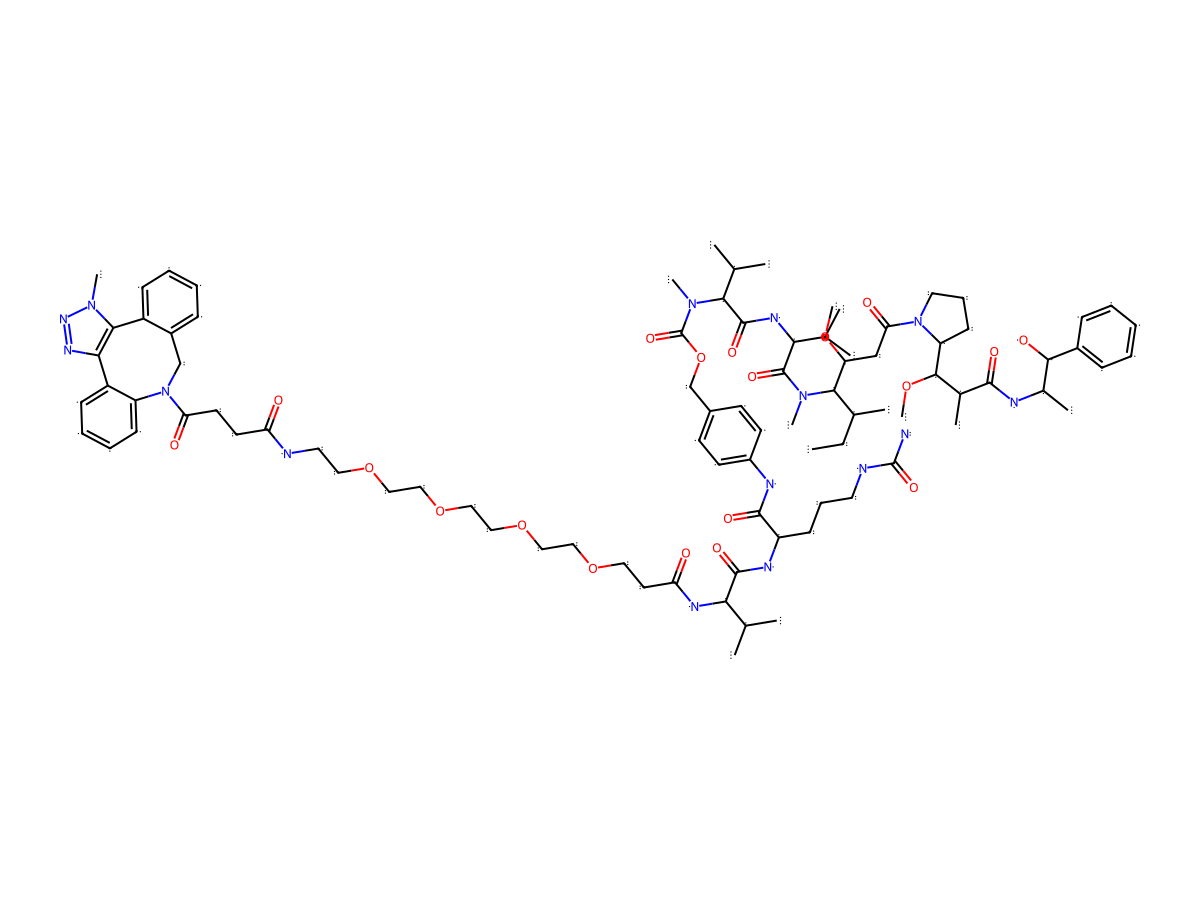
|
|||||
| Antibody Name |
aTCRA6S1
|
Antibody Info | ||||
| Antigen Name |
Transcriptional regulatory protein TcrA (TCRA6)
|
Antigen Info | ||||
| Payload Name |
Monomethyl auristatin E
|
Payload Info | ||||
| Payload Target |
Microtubule (MT)
|
Target Info | ||||
| Linker Name |
DBCO-PEG4-Val-Cit-PABC
|
Linker Info | ||||
| Conjugate Type |
Random conjugation through reduced inter-chain cysteines.
|
|||||
2027 Update
ADC-specific functional property
Bystander Killing Effect
| Bystander Killing Effect | Description | Reference |
|---|---|---|
| yes |
Circumventing such tumor escape mechanisms, an ADC is capable to mediate effects, (mainly) by internalization and degradation in target cells and subsequent permeation and diffusion of free payload, leading to the death of surrounding cells. Investigating the in vitro activity of our aTCRA6 DAR2 and aTCRA6 DAR52-8 ADCs, direct cell killing was measured using Jurkat TCRA6 target cells, and killing was analyzed by transfer of respective cell culture supernatants to Jurkat WT off-target cells. Consistent with our preliminary cytotoxicity data, direct killing of TCRA6-positive Jurkat cells was potently induced by the ADCs post 96 h.
Click to Show/Hide
|
[1]
|
General Information of The Activity Data Related to This ADC
Revealed Based on the Cell Line Data
Full List of Activity Data of This Antibody-drug Conjugate
Revealed Based on the Cell Line Data
| Experiment 1 Reporting the Activity Date of This ADC | [1] | ||||
| Efficacy Data | Half Maximal Effective Concentration (EC50) | 0.1 nM | Positive TCRA6 expression (TCRA6+++/++) | ||
| Method Description |
Cytotoxic effects of aTCRA6 DAR2 and aTCRA6 DAR52-8 ADCs were estimated by exposing Jurkat WT and Jurkat TCRA6 T cells to different ADC concentrations. Cell viability was analyzed 72 h post ADC addition by a fluorometric method using CellTiter-Blue Cell Viability Assay (Promega, Wisconsin, USA) according to the manufacturer's instructions. Briefly, cells were seeded in a 96-well plate (1 × 10⁴ cells/well) with the desired ADC concentrations ranging from 0.06-400 nM (for DAR2 ADC) or 0.005-30 nM (for DAR52-8 ADC) in a serial dilution. After 72 h, redox dye (resazurin) was added to the cells, and the plate was incubated for 3-4 h. Fluorescence of resorufin (560Ex/590Em) was recorded using CLARIOstar plus microplate reader (BMG LABTECH, Offenburg, Germany). Cell proliferation was normalized to untreated control cell fluorescence values. The resulting curves were fitted with a variable slope four-parameter fit, and EC50s were calculated using GraphPad Prism.
Click to Show/Hide
|
||||
| In Vitro Model | Childhood T acute lymphoblastic leukemia, Precursor T-cell acute lymphoblastic leukemia | Jurkat cells (TCRA6+) | CVCL_0065 | ||
| Experiment 2 Reporting the Activity Date of This ADC | [1] | ||||
| Efficacy Data | Half Maximal Effective Concentration (EC50) | > 100 nM | Negative TCRA6 expression (TCRA6-) | ||
| Method Description |
Cytotoxic effects of aTCRA6 DAR2 and aTCRA6 DAR52-8 ADCs were estimated by exposing Jurkat WT and Jurkat TCRA6 T cells to different ADC concentrations. Cell viability was analyzed 72 h post ADC addition by a fluorometric method using CellTiter-Blue Cell Viability Assay (Promega, Wisconsin, USA) according to the manufacturer's instructions. Briefly, cells were seeded in a 96-well plate (1 × 10⁴ cells/well) with the desired ADC concentrations ranging from 0.06-400 nM (for DAR2 ADC) or 0.005-30 nM (for DAR52-8 ADC) in a serial dilution. After 72 h, redox dye (resazurin) was added to the cells, and the plate was incubated for 3-4 h. Fluorescence of resorufin (560Ex/590Em) was recorded using CLARIOstar plus microplate reader (BMG LABTECH, Offenburg, Germany). Cell proliferation was normalized to untreated control cell fluorescence values. The resulting curves were fitted with a variable slope four-parameter fit, and EC50s were calculated using GraphPad Prism.
Click to Show/Hide
|
||||
| In Vitro Model | Childhood T acute lymphoblastic leukemia, Precursor T-cell acute lymphoblastic leukemia | Jurkat cells (TCRA6-) | CVCL_0065 | ||
References