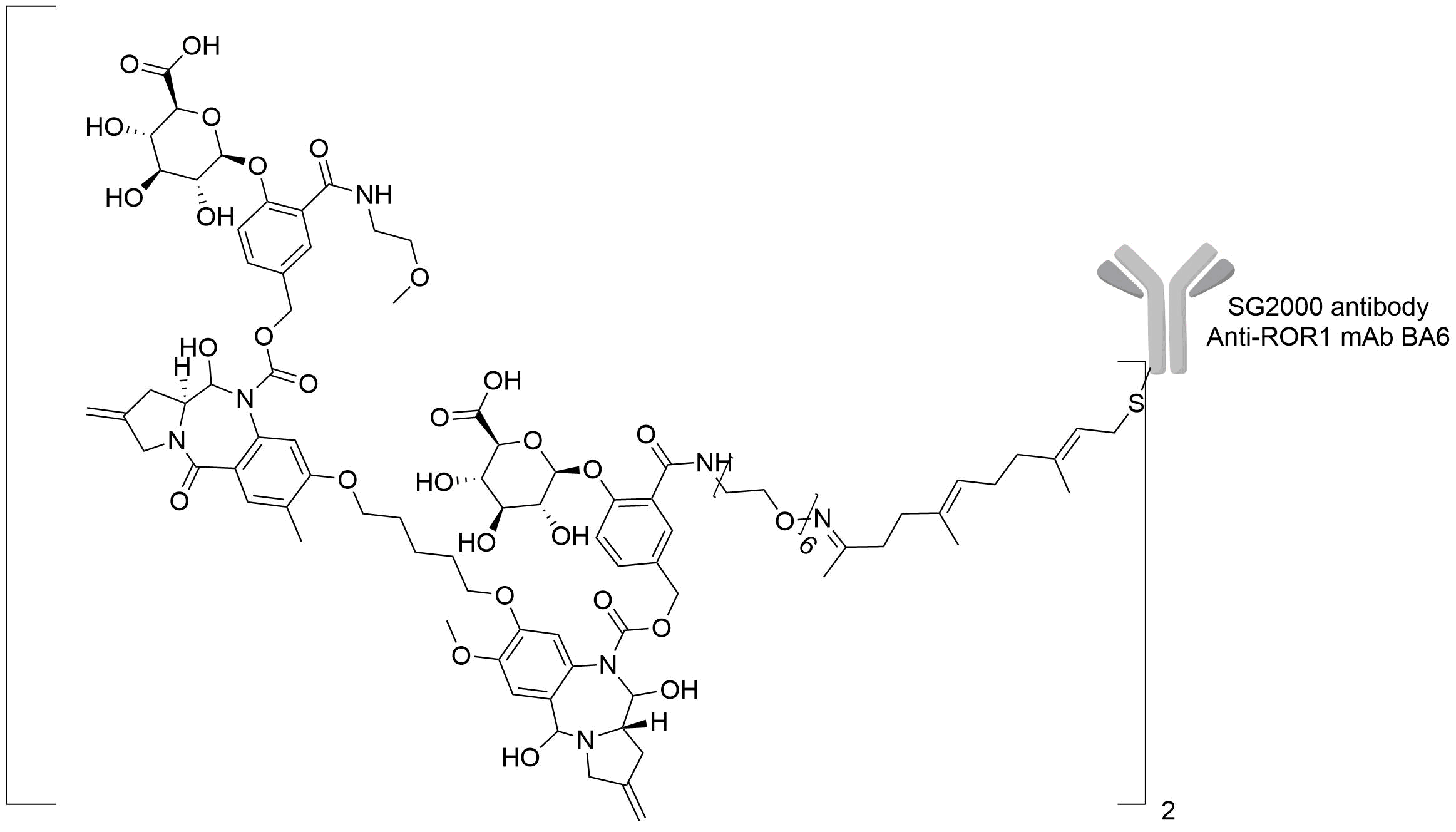
Antibody-drug Conjugate Information
General Information of This Antibody-drug Conjugate (ADC)
| ADC ID |
DRG0XJJWQ
|
|||||
|---|---|---|---|---|---|---|
| ADC Name |
WO2021044208A1 ADC6
|
|||||
| Synonyms |
WO2021044208A1 ADC6
Click to Show/Hide
|
|||||
| Organization |
LegoChem Biosciences, Inc.; ABL Bio, Inc.
|
|||||
| Drug Status |
Investigative
|
|||||
| Indication |
In total 3 Indication(s)
|
|||||
| Drug-to-Antibody Ratio |
2
|
|||||
| Structure |

|
|||||
| Antibody Name |
Anti-ROR1 mAb C2E3
|
Antibody Info | ||||
| Antigen Name |
Inactive tyrosine-protein kinase transmembrane receptor ROR1 (ROR1)
|
Antigen Info | ||||
| Payload Name |
SG2000
|
Payload Info | ||||
| Therapeutic Target |
Human Deoxyribonucleic acid (hDNA)
|
Target Info | ||||
| Linker Name |
WO2021044208A1_ADC6 linker
|
|||||
General Information of The Activity Data Related to This ADC
Discovered Using Cell Line-derived Xenograft Model
Revealed Based on the Cell Line Data
Full List of Activity Data of This Antibody-drug Conjugate
Discovered Using Cell Line-derived Xenograft Model
| Experiment 1 Reporting the Activity Date of This ADC | [1] | ||||
| Efficacy Data | Tumor Growth Inhibition value (TGI) | ≈ 0.00% (Day 41) | Positive ROR1 expression (ROR1+++/++) | ||
| Method Description |
1 x 10⁷ cells/head of human ROR-1 expressing breast cancer cell line HCC1187 were grafted to severe combined immunodeficient (SCID) mice to prepare human cancergrafted mice. After grafting, the mice were grouped when tumor size reached 110 mm3 on average (Day 1), and 1.25 mg/kg of ADC5.
|
||||
| In Vivo Model | HCC1187 CDX model | ||||
| In Vitro Model | Breast ductal carcinoma | HCC1187 cells | CVCL_1247 | ||
| Experiment 2 Reporting the Activity Date of This ADC | [1] | ||||
| Efficacy Data | Tumor Growth Inhibition value (TGI) | ≈ 46.74% (Day 26) | Positive ROR1 expression (ROR1+++/++) | ||
| Method Description |
1 x 10⁷ cells/head of human ROR-1 expressing lung cancer cell line Calu-3 were grafted to severe combined immunodeficient (SCID) mice to prepare human cancergrafted mice. After grafting, the mice were grouped when tumor size reached 111mm3 on average (Day 1), and 1 mg/kg QDx1 of ADC5.
|
||||
| In Vivo Model | Calu-3 CDX model | ||||
| In Vitro Model | Lung adenocarcinoma | Calu-3 cells | CVCL_0609 | ||
| Experiment 3 Reporting the Activity Date of This ADC | [1] | ||||
| Efficacy Data | Tumor Growth Inhibition value (TGI) | ≈ 77.97% (Day 19) | Positive ROR1 expression (ROR1+++/++) | ||
| Method Description |
1 x 10⁷ cells/head of human ROR-1 expressing breast cancer cell line MDA-MB231 were grafted to severe combined immunodeficient (SCID) mice to prepare human cancergrafted mice. After grafting, the mice were grouped when tumor size reached 110 mm3 on average (Day 1), and 0.33 mg/kg ADC5 was intravenously injected twice weekly a total of three times (Days 1, 7 and 14).
Click to Show/Hide
|
||||
| In Vivo Model | MDA-MB-231 CDX model | ||||
| In Vitro Model | Breast adenocarcinoma | MDA-MB-231 cells | CVCL_0062 | ||
| Experiment 4 Reporting the Activity Date of This ADC | [1] | ||||
| Efficacy Data | Tumor Growth Inhibition value (TGI) | ≈ 80.51% (Day 19) | Positive ROR1 expression (ROR1+++/++) | ||
| Method Description |
1 x 10⁷ cells/head of human ROR-1 expressing breast cancer cell line MDA-MB231 were grafted to severe combined immunodeficient (SCID) mice to prepare human cancergrafted mice. After grafting, the mice were grouped when tumor size reached 110 mm3 on average (Day 1), and 0.5 mg/kg of ADC5.
|
||||
| In Vivo Model | MDA-MB-231 CDX model | ||||
| In Vitro Model | Breast adenocarcinoma | MDA-MB-231 cells | CVCL_0062 | ||
| Experiment 5 Reporting the Activity Date of This ADC | [1] | ||||
| Efficacy Data | Tumor Growth Inhibition value (TGI) | ≈ 83.90% (Day 19) | Positive ROR1 expression (ROR1+++/++) | ||
| Method Description |
1 x 10⁷ cells/head of human ROR-1 expressing breast cancer cell line MDA-MB231 were grafted to severe combined immunodeficient (SCID) mice to prepare human cancergrafted mice. After grafting, the mice were grouped when tumor size reached 110 mm3 on average (Day 1), and 1 mg/kg of ADC5.
|
||||
| In Vivo Model | MDA-MB-231 CDX model | ||||
| In Vitro Model | Breast adenocarcinoma | MDA-MB-231 cells | CVCL_0062 | ||
| Experiment 6 Reporting the Activity Date of This ADC | [1] | ||||
| Efficacy Data | Tumor Growth Inhibition value (TGI) | ≈ 86.95% (Day 35) | Positive ROR1 expression (ROR1+++/++) | ||
| Method Description |
1 x 10⁷ cells/head of human ROR-1 expressing lung cancer cell line Calu-3 were grafted to severe combined immunodeficient (SCID) mice to prepare human cancergrafted mice. After grafting, the mice were grouped when tumor size reached 184mm3 on average (Day 1), and 1 mg/kg QDx1 of ADC5.
|
||||
| In Vivo Model | Calu-3 CDX model | ||||
| In Vitro Model | Lung adenocarcinoma | Calu-3 cells | CVCL_0609 | ||
| Experiment 7 Reporting the Activity Date of This ADC | [1] | ||||
| Efficacy Data | Tumor Growth Inhibition value (TGI) | ≈ 88.09% (Day 33) | Positive ROR1 expression (ROR1+++/++) | ||
| Method Description |
1 x 10⁷ cells/head of human ROR-1 expressing mantle cell lymphoma cell line JeKo-1 were grafted to severe combined immunodeficient (SCID) mice to prepare human cancergrafted mice. After grafting, the mice were grouped when tumor size reached 184mm3 on average (Day 1), and 0.25 mg/kg QWx4 of ADC5.
|
||||
| In Vivo Model | JeKo-1 CDX model | ||||
| In Vitro Model | Mantle cell lymphoma | JeKo-1 cells | CVCL_1865 | ||
| Experiment 8 Reporting the Activity Date of This ADC | [1] | ||||
| Efficacy Data | Tumor Growth Inhibition value (TGI) | ≈ 88.16% (Day 41) | Positive ROR1 expression (ROR1+++/++) | ||
| Method Description |
1 x 10⁷ cells/head of human ROR-1 expressing breast cancer cell line HCC1187 were grafted to severe combined immunodeficient (SCID) mice to prepare human cancergrafted mice. After grafting, the mice were grouped when tumor size reached 110 mm3 on average (Day 1), and 3.75 mg/kg QWx4 of ADC2.
|
||||
| In Vivo Model | HCC1187 CDX model | ||||
| In Vitro Model | Breast ductal carcinoma | HCC1187 cells | CVCL_1247 | ||
| Experiment 9 Reporting the Activity Date of This ADC | [1] | ||||
| Efficacy Data | Tumor Growth Inhibition value (TGI) | ≈ 88.98% (Day 19) | Positive ROR1 expression (ROR1+++/++) | ||
| Method Description |
1 x 10⁷ cells/head of human ROR-1 expressing breast cancer cell line MDA-MB231 were grafted to severe combined immunodeficient (SCID) mice to prepare human cancergrafted mice. After grafting, the mice were grouped when tumor size reached 110 mm3 on average (Day 1), and 2 mg/kg of ADC5.
|
||||
| In Vivo Model | MDA-MB-231 CDX model | ||||
| In Vitro Model | Breast adenocarcinoma | MDA-MB-231 cells | CVCL_0062 | ||
| Experiment 10 Reporting the Activity Date of This ADC | [1] | ||||
| Efficacy Data | Tumor Growth Inhibition value (TGI) | ≈ 97.97% (Day 35) | Positive ROR1 expression (ROR1+++/++) | ||
| Method Description |
1 x 10⁷ cells/head of human ROR-1 expressing lung cancer cell line Calu-3 were grafted to severe combined immunodeficient (SCID) mice to prepare human cancergrafted mice. After grafting, the mice were grouped when tumor size reached 184mm3 on average (Day 1), and 0.25 mg/kg QWx4 of ADC5.
|
||||
| In Vivo Model | Calu-3 CDX model | ||||
| In Vitro Model | Lung adenocarcinoma | Calu-3 cells | CVCL_0609 | ||
| Experiment 11 Reporting the Activity Date of This ADC | [1] | ||||
| Efficacy Data | Tumor Growth Inhibition value (TGI) | ≈ 98.79% (Day 56) | Positive ROR1 expression (ROR1+++/++) | ||
| Method Description |
1 x 10⁷ cells/head of human ROR-1 expressing breast cancer cell line MDA-MB-468 were grafted to female Balb/C nude mice to prepare human cancer grafted mice. After grafting.the mice were grouped when tumor size reached 166mm3 on average (Day 1), and 1 mg/kg of ADC5 was intravenously injected into the mice. In the control group, 10ml/kg PBS was intravenously injected into the mice.
Click to Show/Hide
|
||||
| In Vivo Model | MDA-MB-468 CDX model | ||||
| In Vitro Model | Breast adenocarcinoma | MDA-MB-468 cells | CVCL_0419 | ||
| Experiment 12 Reporting the Activity Date of This ADC | [1] | ||||
| Efficacy Data | Tumor Growth Inhibition value (TGI) | ≈ 99.97% (Day 26) | Positive ROR1 expression (ROR1+++/++) | ||
| Method Description |
1 x 10⁷ cells/head of human ROR-1 expressing lung cancer cell line Calu-3 were grafted to severe combined immunodeficient (SCID) mice to prepare human cancergrafted mice. After grafting, the mice were grouped when tumor size reached 111mm3 on average (Day 1), and 4 mg/kg of ADC5.
|
||||
| In Vivo Model | Calu-3 CDX model | ||||
| In Vitro Model | Lung adenocarcinoma | Calu-3 cells | CVCL_0609 | ||
| Experiment 13 Reporting the Activity Date of This ADC | [1] | ||||
| Efficacy Data | Tumor Growth Inhibition value (TGI) | ≈ 100.00% (Day 33) | Positive ROR1 expression (ROR1+++/++) | ||
| Method Description |
1 x 10⁷ cells/head of human ROR-1 expressing mantle cell lymphoma cell line JeKo-1 were grafted to severe combined immunodeficient (SCID) mice to prepare human cancergrafted mice. After grafting, the mice were grouped when tumor size reached 184mm3 on average (Day 1), and 1 mg/kg QDx1 of ADC5.
|
||||
| In Vivo Model | JeKo-1 CDX model | ||||
| In Vitro Model | Mantle cell lymphoma | JeKo-1 cells | CVCL_1865 | ||
Revealed Based on the Cell Line Data
| Experiment 1 Reporting the Activity Date of This ADC | [1] | ||||
| Efficacy Data | Half Maximal Inhibitory Concentration (IC50) | 5.09 nM | Positive ROR1 expression (ROR1+++/++) | ||
| Method Description |
In a 96-well plate, each well was seeded with 4,000 to 5,000 of the respective cancer cell lines. After culturing for 24 hours, they were treated with the ADCs at a concentra-tion of 0.0015 to 10.0 nM (serially diluted threefold). 72 hours later, the number of live cells was measured using WST-8.
|
||||
| In Vitro Model | Breast squamous cell carcinoma | HCC1806 cells | CVCL_1258 | ||
| Experiment 2 Reporting the Activity Date of This ADC | [1] | ||||
| Efficacy Data | Half Maximal Inhibitory Concentration (IC50) | > 10.00 nM | Positive ROR1 expression (ROR1+++/++) | ||
| Method Description |
In a 96-well plate, each well was seeded with 4,000 to 5,000 of the respective cancer cell lines. After culturing for 24 hours, they were treated with the ADCs at a concentra-tion of 0.0015 to 10.0 nM (serially diluted threefold). 72 hours later, the number of live cells was measured using WST-8.
|
||||
| In Vitro Model | Lung adenocarcinoma | NCI-H2228 cells | CVCL_1543 | ||
| Experiment 3 Reporting the Activity Date of This ADC | [1] | ||||
| Efficacy Data | Half Maximal Inhibitory Concentration (IC50) | > 10.00 nM | Positive ROR1 expression (ROR1+++/++) | ||
| Method Description |
In a 96-well plate, each well was seeded with 4,000 to 5,000 of the respective cancer cell lines. After culturing for 24 hours, they were treated with the ADCs at a concentra-tion of 0.0015 to 10.0 nM (serially diluted threefold). 72 hours later, the number of live cells was measured using WST-8.
|
||||
| In Vitro Model | Invasive breast carcinoma | MCF-7 cells | CVCL_0031 | ||
| Experiment 4 Reporting the Activity Date of This ADC | [1] | ||||
| Efficacy Data | Half Maximal Inhibitory Concentration (IC50) | 243 nM | Positive ROR1 expression (ROR1+++/++) | ||
| Method Description |
In a 96-well plate, each well was seeded with 4,000 to 5,000 of the respective cancer cell lines. After culturing for 24 hours, they were treated with the ADCs at a concentra-tion of 0.0015 to 10.0 nM (serially diluted threefold). 72 hours later, the number of live cells was measured using WST-8.
|
||||
| In Vitro Model | Breast adenocarcinoma | MDA-MB-231 cells | CVCL_0062 | ||
| Experiment 5 Reporting the Activity Date of This ADC | [1] | ||||
| Efficacy Data | Half Maximal Inhibitory Concentration (IC50) | 247 nM | Positive ROR1 expression (ROR1+++/++) | ||
| Method Description |
In a 96-well plate, each well was seeded with 4,000 to 5,000 of the respective cancer cell lines. After culturing for 24 hours, they were treated with the ADCs at a concentra-tion of 0.0015 to 10.0 nM (serially diluted threefold). 72 hours later, the number of live cells was measured using WST-8.
|
||||
| In Vitro Model | Mantle cell lymphoma | JeKo-1 cells | CVCL_1865 | ||
References
