
Antibody-drug Conjugate Information
General Information of This Antibody-drug Conjugate (ID: DRG0VUKDB)
| ADC Name |
EP4495597A2 25A-MMAE
|
|||||
|---|---|---|---|---|---|---|
| Synonyms |
EP4495597A2 25A-MMAE
Click to Show/Hide
|
|||||
| Organization |
ICONIC THERAPEUTICS LLC
|
|||||
| Drug Status |
Investigative
|
|||||
| Structure |
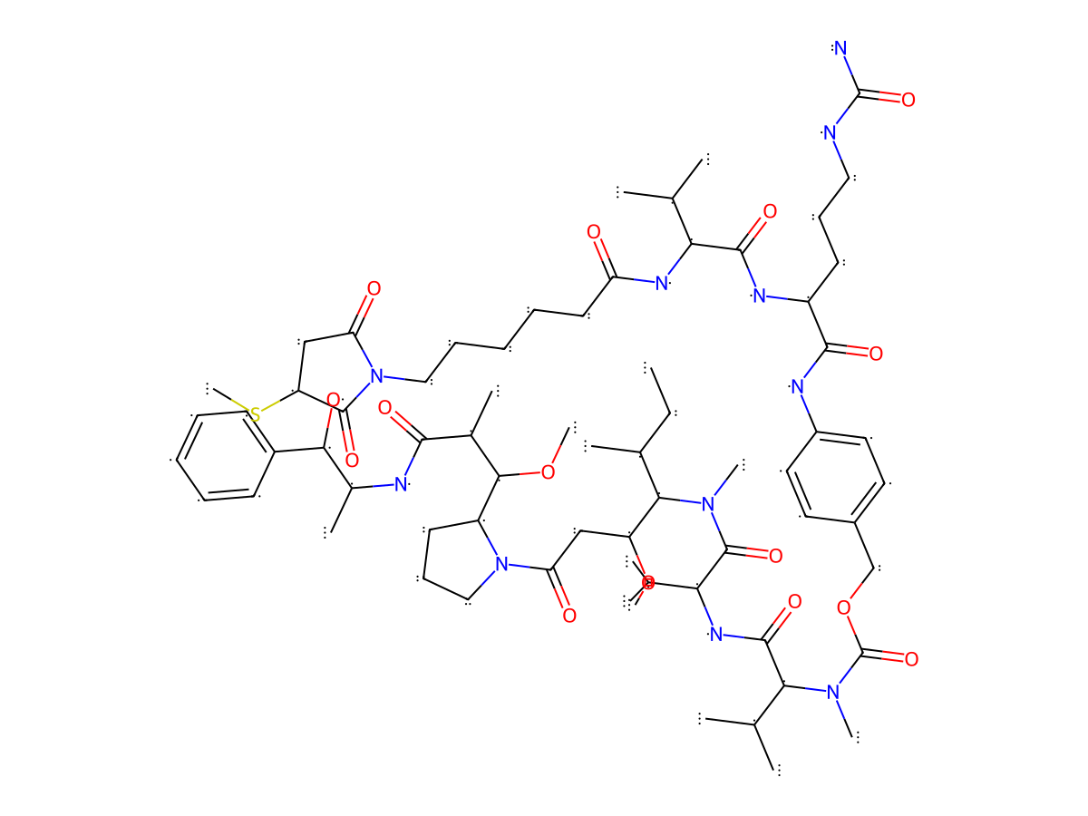
|
|||||
| Antibody Name |
25A
|
Antibody Info | ||||
| Antigen Name |
Tissue factor (F3)
|
Antigen Info | ||||
| Payload Name |
Monomethyl auristatin E
|
Payload Info | ||||
| Payload Target |
Microtubule (MT)
|
Target Info | ||||
| Linker Name |
Mc-Val-Cit-PABC
|
Linker Info | ||||
| Conjugate Type |
Random conjugation through reduced inter-chain cysteines.
|
|||||
| Combination Type |
Vedotin
|
|||||
General Information of The Activity Data Related to This ADC
Discovered Using Cell Line-derived Xenograft Model
Discovered Using Patient-derived Xenograft Model
Revealed Based on the Cell Line Data
Full List of Activity Data of This Antibody-drug Conjugate
Discovered Using Cell Line-derived Xenograft Model
| Experiment 1 Reporting the Activity Date of This ADC | [1] | ||||
| Efficacy Data | Tumor Growth lnhibition value (TGl) | 67% | Positive TF expression (TF+++/++) | ||
| Method Description |
In the MDA-MB-231 xenograft model, the ADCs were administered on day 1 and 8 post-randomization at 2 mg/kg (qwk × 2, IV).
|
||||
| In Vivo Model | MDA-MB-231 xenograft model | ||||
| Experiment 2 Reporting the Activity Date of This ADC | [1] | ||||
| Efficacy Data | Tumor Growth lnhibition value (TGl) | 98% | Positive TF expression (TF+++/++) | ||
| Method Description |
In the MDA-MB-231 xenograft model, the ADCs were administered on day 1 and 8 post-randomization at 4 mg/kg (qwk × 2, IV).
|
||||
| In Vivo Model | MDA-MB-231 xenograft model | ||||
| Experiment 3 Reporting the Activity Date of This ADC | [1] | ||||
| Efficacy Data | Tumor Growth lnhibition value (TGl) | 132% | Positive TF expression (TF+++/++) | ||
| Method Description |
The A431 epidermoid carcinoma cell lines were implanted subcutaneously in the flank of athymic nude mice. Animals were randomized and treated with drugs (5 mg/kg, qwk × 3, IP).
|
||||
| In Vivo Model | A431 xenograft model | ||||
| Experiment 4 Reporting the Activity Date of This ADC | [1] | ||||
| Efficacy Data | Tumor Growth lnhibition value (TGl) | 139% | Positive TF expression (TF+++/++) | ||
| Method Description |
HPAF-II pancreatic carcinoma epidermoid carcinoma cell lines were implanted subcutaneously in the flank of athymic nude mice. Animals were randomized and treated with drugs (2 mg/kg, qwk × 2, IV).
|
||||
| In Vivo Model | HPAF-II pancreatic carcinoma xenograft model | ||||
Discovered Using Patient-derived Xenograft Model
| Experiment 1 Reporting the Activity Date of This ADC | [1] | ||||
| Efficacy Data | Tumor Growth lnhibition value (TGl) | 71% | Positive TF expression (TF+++/++) | ||
| Method Description |
TF-positive PDX models were performed in athymic nude mice (Envigo, Indianapolis, IN) to evaluate the efficacy of the ADCs in vivo. The animals' care was in accordance with institutional guidelines. Study animals were implanted unilaterally on the left flank with tumor fragments. In the head and neck and ovarian PDX model, the mice were treated with drugs (2.5 mg/kg, qwk × 3, IP).
Click to Show/Hide
|
||||
| In Vivo Model | The head and neck and ovarian PDX model | ||||
| Experiment 2 Reporting the Activity Date of This ADC | [1] | ||||
| Efficacy Data | Tumor Growth lnhibition value (TGl) | 112% | Positive TF expression (TF+++/++) | ||
| Method Description |
TF-positive PDX models were performed in athymic nude mice (Envigo, Indianapolis, IN) to evaluate the efficacy of the ADCs in vivo. The animals' care was in accordance with institutional guidelines. Study animals were implanted unilaterally on the left flank with tumor fragments. In the gastric PDX model, the mice were treated with drugs (4 mg/kg, qwk × 2, IV).
Click to Show/Hide
|
||||
| In Vivo Model | The gastric PDX model | ||||
| Experiment 3 Reporting the Activity Date of This ADC | [1] | ||||
| Efficacy Data | Tumor Growth lnhibition value (TGl) | 124% | Positive TF expression (TF+++/++) | ||
| Method Description |
TF-positive PDX models were performed in athymic nude mice (Envigo, Indianapolis, IN) to evaluate the efficacy of the ADCs in vivo. The animals' care was in accordance with institutional guidelines. Study animals were implanted unilaterally on the left flank with tumor fragments. In the head and neck and ovarian PDX model, the mice were treated with drugs (5 mg/kg, qwk × 2, IP).
Click to Show/Hide
|
||||
| In Vivo Model | The head and neck and ovarian PDX model | ||||
Revealed Based on the Cell Line Data
| Experiment 1 Reporting the Activity Date of This ADC | [1] | ||||
| Efficacy Data | Half Maximal inhibitory Concentration (lC50) | 0.06 nM | Positive TF expression (TF+++/++) | ||
| Method Description |
The TF-specific ADCs were added to HPAF-II cells, with either a 72 h incubation or a 4 h incubation followed by removal of excess ADC and culture for another 69 h. HPAF-II cells were lysed in CTG assay reagent after treatment.
|
||||
| In Vitro Model | Pancreatic ductal adenocarcinoma | HPAF-II cells | CVCL_0313 | ||
| Experiment 2 Reporting the Activity Date of This ADC | [1] | ||||
| Efficacy Data | Half Maximal inhibitory Concentration (lC50) | 0.11 nM | Positive TF expression (TF+++/++) | ||
| Method Description |
The TF-specific ADCs were added to A431 cells, with either a 72 h incubation or a 4 h incubation followed by removal of excess ADC and culture for another 68 h. A432cells were lysed in CTG assay reagent after treatment.
|
||||
| In Vitro Model | Skin squamous cell carcinoma | A431 cells | CVCL_0037 | ||
| Experiment 3 Reporting the Activity Date of This ADC | [1] | ||||
| Efficacy Data | Half Maximal inhibitory Concentration (lC50) | 0.14 nM | Positive TF expression (TF+++/++) | ||
| Method Description |
The TF-specific ADCs were added to MDA-MB-231 cells, with either a 72 h incubation or a 4 h incubation followed by removal of excess ADC and culture for another 68 h. MDA-MB-231 cells were lysed in CTG assay reagent after treatment.
|
||||
| In Vitro Model | Breast adenocarcinoma | MDA-MB-231 cells | CVCL_0062 | ||