
Antibody-drug Conjugate Information
General Information of This Antibody-drug Conjugate (ID: DRG0LZDLS)
| ADC Name |
EP3838893B1-ADC-III
|
|||||
|---|---|---|---|---|---|---|
| Synonyms |
MAB-W'-C6-Val-Cit-PAB-MMAE;W'-C6-VC
Click to Show/Hide
|
|||||
| Organization |
Academy of Military Medical Sciences
|
|||||
| Drug Status |
Investigative
|
|||||
| Drug-to-Antibody Ratio |
4;8
|
|||||
| Structure |
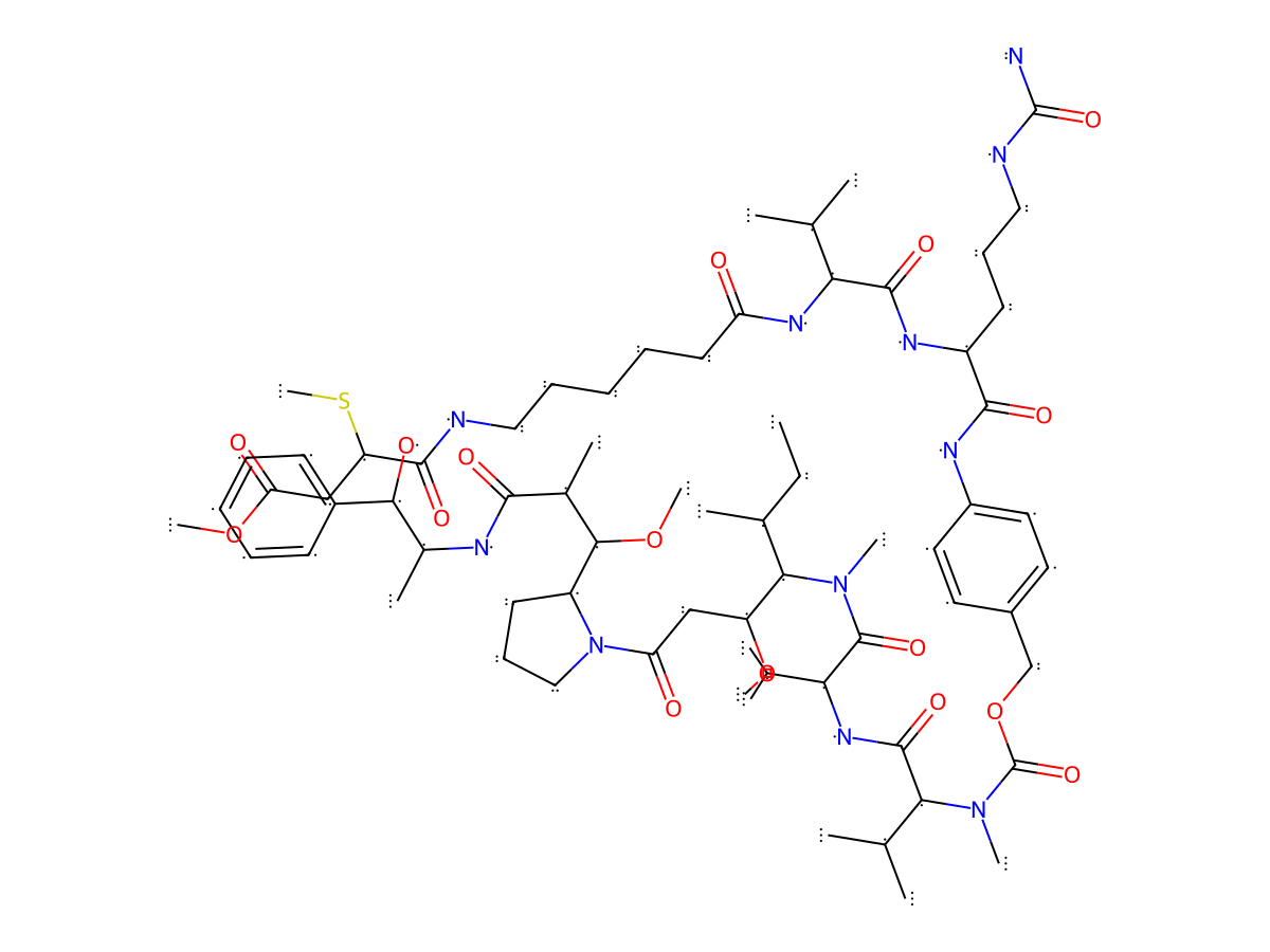
|
|||||
| Antibody Name |
mil40
|
Antibody Info | ||||
| Antigen Name |
Receptor tyrosine-protein kinase erbB-2 (HER2)
|
Antigen Info | ||||
| Payload Name |
Monomethyl auristatin E
|
Payload Info | ||||
| Payload Target |
Microtubule (MT)
|
Target Info | ||||
| Linker Name |
Mm-C6-Val-Cit-PAB
|
Linker Info | ||||
| Conjugate Type |
Random conjugation through reduced inter-chain cysteines.
|
|||||
| Combination Type |
Mm-C6-Val-Cit-PAB-MMAE
|
|||||
General Information of The Activity Data Related to This ADC
Revealed Based on the Cell Line Data
Full List of Activity Data of This Antibody-drug Conjugate
Revealed Based on the Cell Line Data
| Experiment 1 Reporting the Activity Date of This ADC | [1] | ||||
| Efficacy Data | Half Maximal inhibitory Concentration (lC50) | 0.0258 nM | Positive HER2 expression (HER2+++/++) | ||
| Method Description |
HCC 1954, 1000cell/well was added to the designated 384-well cell culture plate in the appropriate growth media. The ADCs stock solution was diluted in series in a ratio of 1:3 to produce 10 diluted solutions with different concentrations (the compound stock solution was an L-His buffer salt solution with a concentration of about 2 mg/mL, which was diluted with PBS, and the initial maximum concentration at test point was about 500 mg/mL). 4ul of the compound diluted solutions with different concentrations was distributed into a 384-well plate. After 96 hours, the plate was taken from the incubator and equilibrated at room temperature for 10 minutes. 40uL of CellTiter-Glo reagent was added to each well to be tested (the ratio of CellTiter-Glo reagent to medium was 1:1).the plate was then placed at room temperature for 30 minutes, and then reading was carried out on an EnSpire reader for cell counting.
Click to Show/Hide
|
||||
| In Vitro Model | Breast ductal carcinoma | HCC1954 cells | CVCL_1259 | ||
| Experiment 2 Reporting the Activity Date of This ADC | [1] | ||||
| Efficacy Data | Half Maximal inhibitory Concentration (lC50) | 0.0536 nM | Positive HER2 expression (HER2+++/++) | ||
| Method Description |
NCI-N87, 1000cell/well was added to the designated 384-well cell culture plate in the appropriate growth media. The ADCs stock solution was diluted in series in a ratio of 1:3 to produce 10 diluted solutions with different concentrations (the compound stock solution was an L-His buffer salt solution with a concentration of about 2 mg/mL, which was diluted with PBS, and the initial maximum concentration at test point was about 500 mg/mL). 4ul of the compound diluted solutions with different concentrations was distributed into a 384-well plate. After 96 hours, the plate was taken from the incubator and equilibrated at room temperature for 10 minutes. 40uL of CellTiter-Glo reagent was added to each well to be tested (the ratio of CellTiter-Glo reagent to medium was 1:1).the plate was then placed at room temperature for 30 minutes, and then reading was carried out on an EnSpire reader for cell counting.
Click to Show/Hide
|
||||
| In Vitro Model | Gastric tubular adenocarcinoma | NCI-N87 cells | CVCL_1603 | ||
| Experiment 3 Reporting the Activity Date of This ADC | [1] | ||||
| Efficacy Data | Half Maximal inhibitory Concentration (lC50) | 0.1026 nM | Positive HER2 expression (HER2+++/++) | ||
| Method Description |
BT-474, 1000cell/well was added to the designated 384-well cell culture plate in the appropriate growth media. The ADCs stock solution was diluted in series in a ratio of 1:3 to produce 10 diluted solutions with different concentrations (the compound stock solution was an L-His buffer salt solution with a concentration of about 2 mg/mL, which was diluted with PBS, and the initial maximum concentration at test point was about 500 mg/mL). 4ul of the compound diluted solutions with different concentrations was distributed into a 384-well plate. After 96 hours, the plate was taken from the incubator and equilibrated at room temperature for 10 minutes. 40uL of CellTiter-Glo reagent was added to each well to be tested (the ratio of CellTiter-Glo reagent to medium was 1:1).the plate was then placed at room temperature for 30 minutes, and then reading was carried out on an EnSpire reader for cell counting.
Click to Show/Hide
|
||||
| In Vitro Model | Invasive breast carcinoma | BT-474 cells | CVCL_0179 | ||
| Experiment 4 Reporting the Activity Date of This ADC | [1] | ||||
| Efficacy Data | Half Maximal inhibitory Concentration (lC50) | 0.2074 nM | Positive HER2 expression (HER2+++/++) | ||
| Method Description |
SK-OV-3, 1000cell/well was added to the designated 384-well cell culture plate in the appropriate growth media. The ADCs stock solution was diluted in series in a ratio of 1:3 to produce 10 diluted solutions with different concentrations (the compound stock solution was an L-His buffer salt solution with a concentration of about 2 mg/mL, which was diluted with PBS, and the initial maximum concentration at test point was about 500 mg/mL). 4ul of the compound diluted solutions with different concentrations was distributed into a 384-well plate. After 96 hours, the plate was taken from the incubator and equilibrated at room temperature for 10 minutes. 40uL of CellTiter-Glo reagent was added to each well to be tested (the ratio of CellTiter-Glo reagent to medium was 1:1).the plate was then placed at room temperature for 30 minutes, and then reading was carried out on an EnSpire reader for cell counting.
Click to Show/Hide
|
||||
| In Vitro Model | Ovarian serous adenocarcinoma | SKOV-3 cells | CVCL_0532 | ||
| Experiment 5 Reporting the Activity Date of This ADC | [1] | ||||
| Efficacy Data | Half Maximal inhibitory Concentration (lC50) | > 1.5316 nM | Negative HER2 expression (HER2-) | ||
| Method Description |
MCF-7, 1000cell/well was added to the designated 384-well cell culture plate in the appropriate growth media. The ADCs stock solution was diluted in series in a ratio of 1:3 to produce 10 diluted solutions with different concentrations (the compound stock solution was an L-His buffer salt solution with a concentration of about 2 mg/mL, which was diluted with PBS, and the initial maximum concentration at test point was about 500 mg/mL). 4ul of the compound diluted solutions with different concentrations was distributed into a 384-well plate. After 96 hours, the plate was taken from the incubator and equilibrated at room temperature for 10 minutes. 40uL of CellTiter-Glo reagent was added to each well to be tested (the ratio of CellTiter-Glo reagent to medium was 1:1).the plate was then placed at room temperature for 30 minutes, and then reading was carried out on an EnSpire reader for cell counting.
Click to Show/Hide
|
||||
| In Vitro Model | Invasive breast carcinoma | MCF-7 cells | CVCL_0031 | ||
| Experiment 6 Reporting the Activity Date of This ADC | [1] | ||||
| Efficacy Data | Half Maximal inhibitory Concentration (lC50) | > 1.5316 nM | Negative HER2 expression (HER2-) | ||
| Method Description |
MDA-MB-468, 1000cell/well was added to the designated 384-well cell culture plate in the appropriate growth media. The ADCs stock solution was diluted in series in a ratio of 1:3 to produce 10 diluted solutions with different concentrations (the compound stock solution was an L-His buffer salt solution with a concentration of about 2 mg/mL, which was diluted with PBS, and the initial maximum concentration at test point was about 500 mg/mL). 4ul of the compound diluted solutions with different concentrations was distributed into a 384-well plate. After 96 hours, the plate was taken from the incubator and equilibrated at room temperature for 10 minutes. 40uL of CellTiter-Glo reagent was added to each well to be tested (the ratio of CellTiter-Glo reagent to medium was 1:1).the plate was then placed at room temperature for 30 minutes, and then reading was carried out on an EnSpire reader for cell counting.
Click to Show/Hide
|
||||
| In Vitro Model | Breast adenocarcinoma | MDA-MB-468 cells | CVCL_0419 | ||