
Antibody-drug Conjugate Information
General Information of This Antibody-drug Conjugate (ID: DRG0LGZNC)
| ADC Name |
Anti-CD276 mAb-MMAF
|
|||||
|---|---|---|---|---|---|---|
| Synonyms |
Anti-CD276 mAb-MMAF
Click to Show/Hide
|
|||||
| Organization |
The Ohio State University (OSU).
|
|||||
| Drug Status |
Investigative
|
|||||
| Drug-to-Antibody Ratio |
3- 4
|
|||||
| Structure |
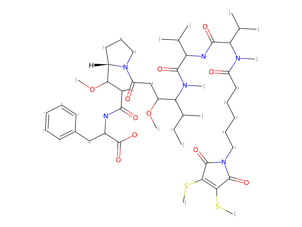
|
|||||
| Antibody Name |
Anti-CD276 mAb
|
Antibody Info | ||||
| Antigen Name |
CD276 antigen (CD276)
|
Antigen Info | ||||
| Payload Name |
Monomethyl auristatin F
|
Payload Info | ||||
| Payload Target |
Microtubule (MT)
|
Target Info | ||||
| Linker Name |
Undisclosed
|
|||||
| Conjugate Type |
Random conjugation through reduced inter-chain cysteines.
|
|||||
General Information of The Activity Data Related to This ADC
Discovered Using Cell Line-derived Xenograft Model
| Standard Type | Value | Units | Cell Line | Disease Model |
|---|---|---|---|---|
| Tumor Growth lnhibition value (TGl) |
64.1
|
%
|
Undisclosed | Undisclosed |
| Tumor Growth lnhibition value (TGl) |
91.9
|
%
|
Undisclosed | Undisclosed |
Revealed Based on the Cell Line Data
Full List of Activity Data of This Antibody-drug Conjugate
Discovered Using Cell Line-derived Xenograft Model
| Experiment 1 Reporting the Activity Date of This ADC | [1] | ||||
| Efficacy Data | Tumor Growth lnhibition value (TGl) |
64.10%
|
|||
| Method Description |
To evaluate the synergetic anti-NSCLC efficacy of CD276 mAb-MMAF, about 3 × 10⁶ mouse LLC-FLuc cells were s.c. injected into C57BL mice to establish immunocompetent models. The mice were i.v. injected with saline (vehicle, control), 12 mg/kg anti-CD276 mAb (control), and 12 and 24 mg/kg anti-CD276 mAb-MMAF (n = 6). (12 mg/kg)
|
||||
| In Vivo Model | H460-FLuc cells into nude mice model | ||||
| Experiment 2 Reporting the Activity Date of This ADC | [1] | ||||
| Efficacy Data | Tumor Growth lnhibition value (TGl) |
91.90%
|
|||
| Method Description |
To evaluate the synergetic anti-NSCLC efficacy of CD276 mAb-MMAF, about 3 × 10⁶ mouse LLC-FLuc cells were s.c. injected into C57BL mice to establish immunocompetent models. The mice were i.v. injected with saline (vehicle, control), 12 mg/kg anti-CD276 mAb (control), and 12 and 24 mg/kg anti-CD276 mAb-MMAF (n = 6). (24 mg/kg)
|
||||
| In Vivo Model | H460-FLuc cells into nude mice model | ||||
Revealed Based on the Cell Line Data
| Experiment 1 Reporting the Activity Date of This ADC | [1] | ||||
| Efficacy Data | Half Maximal inhibitory Concentration (lC50) | 32.5 nM | High CD276 expression (CD276 +++) | ||
| Method Description |
The cytotoxicity assay was performed using human NSCLC cell line H460-FLuc and mouse LLC-FLuc. With different cell division rates, 3000 H460-FLuc cells or 1000 LLC-FLuc cells with 200 uL of RPMI-1640 or a DMEM complete growth medium were seeded into each well in the 96-well plates. The cells were incubated at 37 °C in a CO2 incubator for 24 h, followed by the addition of free MMAF or ADC at dosages of 0, 10, 20, 40, 80, 120, 160, and 200 nM. The 3- (4,5-Dimethylthiazol-2-yl)-2,5-Diphenyltetrazolium Bromide Cell Proliferation Assay (MTT Assay) was performed following the biomanufacturer procedure after 5-day treatment. At the end of treatment, the spent medium was removed and replaced with 100 uL of the fresh culture medium supplemented with 10 uL of the MTT reagent in each well. After 2 h of incubation in the dark, 100 uL of the detergent reagent was added to each well until a purple color was developed. The OD value was read at 590 nm using a SpectraMax iD3 plate reader (San Jose, CA, USA), and IC50 values were calculated.
Click to Show/Hide
|
||||
| In Vitro Model | Lung large cell carcinoma | H460 cells (Human) | CVCL_0459 | ||
| Experiment 2 Reporting the Activity Date of This ADC | [1] | ||||
| Efficacy Data | Half Maximal inhibitory Concentration (lC50) | 60.5 nM | Low CD276 expression (CD276+) | ||
| Method Description |
The cytotoxicity assay was performed using human NSCLC cell line H460-FLuc and mouse LLC-FLuc. With different cell division rates, 3000 H460-FLuc cells or 1000 LLC-FLuc cells with 200 uL of RPMI-1640 or a DMEM complete growth medium were seeded into each well in the 96-well plates. The cells were incubated at 37 °C in a CO2 incubator for 24 h, followed by the addition of free MMAF or ADC at dosages of 0, 10, 20, 40, 80, 120, 160, and 200 nM. The 3- (4,5-Dimethylthiazol-2-yl)-2,5-Diphenyltetrazolium Bromide Cell Proliferation Assay (MTT Assay) was performed following the biomanufacturer procedure after 5-day treatment. At the end of treatment, the spent medium was removed and replaced with 100 uL of the fresh culture medium supplemented with 10 uL of the MTT reagent in each well. After 2 h of incubation in the dark, 100 uL of the detergent reagent was added to each well until a purple color was developed. The OD value was read at 590 nm using a SpectraMax iD3 plate reader (San Jose, CA, USA), and IC50 values were calculated.
Click to Show/Hide
|
||||
| In Vitro Model | Malignant tumors of the mouse pulmonary system | LCC cells (Mouse) | CVCL_5653 | ||