
Antibody-drug Conjugate Information
General Information of This Antibody-drug Conjugate (ID: DRG0ZJKIV)
| ADC Name |
FL1-PNU
|
|||||
|---|---|---|---|---|---|---|
| Synonyms |
FL1-PNU
Click to Show/Hide
|
|||||
| Organization |
The Finsen Laboratory.; Biotech Research & Innovation Centre (BRIC), Faculty of Health and Medical Sciences, University of Copenhagen.
|
|||||
| Drug Status |
Investigative
|
|||||
| Drug-to-Antibody Ratio |
2
|
|||||
| Structure |
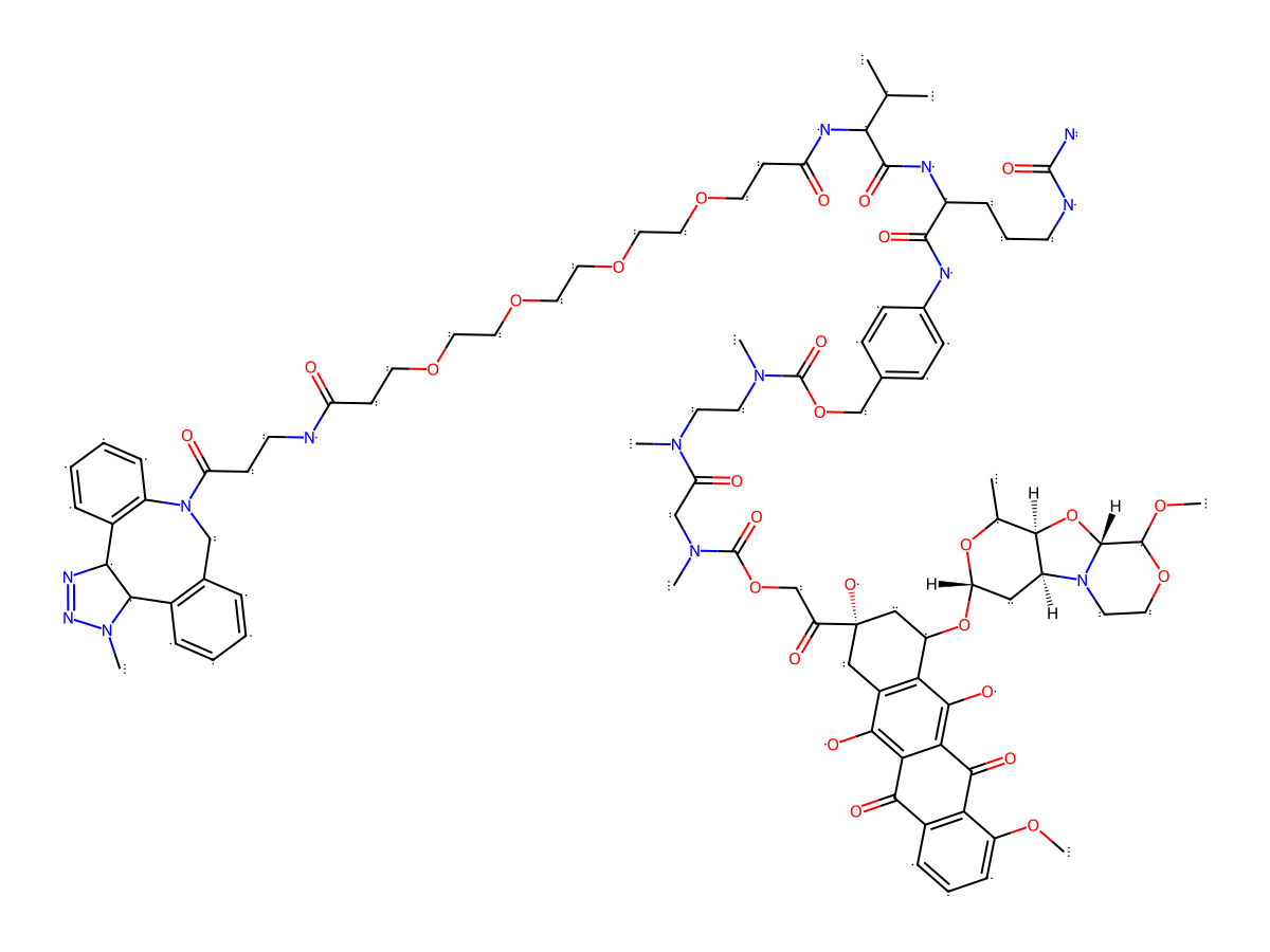
|
|||||
| Antibody Name |
FL1
|
Antibody Info | ||||
| Antigen Name |
Urokinase-type plasminogen activator (Plau)
|
Antigen Info | ||||
| Payload Name |
Undisclosed
|
|||||
| Linker Name |
DBCO-PEG4-Val-Cit-PABC
|
Linker Info | ||||
| Conjugate Type |
Random conjugation through reduced inter-chain cysteines.
|
|||||
General Information of The Activity Data Related to This ADC
Revealed Based on the Cell Line Data
Full List of Activity Data of This Antibody-drug Conjugate
Revealed Based on the Cell Line Data
| Experiment 1 Reporting the Activity Date of This ADC | [1] | ||||
| Efficacy Data | Half Maximal Effective Concentration (EC50) | 0.005 nM | High uPAR expression (uPAR +++) | ||
| Method Description |
Cells, plated in triplicates at 3 × 10³ cells per well in 96-well plates, were dosed with eight series of fourfold serially diluted PNU conjugates, unconjugated FL1, and free toxin (Creative Biolabs, #ADC-P-025) in 100 ul of complete growth media. Control cells were incubated with PBS-supplemented media. Cell viability was measured after 3 or 5 days of incubation, using CellTiter 96 Aqueous one-solution cell-proliferation assay reagent (MTS, Promega, #G358), as per the vendor's recommendations. Plate absorbance was then measured at 490 and 650 nm (for background subtraction) using a Spectra Max Plus plate reader (Molecular Devices). Data were plotted as a function of drug concentration (nanomolar) in GraphPad Prism 9 and fitted to a four-parameter nonlinear regression function, from which EC50 values were derived.
Click to Show/Hide
|
||||
| In Vitro Model | Breast adenocarcinoma | MDA-MB-231 cells | CVCL_0062 | ||
| Experiment 2 Reporting the Activity Date of This ADC | [1] | ||||
| Efficacy Data | Half Maximal Effective Concentration (EC50) | 0.02 nM | High uPAR expression (uPAR +++) | ||
| Method Description |
Cells, plated in triplicates at 3 × 10³ cells per well in 96-well plates, were dosed with eight series of fourfold serially diluted PNU conjugates, unconjugated FL1, and free toxin (Creative Biolabs, #ADC-P-025) in 100 ul of complete growth media. Control cells were incubated with PBS-supplemented media. Cell viability was measured after 3 or 5 days of incubation, using CellTiter 96 Aqueous one-solution cell-proliferation assay reagent (MTS, Promega, #G358), as per the vendor's recommendations. Plate absorbance was then measured at 490 and 650 nm (for background subtraction) using a Spectra Max Plus plate reader (Molecular Devices). Data were plotted as a function of drug concentration (nanomolar) in GraphPad Prism 9 and fitted to a four-parameter nonlinear regression function, from which EC50 values were derived.
Click to Show/Hide
|
||||
| In Vitro Model | Pancreatic ductal adenocarcinoma | KPC2 cells | Mus musculus | ||
| Experiment 3 Reporting the Activity Date of This ADC | [1] | ||||
| Efficacy Data | Half Maximal Effective Concentration (EC50) | 0.03 nM | High uPAR expression (uPAR +++) | ||
| Method Description |
Cells, plated in triplicates at 3 × 10³ cells per well in 96-well plates, were dosed with eight series of fourfold serially diluted PNU conjugates, unconjugated FL1, and free toxin (Creative Biolabs, #ADC-P-025) in 100 ul of complete growth media. Control cells were incubated with PBS-supplemented media. Cell viability was measured after 3 or 5 days of incubation, using CellTiter 96 Aqueous one-solution cell-proliferation assay reagent (MTS, Promega, #G358), as per the vendor's recommendations. Plate absorbance was then measured at 490 and 650 nm (for background subtraction) using a Spectra Max Plus plate reader (Molecular Devices). Data were plotted as a function of drug concentration (nanomolar) in GraphPad Prism 9 and fitted to a four-parameter nonlinear regression function, from which EC50 values were derived.
Click to Show/Hide
|
||||
| In Vitro Model | Pancreatic ductal adenocarcinoma | KPC1 cells | Mus musculus | ||
| Experiment 4 Reporting the Activity Date of This ADC | [1] | ||||
| Efficacy Data | Half Maximal Effective Concentration (EC50) | 0.05 nM | Moderate uPAR expression (uPAR++) | ||
| Method Description |
Cells, plated in triplicates at 3 × 10³ cells per well in 96-well plates, were dosed with eight series of fourfold serially diluted PNU conjugates, unconjugated FL1, and free toxin (Creative Biolabs, #ADC-P-025) in 100 ul of complete growth media. Control cells were incubated with PBS-supplemented media. Cell viability was measured after 3 or 5 days of incubation, using CellTiter 96 Aqueous one-solution cell-proliferation assay reagent (MTS, Promega, #G358), as per the vendor's recommendations. Plate absorbance was then measured at 490 and 650 nm (for background subtraction) using a Spectra Max Plus plate reader (Molecular Devices). Data were plotted as a function of drug concentration (nanomolar) in GraphPad Prism 9 and fitted to a four-parameter nonlinear regression function, from which EC50 values were derived.
Click to Show/Hide
|
||||
| In Vitro Model | Pancreatic ductal adenocarcinoma | BxPC-3 cells | CVCL_0186 | ||
| Experiment 5 Reporting the Activity Date of This ADC | [1] | ||||
| Efficacy Data | Half Maximal Effective Concentration (EC50) | 0.08 nM | High uPAR expression (uPAR +++) | ||
| Method Description |
Cells, plated in triplicates at 3 × 10³ cells per well in 96-well plates, were dosed with eight series of fourfold serially diluted PNU conjugates, unconjugated FL1, and free toxin (Creative Biolabs, #ADC-P-025) in 100 ul of complete growth media. Control cells were incubated with PBS-supplemented media. Cell viability was measured after 3 or 5 days of incubation, using CellTiter 96 Aqueous one-solution cell-proliferation assay reagent (MTS, Promega, #G358), as per the vendor's recommendations. Plate absorbance was then measured at 490 and 650 nm (for background subtraction) using a Spectra Max Plus plate reader (Molecular Devices). Data were plotted as a function of drug concentration (nanomolar) in GraphPad Prism 9 and fitted to a four-parameter nonlinear regression function, from which EC50 values were derived.
Click to Show/Hide
|
||||
| In Vitro Model | Pancreatic ductal adenocarcinoma | AsPc1 cells | CVCL_0152 | ||
| Experiment 6 Reporting the Activity Date of This ADC | [1] | ||||
| Efficacy Data | Half Maximal Effective Concentration (EC50) | 0.3 nM | Moderate uPAR expression (uPAR++) | ||
| Method Description |
Cells, plated in triplicates at 3 × 10³ cells per well in 96-well plates, were dosed with eight series of fourfold serially diluted PNU conjugates, unconjugated FL1, and free toxin (Creative Biolabs, #ADC-P-025) in 100 ul of complete growth media. Control cells were incubated with PBS-supplemented media. Cell viability was measured after 3 or 5 days of incubation, using CellTiter 96 Aqueous one-solution cell-proliferation assay reagent (MTS, Promega, #G358), as per the vendor's recommendations. Plate absorbance was then measured at 490 and 650 nm (for background subtraction) using a Spectra Max Plus plate reader (Molecular Devices). Data were plotted as a function of drug concentration (nanomolar) in GraphPad Prism 9 and fitted to a four-parameter nonlinear regression function, from which EC50 values were derived.
Click to Show/Hide
|
||||
| In Vitro Model | Pancreatic ductal adenocarcinoma | MIA PaCa-2 cells | CVCL_0428 | ||
| Experiment 7 Reporting the Activity Date of This ADC | [1] | ||||
| Efficacy Data | Half Maximal Effective Concentration (EC50) | 12 nM | Negative uPAR expression (uPAR-) | ||
| Method Description |
Cells, plated in triplicates at 3 × 10³ cells per well in 96-well plates, were dosed with eight series of fourfold serially diluted PNU conjugates, unconjugated FL1, and free toxin (Creative Biolabs, #ADC-P-025) in 100 ul of complete growth media. Control cells were incubated with PBS-supplemented media. Cell viability was measured after 3 or 5 days of incubation, using CellTiter 96 Aqueous one-solution cell-proliferation assay reagent (MTS, Promega, #G358), as per the vendor's recommendations. Plate absorbance was then measured at 490 and 650 nm (for background subtraction) using a Spectra Max Plus plate reader (Molecular Devices). Data were plotted as a function of drug concentration (nanomolar) in GraphPad Prism 9 and fitted to a four-parameter nonlinear regression function, from which EC50 values were derived.
Click to Show/Hide
|
||||
| In Vitro Model | Pancreatic ductal adenocarcinoma | KPC2 cells (uPAR KO) | Mus musculus | ||
| Experiment 8 Reporting the Activity Date of This ADC | [1] | ||||
| Efficacy Data | Half Maximal Effective Concentration (EC50) | 17 nM | Negative uPAR expression (uPAR-) | ||
| Method Description |
Cells, plated in triplicates at 3 × 10³ cells per well in 96-well plates, were dosed with eight series of fourfold serially diluted PNU conjugates, unconjugated FL1, and free toxin (Creative Biolabs, #ADC-P-025) in 100 ul of complete growth media. Control cells were incubated with PBS-supplemented media. Cell viability was measured after 3 or 5 days of incubation, using CellTiter 96 Aqueous one-solution cell-proliferation assay reagent (MTS, Promega, #G358), as per the vendor's recommendations. Plate absorbance was then measured at 490 and 650 nm (for background subtraction) using a Spectra Max Plus plate reader (Molecular Devices). Data were plotted as a function of drug concentration (nanomolar) in GraphPad Prism 9 and fitted to a four-parameter nonlinear regression function, from which EC50 values were derived.
Click to Show/Hide
|
||||
| In Vitro Model | Pancreatic ductal adenocarcinoma | KPC1 cells (uPAR KO) | Mus musculus | ||
References