
Antibody-drug Conjugate Information
General Information of This Antibody-drug Conjugate (ID: DRG0XGZGL)
| ADC Name |
CD79b-3
|
|||||
|---|---|---|---|---|---|---|
| Synonyms |
CD79b-3
Click to Show/Hide
|
|||||
| Organization |
Novartis Biomedical Research.
|
|||||
| Drug Status |
Investigative
|
|||||
| Drug-to-Antibody Ratio |
4
|
|||||
| Structure |
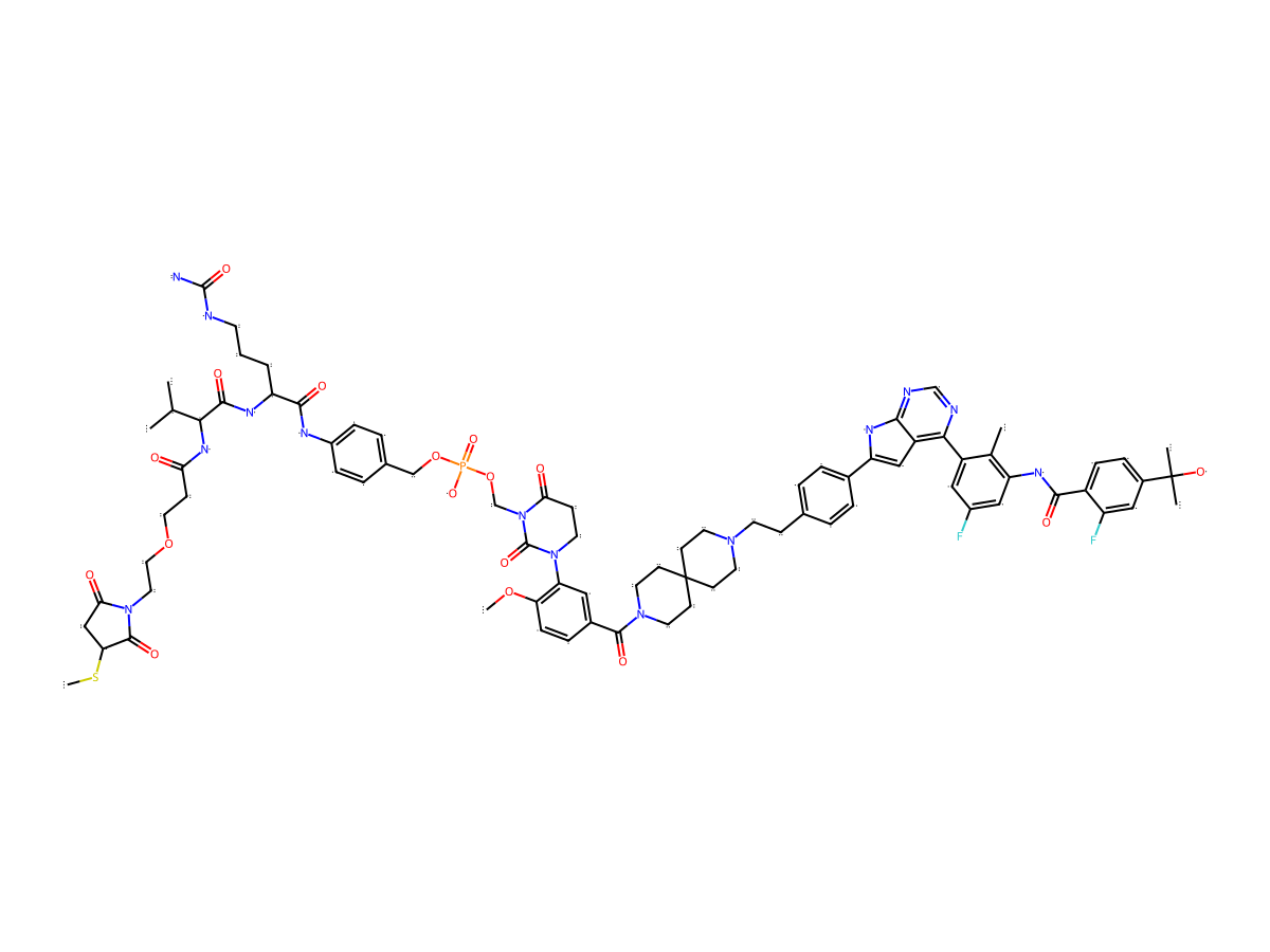
|
|||||
| Antibody Name |
Undisclosed
|
|||||
| Antigen Name |
B-cell antigen receptor complex-associated protein beta chain (CD79B)
|
Antigen Info | ||||
| Payload Name |
BTK PROTAC 1
|
Payload Info | ||||
| Linker Name |
Undisclosed
|
|||||
| Conjugate Type |
Random conjugation through reduced inter-chain cysteines.
|
|||||
General Information of The Activity Data Related to This ADC
Full List of Activity Data of This Antibody-drug Conjugate
Revealed Based on the Cell Line Data
| Experiment 1 Reporting the Activity Date of This ADC | [1] | ||||
| Efficacy Data | Half Maximal Degradation Concentration (DC50) |
0.045 nM
|
|||
| Method Description |
The TMD8 wt cell line was cultured in media that is optimal for their growth at 5% CO 2, 37°C in a tissue culture incubator. Prior to seeding for the growth inhibition assay, the cells were split at least 2 days before the assay to ensure optimal growth density. On the day of seeding, suspension cells were harvested. Cell viability and cell density were determined using a cell counter (Vi-Cell XR Cell Viability Analyzer, Beckman Coulter). Cells with higher than 85% viability were seeded in white clear bottom 96-well TC treated plates (Corning cat. #3903). Cells were seeded at a density of 75,000 cells per well in 70 uL of standard growth media. Plates were incubated at 5% CO 2, 37°C overnight in a tissue culture incubator. On the day of dosing, all ADCs and low molecular weight payloads were prepared at 8X in standard growth media. The prepared treatments were added to the cells, resulting in final concentrations of 9.92e-6 - 100 nM and a final volume of 80 uL per well. Each drug concentration was tested in duplicates. Plates were incubated at 5% CO 2, 37°C for 48 hours in a tissue culture incubator. After 48 hours, cells were lysed with 80 uL of freshly prepared lysis buffer (10X Millipore RIPA Lysis Buffer + 0.5M EDTA + 1x Roche cOmpleteTM Protease Inhibitor Cocktail EDTA-free tablet, diluted in ddH 2O to 2X). Cells were resuspended and incubated on ice for 10 minutes. Lysates were stored until further use at -80°C. Prior to running the assay, MULTI-ARRAY 96 Plate Pack, SECTOR Plates (MSD, L15XA-3) were coated with 50 uL/well of a BTK (D3H5) Rabbit mAb (Cell Signaling Technology, 8547BF) at 1 ug/mL diluted in PBS overnight at 4°C. The next day, MSD plates were washed 3 times with 300 uL/well of PBS + 0.05% Tween. Plates were then blocked with 150 uL/well of PBS + 5% BSA buffer, shaking at 450 rpm for 1 hour at room temperature. Plates were then washed 3 times with 300 uL/well of PBS + 0.05% Tween. Each lysate sample was added to the MSD plates at 25 uL/well, shaking at 450 rpm for 2 hours at room temperature. Plates were then washed 3 times with 300 uL/well of PBS + 0.05% Tween. A Sulfo-tag purified mouse anti-human BTK antibody (BD, clone 53, 624084) was diluted to 0.5 ug/mL and 25 uL was added to each well. Plates were shaken at 450 rpm for 1 hour at room temperature. Plates were then washed 3 times with 300 uL/well of PBS + 0.05% Tween. MSD Read Buffer T (4x) (MSD, R92TC-1) was diluted to 2X with ddH 2O and 150 uL was added to each well. Plates were immediately read on an MSD Sector 600 Imager. To evaluate the effect of the drug treatments, signal levels from wells containing untreated cells (100% viability) were used to normalize treated samples. A variable slope model was applied to fit a nonlinear regression curve to the data in GraphPad PRISM version 10 software. GI50 and Amax values were extrapolated from the resultant curves.
Click to Show/Hide
|
||||
| In Vitro Model | Diffuse large B-cell lymphoma, Diffuse large B-cell lymphoma activated B-cell type | TMD8 cells (wt) | CVCL_A442 | ||
References