
Antibody-drug Conjugate Information
General Information of This Antibody-drug Conjugate (ID: DRG0REROK)
| ADC Name |
BGA9962
|
|||||
|---|---|---|---|---|---|---|
| Synonyms |
BGA9962
Click to Show/Hide
|
|||||
| Organization |
BEIGENE, LTD. | BEIGENE SWITZERLAND GMBH
|
|||||
| Drug Status |
Investigative
|
|||||
| Drug-to-Antibody Ratio |
7.8
|
|||||
| Structure |
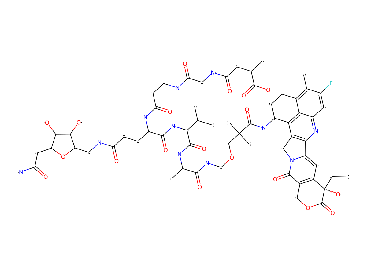
|
|||||
| Antibody Name |
BGA5384
|
Antibody Info | ||||
| Antigen Name |
Carcinoembryonic antigen-related cell adhesion molecule 5 (CEACAM5)
|
Antigen Info | ||||
| Payload Name |
ADC-C3 Payload
|
Payload Info | ||||
| Linker Name |
Linker of ADC2-58
|
Linker Info | ||||
| Conjugate Type |
Random conjugation through reduced inter-chain cysteines.
|
|||||
General Information of The Activity Data Related to This ADC
Discovered Using Patient-derived Xenograft Model
Revealed Based on the Cell Line Data
Full List of Activity Data of This Antibody-drug Conjugate
Discovered Using Patient-derived Xenograft Model
| Experiment 1 Reporting the Activity Date of This ADC | [1] | ||||
| Efficacy Data | Tumor Growth lnhibition value (TGl) | 106% | Positive CEA expression (CEA+++/++) | ||
| Method Description |
Human patient-derived gastric tumors were induced on the right flank by a subcutaneous injection. When tumor volume reached approximately 200 mm3 in size, mice were randomized into 5 groups with 8, 9, and 9 animals in vehicle, BGA7650, and BGA9962 groups on Day 0, respectively. After ensuring all cohorts hadapproximately equal average tumor volumes to start, animals were intravenously
administered vehicle, BGA9962 (2 mg/kg) on treatment Day 1. Animal body weight and tumor volume were measured twice weekly.
Click to Show/Hide
|
||||
| In Vivo Model | Human patient-derived gastric xenograft model | ||||
| Experiment 2 Reporting the Activity Date of This ADC | [1] | ||||
| Efficacy Data | Tumor Growth lnhibition value (TGl) | 107% | Positive CEA expression (CEA+++/++) | ||
| Method Description |
Human patient-derived gastric tumors were induced on the right flank by a subcutaneous injection. When tumor volume reached approximately 200 mm3 in size, mice were randomized into 5 groups with 8, 9, and 9 animals in vehicle, BGA7650, and BGA9962 groups on Day 0, respectively. After ensuring all cohorts hadapproximately equal average tumor volumes to start, animals were intravenously
administered vehicle, BGA9962 (6 mg/kg) on treatment Day 1. Animal body weight and tumor volume were measured twice weekly.
Click to Show/Hide
|
||||
| In Vivo Model | Human patient-derived gastric xenograft model | ||||
Revealed Based on the Cell Line Data
| Experiment 1 Reporting the Activity Date of This ADC | [1] | ||||
| Efficacy Data | Half Maximal Effective Concentration (EC50) | 0.17 nM | Positive CEA expression (CEA+++/++) | ||
| Method Description |
NCI-H2122 cells were plated at a density of 2000 cells per well and the next day were treated with payload compounds (5x dilution) for 6 days.
|
||||
| In Vitro Model | Lung adenocarcinoma | NCI-H2122 cells | CVCL_1531 | ||
| Experiment 2 Reporting the Activity Date of This ADC | [1] | ||||
| Efficacy Data | Half Maximal Effective Concentration (EC50) | 0.39 nM | Positive CEA expression (CEA+++/++) | ||
| Method Description |
MKN45 cells were plated at a density of 5000 cells per well and the next day were treated with payload compounds (5x dilution) for 6 days.
|
||||
| In Vitro Model | Gastric adenocarcinoma | MKN45 cells | CVCL_0434 | ||
| Experiment 3 Reporting the Activity Date of This ADC | [1] | ||||
| Efficacy Data | Half Maximal Effective Concentration (EC50) | 0.83 nM | Positive CEA expression (CEA+++/++) | ||
| Method Description |
Ls147T cells were plated at a density of 5000 cells per well and the next day were treated with payload compounds (5x dilution) for 6 days.
|
||||
| In Vitro Model | Colon adenocarcinoma | Ls147T cells | CVCL_1384 | ||
| Experiment 4 Reporting the Activity Date of This ADC | [1] | ||||
| Efficacy Data | Half Maximal Effective Concentration (EC50) | 27 nM | Negative CEA expression (CEA-) | ||
| Method Description |
MDA-MB-231 cells were plated at a density of 2000 cells per well and the next day were treated with payload compounds (5x dilution) for 6 days.
|
||||
| In Vitro Model | Breast adenocarcinoma | MDA-MB-231 cells | CVCL_0062 | ||