Payload Information
General Information of This Payload
| Payload ID | PAY0UXSPW |
|||||
|---|---|---|---|---|---|---|
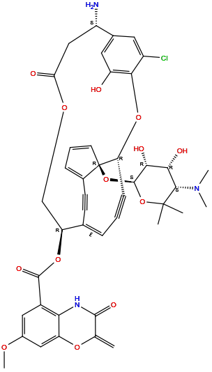
| Name | Lidamycin |
|||||
| Synonyms |
C-1027; CHEBI:68320; lidamycin; CHEMBL506048; Q27136818; [(1R,7S,12R,13E,20R)-7-Amino-4-chloro-20-[(2S,3R,4R,5S)-5-(dimethylamino)-3,4-dihydroxy-6,6-dimethyloxan-2-yl]oxy-25-hydroxy-9-oxo-2,10-dioxatetracyclo[11.7.3.23,6.016,20]pentacosa-3(25),4,6(24),13(23),16,18-hexaen-14,21-diyn-12-yl] 7-methoxy-2-methylidene-3-oxo-4H-1,4-benzoxazine-5-carboxylate; 149438-20-0
Click to Show/Hide
|
|||||
| Target(s) | Human Deoxyribonucleic acid (hDNA) | |||||
| Structure |

|
|||||
| Formula | C43H42ClN3O13 |
|||||
| Isosmiles | CC1([C@H]([C@H]([C@H]([C@@H](O1)O[C@]23C=CC=C2C#C/C/4=C\C#C[C@H]3OC5=C(C=C(C=C5Cl)[C@H](CC(=O)OC[C@@H]4OC(=O)C6=C7C(=CC(=C6)OC)OC(=C)C(=O)N7)N)O)O)O)N(C)C)C |
|||||
| PubChem CID | ||||||
| InChI |
InChI=1S/C43H42ClN3O13/c1-21-39(52)46-34-26(17-25(54-6)18-30(34)56-21)40(53)57-31-20-55-33(49)19-28(45)23-15-27(44)37(29(48)16-23)58-32-11-7-9-22(31)12-13-24-10-8-14-43(24,32)60-41-36(51)35(50)38(47(4)5)42(2,3)59-41/h8-10,14-18,28,31-32,35-36,38,41,48,50-51H,1,19-20,45H2,2-6H3,(H,46,52)/b22-9+/t28-,31-,32+,35-,36+,38-,41-,43+/m0/s1
|
|||||
| InChIKey |
DGGZCXUXASNDAC-QQNGCVSVSA-N
|
|||||
| IUPAC Name |
[(1R,7S,12R,13E,20R)-7-amino-4-chloro-20-[(2S,3R,4R,5S)-5-(dimethylamino)-3,4-dihydroxy-6,6-dimethyloxan-2-yl]oxy-25-hydroxy-9-oxo-2,10-dioxatetracyclo[11.7.3.23,6.016,20]pentacosa-3(25),4,6(24),13(23),16,18-hexaen-14,21-diyn-12-yl] 7-methoxy-2-methylidene-3-oxo-4H-1,4-benzoxazine-5-carboxylate
|
|||||
| Pharmaceutical Properties | Molecule Weight |
844.3 |
Polar area |
218 |
||
Complexity |
1970 |
xlogp Value |
2 |
|||
Heavy Count |
60 |
Rot Bonds |
7 |
|||
Hbond acc |
15 |
Hbond Donor |
5 |
|||
The activity data of This Payload
| Standard Type | Value | Units | Cell line | Disease Model | Cell line ID | Reference |
|---|---|---|---|---|---|---|
| Half Maximal Inhibitory Concentration (IC50) | 20 | nM |
P388 cells
|
Lymphoma
|
[1] |
Each Antibody-drug Conjugate Related to This Payload
Full Information of The Activity Data of The ADC(s) Related to This Payload
Dualtargeting lidamycin ADC [Investigative]
Discovered Using Patient-derived Xenograft Model
| Experiment 1 Reporting the Activity Date of This ADC | [2] | ||||
| Efficacy Data | Tumor Growth Inhibition value (TGI) | ≈ 56.63%±9.71% | Positive EGFR expression (EGFR+++/++) | ||
| Method Description |
PDX mice were administrated vehicle or DTLL at the LDMequivalent dose of 0.1 mg/kg once a week for 3 wk. Tumor volumes were measured after animals were sacrificed on Days 24 and 39, respectively. DTLL was administered via tail vein injection once a week for three weeks.
|
||||
| In Vivo Model | Pancreatic cancer PDX model (PDX: PA1338) | ||||
Fv-LDP-D3-AE [Investigative]
Discovered Using Cell Line-derived Xenograft Model
| Experiment 1 Reporting the Activity Date of This ADC | [3] | ||||
| Efficacy Data | Tumor Growth Inhibition value (TGI) | ≈ 64.00% (Day 28) | High EGFR expression (EGFR+++) | ||
| Method Description |
KYSE150 cells (5 10 6 cells/200 L) suspended in PBS were inoculated subcutaneously into the right armpit of athymic mice. When the tumor volume reached about 80mm3, nude mice were randomized into two groups (n=6, per group), a control group and a Fv-LDP-D3-AE (0.2 mg/kg) group. The drug treatment group was given by tail vein injection once a week for two consecutive weeks.
Click to Show/Hide
|
||||
| In Vivo Model | Esophageal squamous cell carcinoma CDX model | ||||
| In Vitro Model | Esophageal squamous cell carcinoma | KYSE-150 cells | CVCL_1348 | ||
| Experiment 2 Reporting the Activity Date of This ADC | [3] | ||||
| Efficacy Data | Tumor Growth Inhibition value (TGI) | ≈ 73.00% (Day 28) | High EGFR expression (EGFR+++) | ||
| Method Description |
KYSE150 cells (5 10 6 cells/200 L) suspended in PBS were inoculated subcutaneously into the right armpit of athymic mice. When the tumor volume reached about 80mm3, nude mice were randomized into two groups (n=6, per group), a control group and a Fv-LDP-D3-AE (0.25 mg/kg) group. The drug treatment group was given by tail vein injection once a week for two consecutive weeks.
Click to Show/Hide
|
||||
| In Vivo Model | Esophageal squamous cell carcinoma CDX model | ||||
| In Vitro Model | Esophageal squamous cell carcinoma | KYSE-150 cells | CVCL_1348 | ||
| Experiment 3 Reporting the Activity Date of This ADC | [3] | ||||
| Efficacy Data | Tumor Growth Inhibition value (TGI) | ≈ 81.00% (Day 28) | High EGFR expression (EGFR+++) | ||
| Method Description |
KYSE150 cells (5 10 6 cells/200 L) suspended in PBS were inoculated subcutaneously into the right armpit of athymic mice. When the tumor volume reached about 80mm3, nude mice were randomized into two groups (n=6, per group), a control group and a Fv-LDP-D3-AE (0.5 mg/kg) group. The drug treatment group was given by tail vein injection once a week for two consecutive weeks.
Click to Show/Hide
|
||||
| In Vivo Model | Esophageal squamous cell carcinoma CDX model | ||||
| In Vitro Model | Esophageal squamous cell carcinoma | KYSE-150 cells | CVCL_1348 | ||
Revealed Based on the Cell Line Data
| Experiment 1 Reporting the Activity Date of This ADC | [3] | ||||
| Efficacy Data | Half Maximal Inhibitory Concentration (IC50) |
33.00 ng/mL
|
High EGFR expression (EGFR+++) | ||
| Method Description |
The CCK8 method was used to evaluate the cytotoxicity of Fv-LDP-D3 and Fv-LDP-D3-AE in KYSE150, KYSE520, and Eca109 cells.
|
||||
| In Vitro Model | Esophageal squamous cell carcinoma | KYSE-150 cells | CVCL_1348 | ||
| Experiment 2 Reporting the Activity Date of This ADC | [3] | ||||
| Efficacy Data | Half Maximal Inhibitory Concentration (IC50) |
44.00 ng/mL
|
High EGFR expression (EGFR+++) | ||
| Method Description |
The CCK8 method was used to evaluate the cytotoxicity of Fv-LDP-D3 and Fv-LDP-D3-AE in KYSE150, KYSE520, and Eca109 cells.
|
||||
| In Vitro Model | Esophageal squamous cell carcinoma | KYSE-520 cells | CVCL_1355 | ||
| Experiment 3 Reporting the Activity Date of This ADC | [3] | ||||
| Efficacy Data | Half Maximal Inhibitory Concentration (IC50) |
75.00 ng/mL
|
Moderate EGFR expression (EGFR++) | ||
| Method Description |
The CCK8 method was used to evaluate the cytotoxicity of Fv-LDP-D3 and Fv-LDP-D3-AE in KYSE150, KYSE520, and Eca109 cells.
|
||||
| In Vitro Model | Esophageal squamous cell carcinoma | Eca-109 cells | CVCL_6898 | ||
References
