
Payload Information
General Information of This Payload (ID: PAY0MZOZX)
| Name |
Alpha-amanitin
|
|||||
|---|---|---|---|---|---|---|
| Synonyms |
Alpha-amanitin; 23109-05-9; alpha-Amanitine; .alpha.-Amanitin; HSDB 3458; C39H54N10O14S; EINECS 245-432-2; BRN 1071138; C39-H54-N10-O14-S; CHEMBL3792970; SCHEMBL16491633; Cyclic(L-asparaginyl-4-hydroxy-L-prolyl-(R)-4,5-dihydroxy-L-isoleucyl-6-hydroxy-2-mercapto-L-tryptophylglycyl-L-isoleucylglycyl-L-cysteinyl), cyclic (4-8)-sulfide, (R)-S-oxide; AKOS024457936; CS-5321; HY-19610; 11000-43-4; cyclo[L-Asparaginyl-4-hydroxy-L-proly-(R-4,5-dihydroxy-L-isoleucyl-6-hydroxy-2-mercapto-L-tryptophylglycyl-L-isoleucylglycyl-L-cysteinyl]cyclic (4?8)-sulfide (R)-S-oxide
Click to Show/Hide
|
|||||
| Target | DNA-directed RNA polymerase II subunit RPB2 (POLR2B); DNA-directed RNA polymerase III subunit RPC7 (POLR3G) | |||||
| Structure |
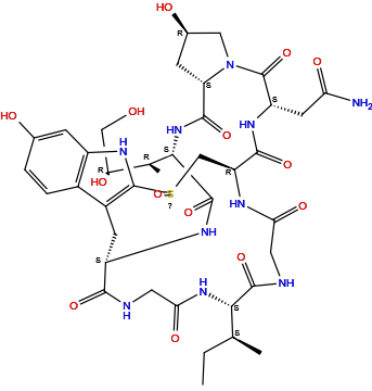
|
|||||
| Formula | C39H54N10O14S |
|||||
| Isosmiles | CC[C@H](C)[C@H]1C(=O)NCC(=O)N[C@H]2CS(=O)C3=C(C[C@@H](C(=O)NCC(=O)N1)NC(=O)[C@@H](NC(=O)[C@@H]4C[C@H](CN4C(=O)[C@@H](NC2=O)CC(=O)N)O)[C@@H](C)[C@H](CO)O)C5=C(N3)C=C(C=C5)O |
|||||
| PubChem CID | ||||||
| InChI |
InChI=1S/C39H54N10O14S/c1-4-16(2)31-36(60)42-11-29(55)43-25-15-64(63)38-21(20-6-5-18(51)7-22(20)46-38)9-23(33(57)41-12-30(56)47-31)44-37(61)32(17(3)27(53)14-50)48-35(59)26-8-19(52)13-49(26)39(62)24(10-28(40)54)45-34(25)58/h5-7,16-17,19,23-27,31-32,46,50-53H,4,8-15H2,1-3H3,(H2,40,54)(H,41,57)(H,42,60)(H,43,55)(H,44,61)(H,45,58)(H,47,56)(H,48,59)/t16-,17-,19+,23-,24-,25-,26-,27-,31-,32-,64?/m0/s1
|
|||||
| InChIKey |
CIORWBWIBBPXCG-JAXJKTSHSA-N
|
|||||
| IUPAC Name |
2-[(1R,4S,8R,10S,13S,16S,34S)-34-[(2S)-butan-2-yl]-13-[(2R,3R)-3,4-dihydroxybutan-2-yl]-8,22-dihydroxy-2,5,11,14,27,30,33,36,39-nonaoxo-27lambda4-thia-3,6,12,15,25,29,32,35,38-nonazapentacyclo[14.12.11.06,10.018,26.019,24]nonatriaconta-18(26),19(24),20,22-tetraen-4-yl]acetamide
|
|||||
| Pharmaceutical Properties | Molecule Weight |
919 |
Polar area |
400 |
||
Complexity |
1840 |
xlogp Value |
-4.4 |
|||
Heavy Count |
64 |
Rot Bonds |
7 |
|||
Hbond acc |
15 |
Hbond Donor |
13 |
|||
The activity data of This Payload
| Standard Type | Value | Units | Cell line | Disease Model | Cell line ID | Reference |
|---|---|---|---|---|---|---|
| Half Maximal Inhibitory Concentration (IC50) | 1 | nM |
HeLa cells
|
Endocervical adenocarcinoma
|
[1] | |
| Half Maximal Inhibitory Concentration (IC50) | 1000 | nM |
HeLa cells
|
Endocervical adenocarcinoma
|
[1] | |
| Half Maximal Inhibitory Concentration (IC50) | 1000 | nM |
HEK293 cells
|
Normal
|
[1] | |
| Half Maximal Inhibitory Concentration (IC50) | 1000 | nM |
CHO cells
|
Normal
|
[1] | |
| Half Maximal Inhibitory Concentration (IC50) | 50 | nM |
HT-29 cells
|
Colon adenocarcinoma
|
[2] | |
| Half Maximal Inhibitory Concentration (IC50) | 560 | nM |
Hep-G2 cells
|
Hepatoblastoma
|
[1] |
Each Antibody-drug Conjugate Related to This Payload
Full Information of The Activity Data of The ADC(s) Related to This Payload
Trastuzumab-alpha-amanitin conjugate 11 [Investigative]
Discovered Using Cell Line-derived Xenograft Model
| Experiment 1 Reporting the Activity Date of This ADC | [3] | ||||
| Efficacy Data | Tumor Growth Inhibition value (TGI) | ≈ 28.53% | High HER2 expression (HER2 +++) | ||
| Method Description |
A mouse tumor xenograft model, wherein 250,000,000 SKOV-3 ovarial carcinoma cellsare implated sub-cutaneously (s.c,) into SCID mice and allowed to grow for 10 days. After 10 days a single dose of 30 ug/kg body weight of various a-amanitin-Herceptin conjugates.
|
||||
| In Vivo Model | SK-OV-3 CDX model | ||||
| In Vitro Model | Ovarian serous cystadenocarcinoma | SK-OV-3 cells | CVCL_0532 | ||
| Experiment 2 Reporting the Activity Date of This ADC | [3] | ||||
| Efficacy Data | Tumor Growth Inhibition value (TGI) | ≈ 94.60% | High HER2 expression (HER2 +++) | ||
| Method Description |
A mouse tumor xenograft model, wherein 250,000,000 SKOV-3 ovarial carcinoma cellsare implated sub-cutaneously (s.c,) into SCID mice and allowed to grow for 10 days. After 10 days a single dose of 150 ug/kg body weight of various a-amanitin-Herceptin conjugates.
|
||||
| In Vivo Model | SK-OV-3 CDX model | ||||
| In Vitro Model | Ovarian serous cystadenocarcinoma | SK-OV-3 cells | CVCL_0532 | ||
Trastuzumab-alpha-amanitin conjugate 4 [Investigative]
Discovered Using Cell Line-derived Xenograft Model
| Experiment 1 Reporting the Activity Date of This ADC | [3] | ||||
| Efficacy Data | Tumor Growth Inhibition value (TGI) | ≈ 37.30% | High HER2 expression (HER2 +++) | ||
| Method Description |
A mouse tumor xenograft model, wherein 250,000,000 SKOV-3 ovarial carcinoma cellsare implated sub-cutaneously (s.c,) into SCID mice and allowed to grow for 10 days. After 10 days a single dose of 30 ug/kg body weight of various a-amanitin-Herceptin conjugates.
|
||||
| In Vivo Model | SK-OV-3 CDX model | ||||
| In Vitro Model | Ovarian serous cystadenocarcinoma | SK-OV-3 cells | CVCL_0532 | ||
| Experiment 2 Reporting the Activity Date of This ADC | [3] | ||||
| Efficacy Data | Tumor Growth Inhibition value (TGI) | ≈ 76.05% | High HER2 expression (HER2 +++) | ||
| Method Description |
A mouse tumor xenograft model, wherein 250,000,000 SKOV-3 ovarial carcinoma cellsare implated sub-cutaneously (s.c,) into SCID mice and allowed to grow for 10 days. After 10 days a single dose of 150 ug/kg body weight of various a-amanitin-Herceptin conjugates.
|
||||
| In Vivo Model | SK-OV-3 CDX model | ||||
| In Vitro Model | Ovarian serous cystadenocarcinoma | SK-OV-3 cells | CVCL_0532 | ||
Trastuzumab-alpha-amanitin conjugate 10 [Investigative]
Discovered Using Cell Line-derived Xenograft Model
| Experiment 1 Reporting the Activity Date of This ADC | [3] | ||||
| Efficacy Data | Tumor Growth Inhibition value (TGI) | ≈ 55.08% | High HER2 expression (HER2 +++) | ||
| Method Description |
A mouse tumor xenograft model, wherein 250,000,000 SKOV-3 ovarial carcinoma cellsare implated sub-cutaneously (s.c,) into SCID mice and allowed to grow for 10 days. After 10 days a single dose of 30 ug/kg body weight of various a-amanitin-Herceptin conjugates.
|
||||
| In Vivo Model | SK-OV-3 CDX model | ||||
| In Vitro Model | Ovarian serous cystadenocarcinoma | SK-OV-3 cells | CVCL_0532 | ||
| Experiment 2 Reporting the Activity Date of This ADC | [3] | ||||
| Efficacy Data | Tumor Growth Inhibition value (TGI) | ≈ 98.16% | High HER2 expression (HER2 +++) | ||
| Method Description |
A mouse tumor xenograft model, wherein 250,000,000 SKOV-3 ovarial carcinoma cellsare implated sub-cutaneously (s.c,) into SCID mice and allowed to grow for 10 days. After 10 days a single dose of 150 ug/kg body weight of various a-amanitin-Herceptin conjugates.
|
||||
| In Vivo Model | SK-OV-3 CDX model | ||||
| In Vitro Model | Ovarian serous cystadenocarcinoma | SK-OV-3 cells | CVCL_0532 | ||
Trastuzumab-alpha-amanitin conjugate 3 [Investigative]
Discovered Using Cell Line-derived Xenograft Model
| Experiment 1 Reporting the Activity Date of This ADC | [3] | ||||
| Efficacy Data | Tumor Growth Inhibition value (TGI) | ≈ 71.25% | High HER2 expression (HER2 +++) | ||
| Method Description |
A mouse tumor xenograft model, wherein 250,000,000 SKOV-3 ovarial carcinoma cellsare implated sub-cutaneously (s.c,) into SCID mice and allowed to grow for 10 days. After 10 days a single dose of 30 ug/kg body weight of various a-amanitin-Herceptin conjugates.
|
||||
| In Vivo Model | SK-OV-3 CDX model | ||||
| In Vitro Model | Ovarian serous cystadenocarcinoma | SK-OV-3 cells | CVCL_0532 | ||
| Experiment 2 Reporting the Activity Date of This ADC | [3] | ||||
| Efficacy Data | Tumor Growth Inhibition value (TGI) | ≈ 85.43% | High HER2 expression (HER2 +++) | ||
| Method Description |
A mouse tumor xenograft model, wherein 250,000,000 SKOV-3 ovarial carcinoma cellsare implated sub-cutaneously (s.c,) into SCID mice and allowed to grow for 10 days. After 10 days a single dose of 150 ug/kg body weight of various a-amanitin-Herceptin conjugates.
|
||||
| In Vivo Model | SK-OV-3 CDX model | ||||
| In Vitro Model | Ovarian serous cystadenocarcinoma | SK-OV-3 cells | CVCL_0532 | ||
OHPAS ADC-1 [Investigative]
Discovered Using Cell Line-derived Xenograft Model
| Experiment 1 Reporting the Activity Date of This ADC | [4] | ||||
| Efficacy Data | Tumor Growth Inhibition value (TGI) | ≈ 74.10% | High HER2 expression (HER2 +++) | ||
| Method Description |
We also tested the OHPAS ADCs in in vivo xenograft mouse models using N87 cell lines. The experiment was followed up to 110 days after administration of ADCs at two different doses (0.5 mg/kg) on day one (initial tumor volume 100 mm3).
|
||||
| In Vivo Model | NCI-N87 CDX model | ||||
| In Vitro Model | Gastric tubular adenocarcinoma | NCI-N87 cells | CVCL_1603 | ||
| Experiment 2 Reporting the Activity Date of This ADC | [4] | ||||
| Efficacy Data | Tumor Growth Inhibition value (TGI) | ≈ 98.50% | High HER2 expression (HER2 +++) | ||
| Method Description |
We also tested the OHPAS ADCs in in vivo xenograft mouse models using N87 cell lines. The experiment was followed up to 110 days after administration of ADCs at two different doses (2 mg/kg) on day one (initial tumor volume 100 mm3).
|
||||
| In Vivo Model | NCI-N87 CDX model | ||||
| In Vitro Model | Gastric tubular adenocarcinoma | NCI-N87 cells | CVCL_1603 | ||
Revealed Based on the Cell Line Data
| Experiment 1 Reporting the Activity Date of This ADC | [4] | ||||
| Efficacy Data | Half Maximal Inhibitory Concentration (IC50) |
0.03 nM
|
High HER2 expression (HER2 +++) | ||
| Method Description |
With OHPAS ADCs 1-4 , we first performed cell-based MTT assays against a series of HER2 positive/negative cell lines using the commercially available HER2 ADC, T-DM1 (average DAR 3.5), which we purchased as a positive control.
|
||||
| In Vitro Model | Breast adenocarcinoma | SK-BR-3 cells | CVCL_0033 | ||
| Experiment 2 Reporting the Activity Date of This ADC | [4] | ||||
| Efficacy Data | Half Maximal Inhibitory Concentration (IC50) |
0.12 nM
|
High HER2 expression (HER2 +++) | ||
| Method Description |
With OHPAS ADCs 1-4 , we first performed cell-based MTT assays against a series of HER2 positive/negative cell lines using the commercially available HER2 ADC, T-DM1 (average DAR 3.5), which we purchased as a positive control.
|
||||
| In Vitro Model | Ovarian serous cystadenocarcinoma | SK-OV-3 cells | CVCL_0532 | ||
| Experiment 3 Reporting the Activity Date of This ADC | [4] | ||||
| Efficacy Data | Half Maximal Inhibitory Concentration (IC50) |
0.6 nM
|
High HER2 expression (HER2 +++) | ||
| Method Description |
With OHPAS ADCs 1-4 , we first performed cell-based MTT assays against a series of HER2 positive/negative cell lines using the commercially available HER2 ADC, T-DM1 (average DAR 3.5), which we purchased as a positive control.
|
||||
| In Vitro Model | Gastric tubular adenocarcinoma | NCI-N87 cells | CVCL_1603 | ||
| Experiment 4 Reporting the Activity Date of This ADC | [4] | ||||
| Efficacy Data | Half Maximal Inhibitory Concentration (IC50) | > 50 nM | Negative HER2 expression (HER2 -) | ||
| Method Description |
With OHPAS ADCs 1-4 , we first performed cell-based MTT assays against a series of HER2 positive/negative cell lines using the commercially available HER2 ADC, T-DM1 (average DAR 3.5), which we purchased as a positive control.
|
||||
| In Vitro Model | Invasive breast carcinoma | MCF-7 cells | CVCL_0031 | ||
| Experiment 5 Reporting the Activity Date of This ADC | [4] | ||||
| Efficacy Data | Half Maximal Inhibitory Concentration (IC50) | > 50 nM | Moderate HER2 expression (HER2 ++) | ||
| Method Description |
With OHPAS ADCs 1-4 , we first performed cell-based MTT assays against a series of HER2 positive/negative cell lines using the commercially available HER2 ADC, T-DM1 (average DAR 3.5), which we purchased as a positive control.
|
||||
| In Vitro Model | Breast ductal carcinoma | JIMT-1 cells | CVCL_2077 | ||
Trastuzumab-alpha-amanitin conjugate 15 [Investigative]
Discovered Using Cell Line-derived Xenograft Model
| Experiment 1 Reporting the Activity Date of This ADC | [3] | ||||
| Efficacy Data | Tumor Growth Inhibition value (TGI) | ≈ 86.62% | High HER2 expression (HER2 +++) | ||
| Method Description |
A mouse tumor xenograft model, wherein 250,000,000 SKOV-3 ovarial carcinoma cellsare implated sub-cutaneously (s.c,) into SCID mice and allowed to grow for 10 days. After 10 days a single dose of 30 ug/kg body weight of various a-amanitin-Herceptin conjugates.
|
||||
| In Vivo Model | SK-OV-3 CDX model | ||||
| In Vitro Model | Ovarian serous cystadenocarcinoma | SK-OV-3 cells | CVCL_0532 | ||
| Experiment 2 Reporting the Activity Date of This ADC | [3] | ||||
| Efficacy Data | Tumor Growth Inhibition value (TGI) | ≈ 98.16% | High HER2 expression (HER2 +++) | ||
| Method Description |
A mouse tumor xenograft model, wherein 250,000,000 SKOV-3 ovarial carcinoma cellsare implated sub-cutaneously (s.c,) into SCID mice and allowed to grow for 10 days. After 10 days a single dose of 150 ug/kg body weight of various a-amanitin-Herceptin conjugates.
|
||||
| In Vivo Model | SK-OV-3 CDX model | ||||
| In Vitro Model | Ovarian serous cystadenocarcinoma | SK-OV-3 cells | CVCL_0532 | ||
Trastuzumab-alpha-amanitin conjugate 7 [Investigative]
Discovered Using Cell Line-derived Xenograft Model
| Experiment 1 Reporting the Activity Date of This ADC | [3] | ||||
| Efficacy Data | Tumor Growth Inhibition value (TGI) | ≈ 90.64% | High HER2 expression (HER2 +++) | ||
| Method Description |
A mouse tumor xenograft model, wherein 250,000,000 SKOV-3 ovarial carcinoma cellsare implated sub-cutaneously (s.c,) into SCID mice and allowed to grow for 10 days. After 10 days a single dose of 30 ug/kg body weight of various a-amanitin-Herceptin conjugates.
|
||||
| In Vivo Model | SK-OV-3 CDX model | ||||
| In Vitro Model | Ovarian serous cystadenocarcinoma | SK-OV-3 cells | CVCL_0532 | ||
| Experiment 2 Reporting the Activity Date of This ADC | [3] | ||||
| Efficacy Data | Tumor Growth Inhibition value (TGI) | ≈ 98.16% | High HER2 expression (HER2 +++) | ||
| Method Description |
A mouse tumor xenograft model, wherein 250,000,000 SKOV-3 ovarial carcinoma cellsare implated sub-cutaneously (s.c,) into SCID mice and allowed to grow for 10 days. After 10 days a single dose of 150 ug/kg body weight of various a-amanitin-Herceptin conjugates.
|
||||
| In Vivo Model | SK-OV-3 CDX model | ||||
| In Vitro Model | Ovarian serous cystadenocarcinoma | SK-OV-3 cells | CVCL_0532 | ||
WO2017089890A1 ADC21 [Investigative]
Revealed Based on the Cell Line Data
| Experiment 1 Reporting the Activity Date of This ADC | [5] | ||||
| Efficacy Data | Half Maximal Inhibitory Concentration (IC50) |
0.09 nM
|
High HER2 expression (HER2 +++) | ||
| Method Description |
Anti-proliferation activities of the antibodies, drugs, and conjugates with regard tothe cancer cell lines were measured. The cells were plated in 96-well, tissue culture platesat 10,000 cells per well. After 24 hour incubation, the antibodies, drugs, and conjugateswere added in various concentrations. The number of viable cells after 72 hours werecounted using SRB assay.
Click to Show/Hide
|
||||
| In Vitro Model | Breast adenocarcinoma | SK-BR-3 cells | CVCL_0033 | ||
| Experiment 2 Reporting the Activity Date of This ADC | [5] | ||||
| Efficacy Data | Half Maximal Inhibitory Concentration (IC50) |
0.25 nM
|
High HER2 expression (HER2+++) | ||
| Method Description |
Anti-proliferation activities of the antibodies, drugs, and conjugates with regard tothe cancer cell lines were measured. The cells were plated in 96-well, tissue culture platesat 10,000 cells per well. After 24 hour incubation, the antibodies, drugs, and conjugateswere added in various concentrations. The number of viable cells after 72 hours werecounted using SRB assay.
Click to Show/Hide
|
||||
| In Vitro Model | Ovarian serous cystadenocarcinoma | SK-OV-3 cells | CVCL_0532 | ||
| Experiment 3 Reporting the Activity Date of This ADC | [5] | ||||
| Efficacy Data | Half Maximal Inhibitory Concentration (IC50) |
0.88 nM
|
High HER2 expression (HER2+++) | ||
| Method Description |
Anti-proliferation activities of the antibodies, drugs, and conjugates with regard tothe cancer cell lines were measured. The cells were plated in 96-well, tissue culture platesat 10,000 cells per well. After 24 hour incubation, the antibodies, drugs, and conjugateswere added in various concentrations. The number of viable cells after 72 hours werecounted using SRB assay.
Click to Show/Hide
|
||||
| In Vitro Model | Gastric tubular adenocarcinoma | NCI-N87 cells | CVCL_1603 | ||
| Experiment 4 Reporting the Activity Date of This ADC | [5] | ||||
| Efficacy Data | Half Maximal Inhibitory Concentration (IC50) | > 33.3 nM | Negative HER2 expression (HER2-) | ||
| Method Description |
Anti-proliferation activities of the antibodies, drugs, and conjugates with regard tothe cancer cell lines were measured. The cells were plated in 96-well, tissue culture platesat 10,000 cells per well. After 24 hour incubation, the antibodies, drugs, and conjugateswere added in various concentrations. The number of viable cells after 72 hours werecounted using SRB assay.
Click to Show/Hide
|
||||
| In Vitro Model | Invasive breast carcinoma | MCF-7 cells | CVCL_0031 | ||
| Experiment 5 Reporting the Activity Date of This ADC | [5] | ||||
| Efficacy Data | Half Maximal Inhibitory Concentration (IC50) | > 33.3 nM | Moderate HER2 expression (HER2++) | ||
| Method Description |
Anti-proliferation activities of the antibodies, drugs, and conjugates with regard tothe cancer cell lines were measured. The cells were plated in 96-well, tissue culture platesat 10,000 cells per well. After 24 hour incubation, the antibodies, drugs, and conjugateswere added in various concentrations. The number of viable cells after 72 hours werecounted using SRB assay.
Click to Show/Hide
|
||||
| In Vitro Model | Breast ductal carcinoma | JIMT-1 cells | CVCL_2077 | ||
WO2017089895A1 ADC21 [Investigative]
Revealed Based on the Cell Line Data
| Experiment 1 Reporting the Activity Date of This ADC | [6] | ||||
| Efficacy Data | Half Maximal Inhibitory Concentration (IC50) |
0.09 nM
|
Positive HER2 expression (HER2+++/++) | ||
| Method Description |
Anti-proliferation activities of the antibodies, drugs, and conjugates with regard tothe cancer cell lines were measured. The cells were plated in 96-well, tissue culture platesat 10,000 cells per well. After 24 hour incubation, the antibodies, drugs, and conjugateswere added in various concentrations. The number of viable cells after 72 hours were counted using SRB assay.
Click to Show/Hide
|
||||
| In Vitro Model | Breast adenocarcinoma | SK-BR-3 cells | CVCL_0033 | ||
| Experiment 2 Reporting the Activity Date of This ADC | [6] | ||||
| Efficacy Data | Half Maximal Inhibitory Concentration (IC50) |
0.25 nM
|
Positive HER2 expression (HER2+++/++) | ||
| Method Description |
Anti-proliferation activities of the antibodies, drugs, and conjugates with regard tothe cancer cell lines were measured. The cells were plated in 96-well, tissue culture platesat 10,000 cells per well. After 24 hour incubation, the antibodies, drugs, and conjugateswere added in various concentrations. The number of viable cells after 72 hours were counted using SRB assay.
Click to Show/Hide
|
||||
| In Vitro Model | Ovarian serous cystadenocarcinoma | SK-OV-3 cells | CVCL_0532 | ||
| Experiment 3 Reporting the Activity Date of This ADC | [6] | ||||
| Efficacy Data | Half Maximal Inhibitory Concentration (IC50) |
0.88 nM
|
Positive HER2 expression (HER2+++/++) | ||
| Method Description |
Anti-proliferation activities of the antibodies, drugs, and conjugates with regard tothe cancer cell lines were measured. The cells were plated in 96-well, tissue culture platesat 10,000 cells per well. After 24 hour incubation, the antibodies, drugs, and conjugateswere added in various concentrations. The number of viable cells after 72 hours were counted using SRB assay.
Click to Show/Hide
|
||||
| In Vitro Model | Gastric tubular adenocarcinoma | NCI-N87 cells | CVCL_1603 | ||
| Experiment 4 Reporting the Activity Date of This ADC | [6] | ||||
| Efficacy Data | Half Maximal Inhibitory Concentration (IC50) | > 33.3 nM | Negative HER2 expression (HER2-) | ||
| Method Description |
Anti-proliferation activities of the antibodies, drugs, and conjugates with regard tothe cancer cell lines were measured. The cells were plated in 96-well, tissue culture platesat 10,000 cells per well. After 24 hour incubation, the antibodies, drugs, and conjugateswere added in various concentrations. The number of viable cells after 72 hours were counted using SRB assay.
Click to Show/Hide
|
||||
| In Vitro Model | Invasive breast carcinoma | MCF-7 cells | CVCL_0031 | ||
| Experiment 5 Reporting the Activity Date of This ADC | [6] | ||||
| Efficacy Data | Half Maximal Inhibitory Concentration (IC50) | > 33.3 nM | Positive HER2 expression (HER2+++/++) | ||
| Method Description |
Anti-proliferation activities of the antibodies, drugs, and conjugates with regard tothe cancer cell lines were measured. The cells were plated in 96-well, tissue culture platesat 10,000 cells per well. After 24 hour incubation, the antibodies, drugs, and conjugateswere added in various concentrations. The number of viable cells after 72 hours were counted using SRB assay.
Click to Show/Hide
|
||||
| In Vitro Model | Breast ductal carcinoma | JIMT-1 cells | CVCL_2077 | ||
hRS7 ATAC 1 [Investigative]
Revealed Based on the Cell Line Data
| Experiment 1 Reporting the Activity Date of This ADC | [7] | ||||
| Efficacy Data | Half Maximal Effective Concentration (EC50) |
0.04 nM
|
Moderate TROP2 expression (TROP2++) | ||
| Method Description |
Cells were seeded at a density of 2 × 10³ cells/well in 90 uL of the respective culture medium. On the following day, cells were exposed to ATACs and incubated at 37°C and 5% CO2 with serial dilutions from 1 × 10⁻⁷ mol/L to 1.28 × 10⁻¹² mol/L of the ATACs for 96 hours. Cell proliferation was assessed by the BrdU chemiluminescent ELISA assay (Roche; Sigma-Aldrich/Merck, Cat. # 1669915001), according to the manufacturer's instructions. Luminescence signal intensities were measured directly in a microplate reader (BMG CLARIOstar). Triplicate wells were used for each treatment, and viability was normalized to untreated cells. Error was calculated as the SEM of three independent experiments.
Click to Show/Hide
|
||||
| In Vitro Model | Pancreatic ductal adenocarcinoma | Capan-2 cells | CVCL_0026 | ||
| Experiment 2 Reporting the Activity Date of This ADC | [7] | ||||
| Efficacy Data | Half Maximal Effective Concentration (EC50) |
0.05 nM
|
High TROP2 expression (TROP2 +++) | ||
| Method Description |
Cells were seeded at a density of 2 × 10³ cells/well in 90 uL of the respective culture medium. On the following day, cells were exposed to ATACs and incubated at 37°C and 5% CO2 with serial dilutions from 1 × 10⁻⁷ mol/L to 1.28 × 10⁻¹² mol/L of the ATACs for 96 hours. Cell proliferation was assessed by the BrdU chemiluminescent ELISA assay (Roche; Sigma-Aldrich/Merck, Cat. # 1669915001), according to the manufacturer's instructions. Luminescence signal intensities were measured directly in a microplate reader (BMG CLARIOstar). Triplicate wells were used for each treatment, and viability was normalized to untreated cells. Error was calculated as the SEM of three independent experiments.
Click to Show/Hide
|
||||
| In Vitro Model | Pancreatic ductal adenocarcinoma | BxPC-3 cells | CVCL_0186 | ||
| Experiment 3 Reporting the Activity Date of This ADC | [7] | ||||
| Efficacy Data | Half Maximal Effective Concentration (EC50) |
0.06 nM
|
High TROP2 expression (TROP2 +++) | ||
| Method Description |
Cells were seeded at a density of 2 × 10³ cells/well in 90 uL of the respective culture medium. On the following day, cells were exposed to ATACs and incubated at 37°C and 5% CO2 with serial dilutions from 1 × 10⁻⁷ mol/L to 1.28 × 10⁻¹² mol/L of the ATACs for 96 hours. Cell proliferation was assessed by the BrdU chemiluminescent ELISA assay (Roche; Sigma-Aldrich/Merck, Cat. # 1669915001), according to the manufacturer's instructions. Luminescence signal intensities were measured directly in a microplate reader (BMG CLARIOstar). Triplicate wells were used for each treatment, and viability was normalized to untreated cells. Error was calculated as the SEM of three independent experiments.
Click to Show/Hide
|
||||
| In Vitro Model | Pancreatic ductal adenocarcinoma | Capan-1 cells | CVCL_0237 | ||
| Experiment 4 Reporting the Activity Date of This ADC | [7] | ||||
| Efficacy Data | Half Maximal Effective Concentration (EC50) | > 100 nM | Moderate TROP2 expression (TROP2++) | ||
| Method Description |
Cells were seeded at a density of 2 × 10³ cells/well in 90 uL of the respective culture medium. On the following day, cells were exposed to ATACs and incubated at 37°C and 5% CO2 with serial dilutions from 1 × 10⁻⁷ mol/L to 1.28 × 10⁻¹² mol/L of the ATACs for 96 hours. Cell proliferation was assessed by the BrdU chemiluminescent ELISA assay (Roche; Sigma-Aldrich/Merck, Cat. # 1669915001), according to the manufacturer's instructions. Luminescence signal intensities were measured directly in a microplate reader (BMG CLARIOstar). Triplicate wells were used for each treatment, and viability was normalized to untreated cells. Error was calculated as the SEM of three independent experiments.
Click to Show/Hide
|
||||
| In Vitro Model | Pancreatic ductal adenocarcinoma | Capan-2 cells | CVCL_0026 | ||
hRS7 ATAC 2 [Investigative]
Revealed Based on the Cell Line Data
| Experiment 1 Reporting the Activity Date of This ADC | [7] | ||||
| Efficacy Data | Half Maximal Effective Concentration (EC50) |
0.1 nM
|
Moderate TROP2 expression (TROP2++) | ||
| Method Description |
Cells were seeded at a density of 2 × 10³ cells/well in 90 uL of the respective culture medium. On the following day, cells were exposed to ATACs and incubated at 37°C and 5% CO2 with serial dilutions from 1 × 10⁻⁷ mol/L to 1.28 × 10⁻¹² mol/L of the ATACs for 96 hours. Cell proliferation was assessed by the BrdU chemiluminescent ELISA assay (Roche; Sigma-Aldrich/Merck, Cat. # 1669915001), according to the manufacturer's instructions. Luminescence signal intensities were measured directly in a microplate reader (BMG CLARIOstar). Triplicate wells were used for each treatment, and viability was normalized to untreated cells. Error was calculated as the SEM of three independent experiments.
Click to Show/Hide
|
||||
| In Vitro Model | Pancreatic ductal adenocarcinoma | Capan-2 cells | CVCL_0026 | ||
| Experiment 2 Reporting the Activity Date of This ADC | [7] | ||||
| Efficacy Data | Half Maximal Effective Concentration (EC50) |
0.2 nM
|
High TROP2 expression (TROP2 +++) | ||
| Method Description |
Cells were seeded at a density of 2 × 10³ cells/well in 90 uL of the respective culture medium. On the following day, cells were exposed to ATACs and incubated at 37°C and 5% CO2 with serial dilutions from 1 × 10⁻⁷ mol/L to 1.28 × 10⁻¹² mol/L of the ATACs for 96 hours. Cell proliferation was assessed by the BrdU chemiluminescent ELISA assay (Roche; Sigma-Aldrich/Merck, Cat. # 1669915001), according to the manufacturer's instructions. Luminescence signal intensities were measured directly in a microplate reader (BMG CLARIOstar). Triplicate wells were used for each treatment, and viability was normalized to untreated cells. Error was calculated as the SEM of three independent experiments.
Click to Show/Hide
|
||||
| In Vitro Model | Pancreatic ductal adenocarcinoma | BxPC-3 cells | CVCL_0186 | ||
| Experiment 3 Reporting the Activity Date of This ADC | [7] | ||||
| Efficacy Data | Half Maximal Effective Concentration (EC50) |
0.2 nM
|
High TROP2 expression (TROP2 +++) | ||
| Method Description |
Cells were seeded at a density of 2 × 10³ cells/well in 90 uL of the respective culture medium. On the following day, cells were exposed to ATACs and incubated at 37°C and 5% CO2 with serial dilutions from 1 × 10⁻⁷ mol/L to 1.28 × 10⁻¹² mol/L of the ATACs for 96 hours. Cell proliferation was assessed by the BrdU chemiluminescent ELISA assay (Roche; Sigma-Aldrich/Merck, Cat. # 1669915001), according to the manufacturer's instructions. Luminescence signal intensities were measured directly in a microplate reader (BMG CLARIOstar). Triplicate wells were used for each treatment, and viability was normalized to untreated cells. Error was calculated as the SEM of three independent experiments.
Click to Show/Hide
|
||||
| In Vitro Model | Pancreatic ductal adenocarcinoma | Capan-1 cells | CVCL_0237 | ||
| Experiment 4 Reporting the Activity Date of This ADC | [7] | ||||
| Efficacy Data | Half Maximal Effective Concentration (EC50) |
138 nM
|
Moderate TROP2 expression (TROP2++) | ||
| Method Description |
Cells were seeded at a density of 2 × 10³ cells/well in 90 uL of the respective culture medium. On the following day, cells were exposed to ATACs and incubated at 37°C and 5% CO2 with serial dilutions from 1 × 10⁻⁷ mol/L to 1.28 × 10⁻¹² mol/L of the ATACs for 96 hours. Cell proliferation was assessed by the BrdU chemiluminescent ELISA assay (Roche; Sigma-Aldrich/Merck, Cat. # 1669915001), according to the manufacturer's instructions. Luminescence signal intensities were measured directly in a microplate reader (BMG CLARIOstar). Triplicate wells were used for each treatment, and viability was normalized to untreated cells. Error was calculated as the SEM of three independent experiments.
Click to Show/Hide
|
||||
| In Vitro Model | Pancreatic ductal adenocarcinoma | Capan-2 cells | CVCL_0026 | ||
References