
Antibody-drug Conjugate Information
General Information of This Antibody-drug Conjugate (ADC)
| ADC ID |
DRG0LCFYU
|
|||||
|---|---|---|---|---|---|---|
| ADC Name |
38751786 5D3 (CC-MLB8237)3.2-AF488
|
|||||
| Synonyms |
5D3 (CC-MLB8237)3.2-AF488 ADC
Click to Show/Hide
|
|||||
| Organization |
The Johns Hopkins University School of Medicine; Johns Hopkins University; Institute of Biotechnology of the Czech Academy of Sciences; University Medical Center Utrecht.
|
|||||
| Drug Status |
Investigative
|
|||||
| Drug-to-Antibody Ratio |
3.2
|
|||||
| Structure |
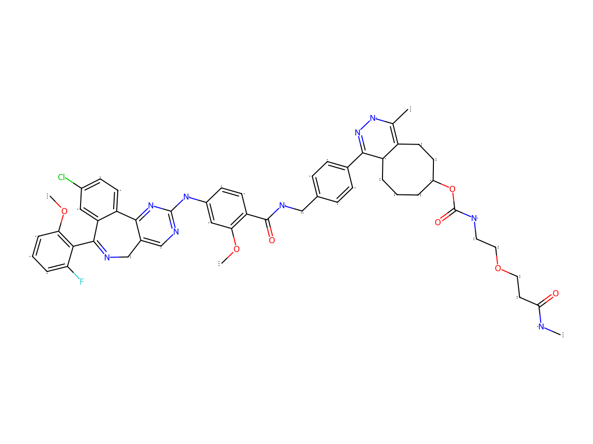
|
|||||
| Antibody Name |
5D3 mAb-AF488
|
Antibody Info | ||||
| Antigen Name |
Glutamate carboxypeptidase 2 (FOLH1)
|
Antigen Info | ||||
| Payload Name |
MLN8237
|
Payload Info | ||||
| Therapeutic Target |
Aurora kinase A (AURKA)
|
Target Info | ||||
| Linker Name |
NHS-PEG-TCO-Tz
|
Linker Info | ||||
| Conjugate Type |
Random conjugation through nucleophilic lysines.
|
|||||
The indication landscape of This ADC(2027 Update)
General Information of The Activity Data Related to This ADC
Full List of Activity Data of This Antibody-drug Conjugate
Revealed Based on the Cell Line Data
| Experiment 1 Reporting the Activity Date of This ADC | [1] | ||||
| Efficacy Data | Half Maximal inhibitory Concentration (lC50) | 8.17 nM | Negative PSMA expression (PSMA-) | ||
| Method Description |
The cytotoxicity of 5D3 (CC-MLN8237)3.2-AF488 was determined by treating PC3-PIP and PC3-Flu (control) cells in 96-well plates in RPMI 1640 culture media in the presence of 5D3- (CC-MLN8237)3.2-AF488 over a series of concentrations. Cells were seeded in a 96-well plate (2000 cells/well) and grown for 24 h to achieve 30%-40% confluency. Then, cells were treated with increasing concentrations of 5D3 (CC-MLN8237)3.2-AF488 or pure MLN8237 equivalent to the drug concentration in 5D3- (CC-MLN8237)3.2-AF555 (1.0 ng/mL-1.0 mg/mL by 10-fold increment). After the incubation for 72 h, IC50 values of 5D3 (CC-MLN8237)3.2-AF488 ATC and corresponding free MLN8237 were determined using the WST-8 assay (CCK-8 assay, Dojindo Molecular Technologies), following the manufacturer's protocol. Briefly, media in each well were replaced by 100 uL of the fresh media. Then, cells in each well were treated with 10 uL of WST-8 reagent and incubated at 37°C for 3 h. During incubation, WST-8 tetrazolium salt is reduced by dehydrogenase in living cells, forming a yellow formazan dye with λmax at 450 nm. After 3 h, the absorbance in wells was measured at 450 nm using a BioTek Epoch microplate spectrophotometer (Agilent Technologies). The concentration of the formazan dye in the media produced by dehydrogenases is directly proportional to the density of viable cells per well. The cell viability of treated cells was normalized to readings in untreated control cells with 100% viability. Data were fitted, and IC50 values for free MLN8237 and 5D3 (CC-MLN8237)3.2-AF488 were calculated using Prism 9 software (GraphPad, San Diego CA).
Click to Show/Hide
|
||||
| In Vitro Model | Prostate carcinoma | PC3-PIP cells | CVCL_E9XK | ||
| Experiment 2 Reporting the Activity Date of This ADC | [1] | ||||
| Efficacy Data | Half Maximal inhibitory Concentration (lC50) | 161.9 nM | Positive PSMA expression (PSMA+++/++) | ||
| Method Description |
The cytotoxicity of 5D3 (CC-MLN8237)3.2-AF488 was determined by treating PC3-PIP and PC3-Flu (control) cells in 96-well plates in RPMI 1640 culture media in the presence of 5D3- (CC-MLN8237)3.2-AF488 over a series of concentrations. Cells were seeded in a 96-well plate (2000 cells/well) and grown for 24 h to achieve 30%-40% confluency. Then, cells were treated with increasing concentrations of 5D3 (CC-MLN8237)3.2-AF488 or pure MLN8237 equivalent to the drug concentration in 5D3- (CC-MLN8237)3.2-AF555 (1.0 ng/mL-1.0 mg/mL by 10-fold increment). After the incubation for 72 h, IC50 values of 5D3 (CC-MLN8237)3.2-AF488 ATC and corresponding free MLN8237 were determined using the WST-8 assay (CCK-8 assay, Dojindo Molecular Technologies), following the manufacturer's protocol. Briefly, media in each well were replaced by 100 uL of the fresh media. Then, cells in each well were treated with 10 uL of WST-8 reagent and incubated at 37°C for 3 h. During incubation, WST-8 tetrazolium salt is reduced by dehydrogenase in living cells, forming a yellow formazan dye with λmax at 450 nm. After 3 h, the absorbance in wells was measured at 450 nm using a BioTek Epoch microplate spectrophotometer (Agilent Technologies). The concentration of the formazan dye in the media produced by dehydrogenases is directly proportional to the density of viable cells per well. The cell viability of treated cells was normalized to readings in untreated control cells with 100% viability. Data were fitted, and IC50 values for free MLN8237 and 5D3 (CC-MLN8237)3.2-AF488 were calculated using Prism 9 software (GraphPad, San Diego CA).
Click to Show/Hide
|
||||
| In Vitro Model | Prostate carcinoma | PC3-Flu cells | Homo sapiens | ||
References