
Payload Information
General Information of This Payload (ID: PAY0DPMMV)
| Name |
3-aminophenyl-hemiasterlin
|
|||||
|---|---|---|---|---|---|---|
| Synonyms |
3-aminophenyl-hemiasterlin
Click to Show/Hide
|
|||||
| Structure |
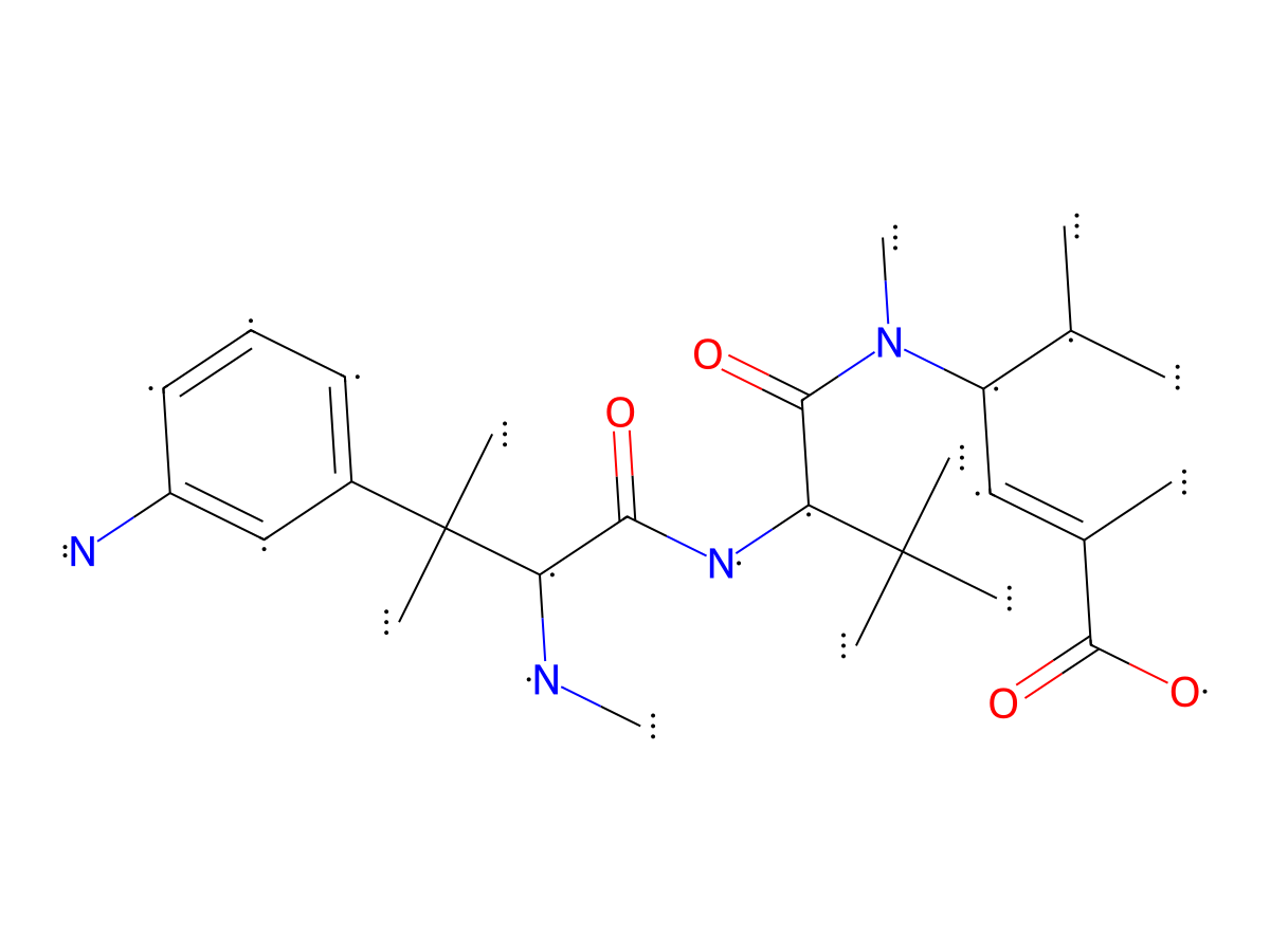
|
|||||
| Formula | C27H44N4O4 |
|||||
| Isosmiles | CC(C)([C@H](NC([C@@H](NC)C(C)(C1=CC=CC(N)=C1)C)=O)C(N([C@@H](C(C)C)/C=C(C(O)=O)\C)C)=O)C |
|||||
| PubChem CID | ||||||
| InChI |
InChI=1S/C27H44N4O4/c1-16(2)20(14-17(3)25(34)35)31(10)24(33)22(26(4,5)6)30-23(32)21(29-9)27(7,8)18-12-11-13-19(28)15-18/h11-16,20-22,29H,28H2,1-10H3,(H,30,32)(H,34,35)/t20-,21-,22-/m1/s1
|
|||||
| InChIKey |
ILRXQTICSGMLFH-YPAWHYETSA-N
|
|||||
| Pharmaceutical Properties | Molecule Weight |
488.673 |
Polar area |
124.76 |
||
Complexity |
911.601856 |
xlogp Value |
3.1792 |
|||
Heavy Count |
35 |
Rot Bonds |
10 |
|||
Hbond acc |
5 |
Hbond Donor |
4 |
|||
Each Antibody-drug Conjugate Related to This Payload
Full Information of The Activity Data of The ADC(s) Related to This Payload
Anti-MUC1/EGFR ADC [Investigative]
Discovered Using Cell Line-derived Xenograft Model
| Experiment 1 Reporting the Activity Date of This ADC | [1] | ||||
| Efficacy Data | Tumor Growth lnhibition value (TGl) |
0%
|
High MUC1 expression (MUC1 +++);Low EGFR expression (EGFR+) | ||
| Method Description |
Female athymic nude mice with established WISH tumors (3- 150 mm3) were treated with a single intravenous (IV) injection 0.1 mg/kg ADC.The effects of treatment on WISH tumor growth and the individual tumor sizes on the day the vehicle control treated tumors reached the study endpoint (> 1,200 mm3) are illustrated.
|
||||
| In Vivo Model | WISH xenograft model | ||||
| Experiment 2 Reporting the Activity Date of This ADC | [1] | ||||
| Efficacy Data | Tumor Growth lnhibition value (TGl) |
12%
|
High MUC1 expression (MUC1 +++);Low EGFR expression (EGFR+) | ||
| Method Description |
Female athymic nude mice with established WISH tumors (3- 150 mm3) were treated with a single intravenous (IV) injection 0.3 mg/kg ADC.The effects of treatment on WISH tumor growth and the individual tumor sizes on the day the vehicle control treated tumors reached the study endpoint (> 1,200 mm3) are illustrated.
|
||||
| In Vivo Model | WISH xenograft model | ||||
| Experiment 3 Reporting the Activity Date of This ADC | [1] | ||||
| Efficacy Data | Tumor Growth lnhibition value (TGl) |
47%
|
High MUC1 expression (MUC1 +++);Low EGFR expression (EGFR+) | ||
| Method Description |
Female athymic nude mice with established WISH tumors (3- 150 mm3) were treated with a single intravenous (IV) injection 0.75 mg/kg ADC.The effects of treatment on WISH tumor growth and the individual tumor sizes on the day the vehicle control treated tumors reached the study endpoint (> 1,200 mm3) are illustrated.
|
||||
| In Vivo Model | WISH xenograft model | ||||
| Experiment 4 Reporting the Activity Date of This ADC | [1] | ||||
| Efficacy Data | Tumor Growth lnhibition value (TGl) |
81%
|
High MUC1 expression (MUC1 +++);Low EGFR expression (EGFR+) | ||
| Method Description |
Female athymic nude mice with established WISH tumors (3- 150 mm3) were treated with a single intravenous (IV) injection 1.5 mg/kg ADC.The effects of treatment on WISH tumor growth and the individual tumor sizes on the day the vehicle control treated tumors reached the study endpoint (> 1,200 mm3) are illustrated. Determined tumor volume after the experiment, measured at day 21.
Click to Show/Hide
|
||||
| In Vivo Model | WISH xenograft model | ||||
| Experiment 5 Reporting the Activity Date of This ADC | [1] | ||||
| Efficacy Data | Tumor Growth lnhibition value (TGl) | > 100% | Moderate MUC1 expression (MUC1++);Low EGFR expression (EGFR+) | ||
| Method Description |
Female CB17 SCID mice with established OVCAR-3 tumors (3- 100 mm3) were treated with a single IV injection of 2.5 mg/kg ADC. Analysis of tumor size on day 28.
|
||||
| In Vivo Model | OVCAR-3 xenograft model | ||||
| Experiment 6 Reporting the Activity Date of This ADC | [1] | ||||
| Efficacy Data | Tumor Growth lnhibition value (TGl) | > 100% | Moderate MUC1 expression (MUC1++);Low EGFR expression (EGFR+) | ||
| Method Description |
Female CB17 SCID mice with established OVCAR-3 tumors (3- 100 mm3) were treated with a single IV injection of 5 mg/kg ADC. Analysis of tumor size on day 28.
|
||||
| In Vivo Model | OVCAR-3 xenograft model | ||||
| Experiment 7 Reporting the Activity Date of This ADC | [1] | ||||
| Efficacy Data | Tumor Growth lnhibition value (TGl) | > 100% | Moderate MUC1 expression (MUC1++);Low EGFR expression (EGFR+) | ||
| Method Description |
Female CB17 SCID mice with established OVCAR-3 tumors (3- 100 mm3) were treated with a single IV injection of 10mg/kg ADC. Analysis of tumor size on day 28.
|
||||
| In Vivo Model | OVCAR-3 xenograft model | ||||
Discovered Using Patient-derived Xenograft Model
| Experiment 1 Reporting the Activity Date of This ADC | [1] | ||||
| Efficacy Data | Tumor Growth lnhibition value (TGl) |
24%
|
|||
| Method Description |
Female nude mice with established LUX089 tumors (3- 150 mm3) were treated with a single IV injection at day 0 at 10mg/kg ADC. In three ofthe five animals treated in the 10 mg/kg dose group, no tumor was measurable up to day 60, when the experiment was finished.
|
||||
| In Vivo Model | NSCLC Patient-derived xenograft (PDX) model LUX089 | ||||
| Experiment 2 Reporting the Activity Date of This ADC | [1] | ||||
| Efficacy Data | Tumor Growth lnhibition value (TGl) |
69%
|
|||
| Method Description |
Female nude mice with established LUX089 tumors (3- 150 mm3) were treated with a single IV injection at day 0 at 10mg/kg ADC. In three ofthe five animals treated in the 10 mg/kg dose group, no tumor was measurable up to day 60, when the experiment was finished.
|
||||
| In Vivo Model | NSCLC Patient-derived xenograft (PDX) model LUX089 | ||||
| Experiment 3 Reporting the Activity Date of This ADC | [1] | ||||
| Efficacy Data | Tumor Growth lnhibition value (TGl) | > 100% | |||
| Method Description |
Female nude mice with established LUX089 tumors (3- 150 mm3) were treated with a single IV injection at day 0 at 10mg/kg ADC. In three ofthe five animals treated in the 10 mg/kg dose group, no tumor was measurable up to day 60, when the experiment was finished.
|
||||
| In Vivo Model | NSCLC Patient-derived xenograft (PDX) model LUX089 | ||||
Revealed Based on the Cell Line Data
| Experiment 1 Reporting the Activity Date of This ADC | [1] | ||||
| Efficacy Data | Half Maximal inhibitory Concentration (lC50) |
0.09 nM
|
Low MUC1 expression (MUC1+);High EGFR expression (EGFR+++) | ||
| Method Description |
A total of 625 cells (MDA-MB-468) in a volume of 25 uL were seeded in a 384-well flat bottom white polystyrene plate the day before the actual assay started. ADC and free drugs were formulated at 2x starting concentration in cell culture medium and filtered through 0.22um cellulose acetate filtered 2 ml centrifuge tubes. Filter sterilized samples were serial diluted (1:3) under sterile conditions and 25 uL of each dilution was added onto cells in triplicates. Plates were cultured at 37 °C in a C02 incubator for 120 hours. For cell viability measurement, 30 uL of Cell Titer-Glo@ reagent (PromegaT M Corp, Madison, WI) was added into each well, and plates processed as per product instructions. Relative luminescence was measured.
Click to Show/Hide
|
||||
| In Vitro Model | Breast adenocarcinoma | MDA-MB-468 cells | CVCL_0419 | ||
| Experiment 2 Reporting the Activity Date of This ADC | [1] | ||||
| Efficacy Data | Half Maximal inhibitory Concentration (lC50) |
0.23 nM
|
Low MUC1 expression (MUC1+);High EGFR expression (EGFR+++) | ||
| Method Description |
A total of 625 cells (NCI-H292) were plated in 90 ul cell culture medium per well of a 96-well black/clear flat bottom TC-treated imaging microplate and cultured overnight. A ten-fold starting concentration of ADCs, a respective serial dilution (1:4) were prepared in cell culture medium briefly before use. Wells were supplied with 10 ul ADC or compound solution. Treatment was performed in technical triplicates. The plates were subsequently incubated for 144h. For subsequent cell viability measurement, 100 ul Cell Titer-Glo reagent was pipetted in each well, and plates were further processed according the manufacturer's instructions.
Click to Show/Hide
|
||||
| In Vitro Model | Lung adenocarcinoma | HCC827 cells | CVCL_2063 | ||
| Experiment 3 Reporting the Activity Date of This ADC | [1] | ||||
| Efficacy Data | Half Maximal inhibitory Concentration (lC50) |
0.41 nM
|
Moderate MUC1 expression (MUC1++);Low EGFR expression (EGFR+) | ||
| Method Description |
A total of 625 cells (OVCAR-3) in a volume of 25 uL were seeded in a 384-well flat bottom white polystyrene plate the day before the actual assay started. ADC and free drugs were formulated at 2x starting concentration in cell culture medium and filtered through 0.22um cellulose acetate filtered 2 ml centrifuge tubes. Filter sterilized samples were serial diluted (1:3) under sterile conditions and 25 uL of each dilution was added onto cells in triplicates. Plates were cultured at 37 °C in a C02 incubator for 120 hours. For cell viability measurement, 30 uL of Cell Titer-Glo@ reagent (PromegaT M Corp, Madison, WI) was added into each well, and plates processed as per product instructions. Relative luminescence was measured.
Click to Show/Hide
|
||||
| In Vitro Model | Ovarian serous adenocarcinoma | OVCAR-3 cells | CVCL_0465 | ||
| Experiment 4 Reporting the Activity Date of This ADC | [1] | ||||
| Efficacy Data | Half Maximal inhibitory Concentration (lC50) |
0.83 nM
|
Low MUC1 expression (MUC1+);Low EGFR expression (EGFR+) | ||
| Method Description |
A total of 625 cells (NCI-H292) were plated in 90 ul cell culture medium per well of a 96-well black/clear flat bottom TC-treated imaging microplate and cultured overnight. A ten-fold starting concentration of ADCs, a respective serial dilution (1:4) were prepared in cell culture medium briefly before use. Wells were supplied with 10 ul ADC or compound solution. Treatment was performed in technical triplicates. The plates were subsequently incubated for 144h. For subsequent cell viability measurement, 100 ul Cell Titer-Glo reagent was pipetted in each well, and plates were further processed according the manufacturer's instructions.
Click to Show/Hide
|
||||
| In Vitro Model | Lung adenocarcinoma | NCI-H1975 cells | CVCL_1511 | ||
| Experiment 5 Reporting the Activity Date of This ADC | [1] | ||||
| Efficacy Data | Half Maximal inhibitory Concentration (lC50) |
1.1 nM
|
Low MUC1 expression (MUC1+);Moderate EGFR expression (EGFR++) | ||
| Method Description |
A total of 625 cells (NCI-H292) were plated in 90 ul cell culture medium per well of a 96-well black/clear flat bottom TC-treated imaging microplate and cultured overnight. A ten-fold starting concentration of ADCs, a respective serial dilution (1:4) were prepared in cell culture medium briefly before use. Wells were supplied with 10 ul ADC or compound solution. Treatment was performed in technical triplicates. The plates were subsequently incubated for 144h. For subsequent cell viability measurement, 100 ul Cell Titer-Glo reagent was pipetted in each well, and plates were further processed according the manufacturer's instructions.
Click to Show/Hide
|
||||
| In Vitro Model | Lung mucoepidermoid carcinoma | NCI-H292 cells | CVCL_0455 | ||
| Experiment 6 Reporting the Activity Date of This ADC | [1] | ||||
| Efficacy Data | Half Maximal inhibitory Concentration (lC50) |
1.4 nM
|
High MUC1 expression (MUC1 +++);Low EGFR expression (EGFR+) | ||
| Method Description |
A total of 625 cells (WISH) in a volume of 25 uL were seeded in a 384-well flat bottom white polystyrene plate the day before the actual assay started. ADC and free drugs were formulated at 2x starting concentration in cell culture medium and filtered through 0.22um cellulose acetate filtered 2 ml centrifuge tubes. Filter sterilized samples were serial diluted (1:3) under sterile conditions and 25 uL of each dilution was added onto cells in triplicates. Plates were cultured at 37 °C in a C02 incubator for 120 hours. For cell viability measurement, 30 uL of Cell Titer-Glo@ reagent (PromegaT M Corp, Madison, WI) was added into each well, and plates processed as per product instructions. Relative luminescence was measured.
Click to Show/Hide
|
||||
| In Vitro Model | Endocervical adenocarcinoma | WISH cells | CVCL_1909 | ||
Anti-EGFR ADC [Investigative]
Revealed Based on the Cell Line Data
| Experiment 1 Reporting the Activity Date of This ADC | [1] | ||||
| Efficacy Data | Half Maximal inhibitory Concentration (lC50) |
0.04 nM
|
Low MUC1 expression (MUC1+);Low EGFR expression (EGFR+) | ||
| Method Description |
A total of 625 cells (NCI-H292) were plated in 90 ul cell culture medium per well of a 96-well black/clear flat bottom TC-treated imaging microplate and cultured overnight. A ten-fold starting concentration of ADCs, a respective serial dilution (1:4) were prepared in cell culture medium briefly before use. Wells were supplied with 10 ul ADC or compound solution. Treatment was performed in technical triplicates. The plates were subsequently incubated for 144h. For subsequent cell viability measurement, 100 ul Cell Titer-Glo reagent was pipetted in each well, and plates were further processed according the manufacturer's instructions.
Click to Show/Hide
|
||||
| In Vitro Model | Lung adenocarcinoma | NCI-H1975 cells | CVCL_1511 | ||
| Experiment 2 Reporting the Activity Date of This ADC | [1] | ||||
| Efficacy Data | Half Maximal inhibitory Concentration (lC50) |
0.06 nM
|
Low MUC1 expression (MUC1+);High EGFR expression (EGFR+++) | ||
| Method Description |
A total of 625 cells (NCI-H292) were plated in 90 ul cell culture medium per well of a 96-well black/clear flat bottom TC-treated imaging microplate and cultured overnight. A ten-fold starting concentration of ADCs, a respective serial dilution (1:4) were prepared in cell culture medium briefly before use. Wells were supplied with 10 ul ADC or compound solution. Treatment was performed in technical triplicates. The plates were subsequently incubated for 144h. For subsequent cell viability measurement, 100 ul Cell Titer-Glo reagent was pipetted in each well, and plates were further processed according the manufacturer's instructions.
Click to Show/Hide
|
||||
| In Vitro Model | Lung adenocarcinoma | HCC827 cells | CVCL_2063 | ||
| Experiment 3 Reporting the Activity Date of This ADC | [1] | ||||
| Efficacy Data | Half Maximal inhibitory Concentration (lC50) |
0.13 nM
|
Moderate MUC1 expression (MUC1++);Low EGFR expression (EGFR+) | ||
| Method Description |
A total of 625 cells (OVCAR-3) in a volume of 25 uL were seeded in a 384-well flat bottom white polystyrene plate the day before the actual assay started. ADC and free drugs were formulated at 2x starting concentration in cell culture medium and filtered through 0.22um cellulose acetate filtered 2 ml centrifuge tubes. Filter sterilized samples were serial diluted (1:3) under sterile conditions and 25 uL of each dilution was added onto cells in triplicates. Plates were cultured at 37 °C in a C02 incubator for 120 hours. For cell viability measurement, 30 uL of Cell Titer-Glo@ reagent (PromegaT M Corp, Madison, WI) was added into each well, and plates processed as per product instructions. Relative luminescence was measured.
Click to Show/Hide
|
||||
| In Vitro Model | Ovarian serous adenocarcinoma | OVCAR-3 cells | CVCL_0465 | ||
| Experiment 4 Reporting the Activity Date of This ADC | [1] | ||||
| Efficacy Data | Half Maximal inhibitory Concentration (lC50) |
0.15 nM
|
Low MUC1 expression (MUC1+);High EGFR expression (EGFR+++) | ||
| Method Description |
A total of 625 cells (MDA-MB-468) in a volume of 25 uL were seeded in a 384-well flat bottom white polystyrene plate the day before the actual assay started. ADC and free drugs were formulated at 2x starting concentration in cell culture medium and filtered through 0.22um cellulose acetate filtered 2 ml centrifuge tubes. Filter sterilized samples were serial diluted (1:3) under sterile conditions and 25 uL of each dilution was added onto cells in triplicates. Plates were cultured at 37 °C in a C02 incubator for 120 hours. For cell viability measurement, 30 uL of Cell Titer-Glo@ reagent (PromegaT M Corp, Madison, WI) was added into each well, and plates processed as per product instructions. Relative luminescence was measured.
Click to Show/Hide
|
||||
| In Vitro Model | Breast adenocarcinoma | MDA-MB-468 cells | CVCL_0419 | ||
| Experiment 5 Reporting the Activity Date of This ADC | [1] | ||||
| Efficacy Data | Half Maximal inhibitory Concentration (lC50) |
0.17 nM
|
Low MUC1 expression (MUC1+);Moderate EGFR expression (EGFR++) | ||
| Method Description |
A total of 625 cells (NCI-H292) were plated in 90 ul cell culture medium per well of a 96-well black/clear flat bottom TC-treated imaging microplate and cultured overnight. A ten-fold starting concentration of ADCs, a respective serial dilution (1:4) were prepared in cell culture medium briefly before use. Wells were supplied with 10 ul ADC or compound solution. Treatment was performed in technical triplicates. The plates were subsequently incubated for 144h. For subsequent cell viability measurement, 100 ul Cell Titer-Glo reagent was pipetted in each well, and plates were further processed according the manufacturer's instructions.
Click to Show/Hide
|
||||
| In Vitro Model | Lung mucoepidermoid carcinoma | NCI-H292 cells | CVCL_0455 | ||
Anti-MUC1 ADC [Investigative]
Revealed Based on the Cell Line Data
| Experiment 1 Reporting the Activity Date of This ADC | [1] | ||||
| Efficacy Data | Half Maximal inhibitory Concentration (lC50) |
0.65 nM
|
High MUC1 expression (MUC1 +++);Low EGFR expression (EGFR+) | ||
| Method Description |
A total of 625 cells (WISH) in a volume of 25 uL were seeded in a 384-well flat bottom white polystyrene plate the day before the actual assay started. ADC and free drugs were formulated at 2x starting concentration in cell culture medium and filtered through 0.22um cellulose acetate filtered 2 ml centrifuge tubes. Filter sterilized samples were serial diluted (1:3) under sterile conditions and 25 uL of each dilution was added onto cells in triplicates. Plates were cultured at 37 °C in a C02 incubator for 120 hours. For cell viability measurement, 30 uL of Cell Titer-Glo@ reagent (PromegaT M Corp, Madison, WI) was added into each well, and plates processed as per product instructions. Relative luminescence was measured.
Click to Show/Hide
|
||||
| In Vitro Model | Endocervical adenocarcinoma | WISH cells | CVCL_1909 | ||
| Experiment 2 Reporting the Activity Date of This ADC | [1] | ||||
| Efficacy Data | Half Maximal inhibitory Concentration (lC50) |
2.7 nM
|
Low MUC1 expression (MUC1+);High EGFR expression (EGFR+++) | ||
| Method Description |
A total of 625 cells (MDA-MB-468) in a volume of 25 uL were seeded in a 384-well flat bottom white polystyrene plate the day before the actual assay started. ADC and free drugs were formulated at 2x starting concentration in cell culture medium and filtered through 0.22um cellulose acetate filtered 2 ml centrifuge tubes. Filter sterilized samples were serial diluted (1:3) under sterile conditions and 25 uL of each dilution was added onto cells in triplicates. Plates were cultured at 37 °C in a C02 incubator for 120 hours. For cell viability measurement, 30 uL of Cell Titer-Glo@ reagent (PromegaT M Corp, Madison, WI) was added into each well, and plates processed as per product instructions. Relative luminescence was measured.
Click to Show/Hide
|
||||
| In Vitro Model | Breast adenocarcinoma | MDA-MB-468 cells | CVCL_0419 | ||
| Experiment 3 Reporting the Activity Date of This ADC | [1] | ||||
| Efficacy Data | Half Maximal inhibitory Concentration (lC50) |
3.6 nM
|
Moderate MUC1 expression (MUC1++);Low EGFR expression (EGFR+) | ||
| Method Description |
A total of 625 cells (OVCAR-3) in a volume of 25 uL were seeded in a 384-well flat bottom white polystyrene plate the day before the actual assay started. ADC and free drugs were formulated at 2x starting concentration in cell culture medium and filtered through 0.22um cellulose acetate filtered 2 ml centrifuge tubes. Filter sterilized samples were serial diluted (1:3) under sterile conditions and 25 uL of each dilution was added onto cells in triplicates. Plates were cultured at 37 °C in a C02 incubator for 120 hours. For cell viability measurement, 30 uL of Cell Titer-Glo@ reagent (PromegaT M Corp, Madison, WI) was added into each well, and plates processed as per product instructions. Relative luminescence was measured.
Click to Show/Hide
|
||||
| In Vitro Model | Ovarian serous adenocarcinoma | OVCAR-3 cells | CVCL_0465 | ||
| Experiment 4 Reporting the Activity Date of This ADC | [1] | ||||
| Efficacy Data | Half Maximal inhibitory Concentration (lC50) |
4.5 nM
|
Low MUC1 expression (MUC1+);High EGFR expression (EGFR+++) | ||
| Method Description |
A total of 625 cells (NCI-H292) were plated in 90 ul cell culture medium per well of a 96-well black/clear flat bottom TC-treated imaging microplate and cultured overnight. A ten-fold starting concentration of ADCs, a respective serial dilution (1:4) were prepared in cell culture medium briefly before use. Wells were supplied with 10 ul ADC or compound solution. Treatment was performed in technical triplicates. The plates were subsequently incubated for 144h. For subsequent cell viability measurement, 100 ul Cell Titer-Glo reagent was pipetted in each well, and plates were further processed according the manufacturer's instructions.
Click to Show/Hide
|
||||
| In Vitro Model | Lung adenocarcinoma | HCC827 cells | CVCL_2063 | ||
| Experiment 5 Reporting the Activity Date of This ADC | [1] | ||||
| Efficacy Data | Half Maximal inhibitory Concentration (lC50) |
20 nM
|
Low MUC1 expression (MUC1+);Moderate EGFR expression (EGFR++) | ||
| Method Description |
A total of 625 cells (NCI-H292) were plated in 90 ul cell culture medium per well of a 96-well black/clear flat bottom TC-treated imaging microplate and cultured overnight. A ten-fold starting concentration of ADCs, a respective serial dilution (1:4) were prepared in cell culture medium briefly before use. Wells were supplied with 10 ul ADC or compound solution. Treatment was performed in technical triplicates. The plates were subsequently incubated for 144h. For subsequent cell viability measurement, 100 ul Cell Titer-Glo reagent was pipetted in each well, and plates were further processed according the manufacturer's instructions.
Click to Show/Hide
|
||||
| In Vitro Model | Lung mucoepidermoid carcinoma | NCI-H292 cells | CVCL_0455 | ||
| Experiment 6 Reporting the Activity Date of This ADC | [1] | ||||
| Efficacy Data | Half Maximal inhibitory Concentration (lC50) |
310 nM
|
Low MUC1 expression (MUC1+);Low EGFR expression (EGFR+) | ||
| Method Description |
A total of 625 cells (NCI-H292) were plated in 90 ul cell culture medium per well of a 96-well black/clear flat bottom TC-treated imaging microplate and cultured overnight. A ten-fold starting concentration of ADCs, a respective serial dilution (1:4) were prepared in cell culture medium briefly before use. Wells were supplied with 10 ul ADC or compound solution. Treatment was performed in technical triplicates. The plates were subsequently incubated for 144h. For subsequent cell viability measurement, 100 ul Cell Titer-Glo reagent was pipetted in each well, and plates were further processed according the manufacturer's instructions.
Click to Show/Hide
|
||||
| In Vitro Model | Lung adenocarcinoma | NCI-H1975 cells | CVCL_1511 | ||