
Linker Information
General Information of This Linker (ID: LIN0XUHTG)
| Name |
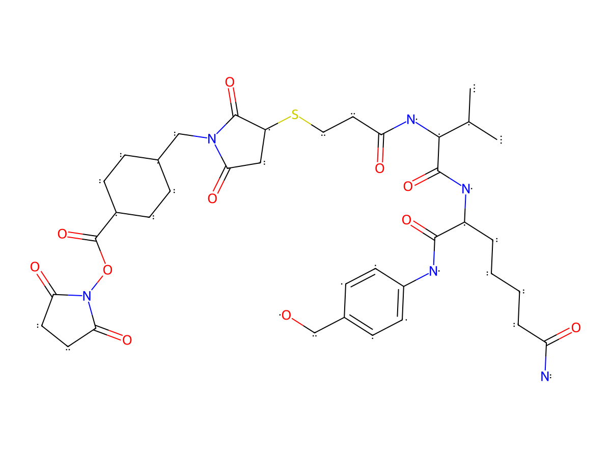
NHS-VC-PAB
|
|||||
|---|---|---|---|---|---|---|
| Linker Type |
Cathepsin-cleavable linker
|
|||||
| Antibody-Linker Relation |
Cleavable
|
|||||
| Structure |
|
|||||
| Formula |
C38H52N6O11S
|
|||||
| Isosmiles |
CC([C@H](NC(CCSC(C1=O)CC(N1CC2CCC(CC2)C(ON3C(CCC3=O)=O)=O)=O)=O)C(N[C@@H](CCCCC(N)=O)C(NC4=CC=C(C=C4)CO)=O)=O)C
|
|||||
| InChI |
InChI=1S/C38H52N6O11S/c1-22(2)34(36(52)41-27(5-3-4-6-29(39)46)35(51)40-26-13-9-24(21-45)10-14-26)42-30(47)17-18-56-28-19-33(50)43(37(28)53)20-23-7-11-25(12-8-23)38(54)55-44-31(48)15-16-32(44)49/h9-10,13-14,22-23,25,27-28,34,45H,3-8,11-12,15-21H2,1-2H3,(H2,39,46)(H,40,51)(H,41,52)(H,42,47)/t23?,25?,27-,28?,34-/m0/s1
|
|||||
| InChIKey |
ITMJGMCIFQTQEJ-ASVOSAJJSA-N
|
|||||
| Pharmaceutical Properties |
Molecule Weight
|
800.932
|
Polar area
|
251.68
|
||
|
Complexity
|
1558.109774
|
xlogp Value
|
1.4531
|
|||
|
Heavy Count
|
56
|
Rot Bonds
|
20
|
|||
|
Hbond acc
|
12
|
Hbond Donor
|
5
|
|||
Each Antibody-drug Conjugate Related to This Linker
Full Information of The Activity Data of The ADC(s) Related to This Linker
F12-MMAE [Investigative]
Discovered Using Cell Line-derived Xenograft Model
| Experiment 1 Reporting the Activity Date of This ADC | [1] | ||||
| Efficacy Data | Tumor Growth lnhibition value (TGl) |
98.30%
|
|||
| Method Description |
Next, we further increased the dose to 6 mg/kg, 4XQ1W and found that F12-MMAE almost eradicated tumors (complete remission, CR; %TGI = 98.3%) : For evaluation of in vivo ADCC effects of IgG1 F12, PC-3-PSCA cells (5 × 10⁶ cells/mouse) in 200 ul of 1:1 PBS/Matrigel (BD Biosciences, Cat# 354234) were injected subcutaneously (s.c.) into the right flank of the Balb/c scid mice. When tumor volume reached 100 mm3, mice were divided randomly into IgG1 F12 treatment or vehicle group and i.p. administered four times total with a four-day interval. For evaluation of the ADC (F12-MMAE or Ab1-MMAE) efficacy in vivo, PC-3-PSCA cells (5 × 10⁶ cells/mouse, 5 mice per group) in 200 ul of 1:1 PBS/Matrigel were injected s.c. into the right flank of the NSG mice. Mice were randomly divided into the different treatment groups when tumors reached 100 mm3. We conducted ADC treatment studies at three different dosing regimens, comparing PSCA-ADC with the isotype control Ab1-ADC. The regimens design were as follows: dose 1): 1 mg/kg and 3 mg/kg, administered twice weekly (2XQ1W); dose 2): 6 mg/kg, administered twice weekly (2XQ1W), followed by a single injection of 3 mg/kg when tumors regrew to 140 mm3; dose 3): 6 mg/kg, administered four times weekly (4XQ1W). All ADCs were i.p. administered. Tumor dimensions and mouse weight were measured periodically with a slide caliper and scale. Tumor volume was calculated by the formula: V = 0.5×length × (width)2. Tumor growth inhibition was calculated by the formula: %TGI = [1- (mean tumor volume (MTV)ADC treated/MTVcontrol)] × 100. Mice were euthanized when the tumor volume exceeded 1000 mm3 or if the animals showed any signs of suffering. At the end of the experiment time point, tumors were isolated and weighed. In the dose 2 experiment, one week after two 6 mg/kg ADC treatments, ~50 ul blood were collected to the purple-topped K+/EDTA tubes, and subjected to complete blood counts using the Abaxis HM5 machine from In vivo Imaging Facilities in Hillman Cancer Center of the University of Pittsburgh. In the dose 3 experiment (6 mg/kg, 4×Q1W), mouse spleen, kidney, heart, lung, and liver were collected and fixed in formalin. The H&E staining were completed by the Biospecimen Core of the University of Pittsburgh.
Click to Show/Hide
|
||||
| In Vivo Model | PC-3-PSCA xenograft mouse model | ||||
Revealed Based on the Cell Line Data
| Experiment 1 Reporting the Activity Date of This ADC | [1] | ||||
| Efficacy Data | Half Maximal Effective Concentration (EC50) |
5.41 nM
|
High PSCA expression (PSCA +++) | ||
| Method Description |
The PC-3 or PC-3-PSCA cells (2.5 × 10⁴ cells) were pre-seeded in 96-well plates overnight. The cells were then incubated with serially diluted ADC, or naked IgG1 antibody or linker-payload combination compound for 4-5 days at 37°C, 8% CO2 with 95% humidity. Then the cell viability was detected by the Promega CellTiter-Glo® Luminescent Cell Viability Assay (CAT# G7570), which is based on quantitation of the ATP present, an indicator of metabolically active cells. The Luminescence was recorded by the BioTek synergy multi-mode reader (Winooski, VT). The killing percentage was calculated by the formula: (1-Lumi with compounds/Lumi without compounds) ×100. The GraphPad Prism9 for was used for calculation of killing IC50. All experiments were performed in duplicate and the error bars denote ±1 SD.
Click to Show/Hide
|
||||
| In Vitro Model | Prostate carcinoma | PC-3-PSCA cells | CVCL_0035 | ||
38981219 ADC 12b [Investigative]
Revealed Based on the Cell Line Data
| Experiment 1 Reporting the Activity Date of This ADC | [2] | ||||
| Efficacy Data | Half Maximal inhibitory Concentration (lC50) |
0.006 nM
|
High TROP2 expression (TROP2 +++) | ||
| Method Description |
The Cell Counting Kit-8 (CCK-8) assay was performed to measure the in vitro efficiency of ADCs. TROP2-expression tumor cell lines (BXPC-3, HCI-441, Fadu, HCC1806, NCI-N87) and TROP2 negative tumor cell lines (Calu-6) were cultured in a 10 % FBS-containing RPMI 1640 medium and were planted into 96-well plates with 6000 cells per well. The plates were incubated overnight at 37 °C in a 5 % CO2 cell incubator. All ADCs were diluted 5-fold with RPMI 1640 medium from an initial concentration of 100 nmol/L, a total of nine concentration gradients. The ADC samples were added to three wells with every single concentration and the 96-well plates were cultured at 37 °C in a 5 % CO2 cell incubator for six days. Then CCK8 solution was added and incubated at 37 °C for 4 h. Optical density (OD) value was measured at 450 nm using a BioTek Epoch. IC50 values and the cell viability curve were calculated by GraphPad software.
Click to Show/Hide
|
||||
| In Vitro Model | Lung papillary adenocarcinoma | NCI-H441 cells | CVCL_1561 | ||
| Experiment 2 Reporting the Activity Date of This ADC | [2] | ||||
| Efficacy Data | Half Maximal inhibitory Concentration (lC50) |
0.017 nM
|
High TROP2 expression (TROP2 +++) | ||
| Method Description |
The Cell Counting Kit-8 (CCK-8) assay was performed to measure the in vitro efficiency of ADCs. TROP2-expression tumor cell lines (BXPC-3, HCI-441, Fadu, HCC1806, NCI-N87) and TROP2 negative tumor cell lines (Calu-6) were cultured in a 10 % FBS-containing RPMI 1640 medium and were planted into 96-well plates with 6000 cells per well. The plates were incubated overnight at 37 °C in a 5 % CO2 cell incubator. All ADCs were diluted 5-fold with RPMI 1640 medium from an initial concentration of 100 nmol/L, a total of nine concentration gradients. The ADC samples were added to three wells with every single concentration and the 96-well plates were cultured at 37 °C in a 5 % CO2 cell incubator for six days. Then CCK8 solution was added and incubated at 37 °C for 4 h. Optical density (OD) value was measured at 450 nm using a BioTek Epoch. IC50 values and the cell viability curve were calculated by GraphPad software.
Click to Show/Hide
|
||||
| In Vitro Model | Gastric tubular adenocarcinoma | NCI-N87 cells | CVCL_1603 | ||
| Experiment 3 Reporting the Activity Date of This ADC | [2] | ||||
| Efficacy Data | Half Maximal inhibitory Concentration (lC50) |
0.019 nM
|
High TROP2 expression (TROP2 +++) | ||
| Method Description |
The Cell Counting Kit-8 (CCK-8) assay was performed to measure the in vitro efficiency of ADCs. TROP2-expression tumor cell lines (BXPC-3, HCI-441, Fadu, HCC1806, NCI-N87) and TROP2 negative tumor cell lines (Calu-6) were cultured in a 10 % FBS-containing RPMI 1640 medium and were planted into 96-well plates with 6000 cells per well. The plates were incubated overnight at 37 °C in a 5 % CO2 cell incubator. All ADCs were diluted 5-fold with RPMI 1640 medium from an initial concentration of 100 nmol/L, a total of nine concentration gradients. The ADC samples were added to three wells with every single concentration and the 96-well plates were cultured at 37 °C in a 5 % CO2 cell incubator for six days. Then CCK8 solution was added and incubated at 37 °C for 4 h. Optical density (OD) value was measured at 450 nm using a BioTek Epoch. IC50 values and the cell viability curve were calculated by GraphPad software.
Click to Show/Hide
|
||||
| In Vitro Model | Pancreatic ductal adenocarcinoma | BxPC-3 cells | CVCL_0186 | ||
| Experiment 4 Reporting the Activity Date of This ADC | [2] | ||||
| Efficacy Data | Half Maximal inhibitory Concentration (lC50) |
0.066 nM
|
High TROP2 expression (TROP2 +++) | ||
| Method Description |
The Cell Counting Kit-8 (CCK-8) assay was performed to measure the in vitro efficiency of ADCs. TROP2-expression tumor cell lines (BXPC-3, HCI-441, Fadu, HCC1806, NCI-N87) and TROP2 negative tumor cell lines (Calu-6) were cultured in a 10 % FBS-containing RPMI 1640 medium and were planted into 96-well plates with 6000 cells per well. The plates were incubated overnight at 37 °C in a 5 % CO2 cell incubator. All ADCs were diluted 5-fold with RPMI 1640 medium from an initial concentration of 100 nmol/L, a total of nine concentration gradients. The ADC samples were added to three wells with every single concentration and the 96-well plates were cultured at 37 °C in a 5 % CO2 cell incubator for six days. Then CCK8 solution was added and incubated at 37 °C for 4 h. Optical density (OD) value was measured at 450 nm using a BioTek Epoch. IC50 values and the cell viability curve were calculated by GraphPad software.
Click to Show/Hide
|
||||
| In Vitro Model | Hypopharyngeal squamous cell carcinoma | FaDu cells | CVCL_1218 | ||
| Experiment 5 Reporting the Activity Date of This ADC | [2] | ||||
| Efficacy Data | Half Maximal inhibitory Concentration (lC50) |
0.086 nM
|
High TROP2 expression (TROP2 +++) | ||
| Method Description |
The Cell Counting Kit-8 (CCK-8) assay was performed to measure the in vitro efficiency of ADCs. TROP2-expression tumor cell lines (BXPC-3, HCI-441, Fadu, HCC1806, NCI-N87) and TROP2 negative tumor cell lines (Calu-6) were cultured in a 10 % FBS-containing RPMI 1640 medium and were planted into 96-well plates with 6000 cells per well. The plates were incubated overnight at 37 °C in a 5 % CO2 cell incubator. All ADCs were diluted 5-fold with RPMI 1640 medium from an initial concentration of 100 nmol/L, a total of nine concentration gradients. The ADC samples were added to three wells with every single concentration and the 96-well plates were cultured at 37 °C in a 5 % CO2 cell incubator for six days. Then CCK8 solution was added and incubated at 37 °C for 4 h. Optical density (OD) value was measured at 450 nm using a BioTek Epoch. IC50 values and the cell viability curve were calculated by GraphPad software.
Click to Show/Hide
|
||||
| In Vitro Model | Breast acantholytic squamous cell carcinoma | HCC-1806 cells | CVCL_1258 | ||
| Experiment 6 Reporting the Activity Date of This ADC | [2] | ||||
| Efficacy Data | Half Maximal inhibitory Concentration (lC50) | > 100 nM | Negative TROP2 expression (TROP2-) | ||
| Method Description |
The Cell Counting Kit-8 (CCK-8) assay was performed to measure the in vitro efficiency of ADCs. TROP2-expression tumor cell lines (BXPC-3, HCI-441, Fadu, HCC1806, NCI-N87) and TROP2 negative tumor cell lines (Calu-6) were cultured in a 10 % FBS-containing RPMI 1640 medium and were planted into 96-well plates with 6000 cells per well. The plates were incubated overnight at 37 °C in a 5 % CO2 cell incubator. All ADCs were diluted 5-fold with RPMI 1640 medium from an initial concentration of 100 nmol/L, a total of nine concentration gradients. The ADC samples were added to three wells with every single concentration and the 96-well plates were cultured at 37 °C in a 5 % CO2 cell incubator for six days. Then CCK8 solution was added and incubated at 37 °C for 4 h. Optical density (OD) value was measured at 450 nm using a BioTek Epoch. IC50 values and the cell viability curve were calculated by GraphPad software.
Click to Show/Hide
|
||||
| In Vitro Model | Lung adenocarcinoma | Calu-6 cells | CVCL_0236 | ||
38981219 ADC 12a [Investigative]
Revealed Based on the Cell Line Data
| Experiment 1 Reporting the Activity Date of This ADC | [2] | ||||
| Efficacy Data | Half Maximal inhibitory Concentration (lC50) |
0.018 nM
|
High TROP2 expression (TROP2 +++) | ||
| Method Description |
The Cell Counting Kit-8 (CCK-8) assay was performed to measure the in vitro efficiency of ADCs. TROP2-expression tumor cell lines (BXPC-3, HCI-441, Fadu, HCC1806, NCI-N87) and TROP2 negative tumor cell lines (Calu-6) were cultured in a 10 % FBS-containing RPMI 1640 medium and were planted into 96-well plates with 6000 cells per well. The plates were incubated overnight at 37 °C in a 5 % CO2 cell incubator. All ADCs were diluted 5-fold with RPMI 1640 medium from an initial concentration of 100 nmol/L, a total of nine concentration gradients. The ADC samples were added to three wells with every single concentration and the 96-well plates were cultured at 37 °C in a 5 % CO2 cell incubator for six days. Then CCK8 solution was added and incubated at 37 °C for 4 h. Optical density (OD) value was measured at 450 nm using a BioTek Epoch. IC50 values and the cell viability curve were calculated by GraphPad software.
Click to Show/Hide
|
||||
| In Vitro Model | Lung papillary adenocarcinoma | NCI-H441 cells | CVCL_1561 | ||
| Experiment 2 Reporting the Activity Date of This ADC | [2] | ||||
| Efficacy Data | Half Maximal inhibitory Concentration (lC50) |
0.028 nM
|
High TROP2 expression (TROP2 +++) | ||
| Method Description |
The Cell Counting Kit-8 (CCK-8) assay was performed to measure the in vitro efficiency of ADCs. TROP2-expression tumor cell lines (BXPC-3, HCI-441, Fadu, HCC1806, NCI-N87) and TROP2 negative tumor cell lines (Calu-6) were cultured in a 10 % FBS-containing RPMI 1640 medium and were planted into 96-well plates with 6000 cells per well. The plates were incubated overnight at 37 °C in a 5 % CO2 cell incubator. All ADCs were diluted 5-fold with RPMI 1640 medium from an initial concentration of 100 nmol/L, a total of nine concentration gradients. The ADC samples were added to three wells with every single concentration and the 96-well plates were cultured at 37 °C in a 5 % CO2 cell incubator for six days. Then CCK8 solution was added and incubated at 37 °C for 4 h. Optical density (OD) value was measured at 450 nm using a BioTek Epoch. IC50 values and the cell viability curve were calculated by GraphPad software.
Click to Show/Hide
|
||||
| In Vitro Model | Gastric tubular adenocarcinoma | NCI-N87 cells | CVCL_1603 | ||
| Experiment 3 Reporting the Activity Date of This ADC | [2] | ||||
| Efficacy Data | Half Maximal inhibitory Concentration (lC50) |
0.034 nM
|
High TROP2 expression (TROP2 +++) | ||
| Method Description |
The Cell Counting Kit-8 (CCK-8) assay was performed to measure the in vitro efficiency of ADCs. TROP2-expression tumor cell lines (BXPC-3, HCI-441, Fadu, HCC1806, NCI-N87) and TROP2 negative tumor cell lines (Calu-6) were cultured in a 10 % FBS-containing RPMI 1640 medium and were planted into 96-well plates with 6000 cells per well. The plates were incubated overnight at 37 °C in a 5 % CO2 cell incubator. All ADCs were diluted 5-fold with RPMI 1640 medium from an initial concentration of 100 nmol/L, a total of nine concentration gradients. The ADC samples were added to three wells with every single concentration and the 96-well plates were cultured at 37 °C in a 5 % CO2 cell incubator for six days. Then CCK8 solution was added and incubated at 37 °C for 4 h. Optical density (OD) value was measured at 450 nm using a BioTek Epoch. IC50 values and the cell viability curve were calculated by GraphPad software.
Click to Show/Hide
|
||||
| In Vitro Model | Pancreatic ductal adenocarcinoma | BxPC-3 cells | CVCL_0186 | ||
| Experiment 4 Reporting the Activity Date of This ADC | [2] | ||||
| Efficacy Data | Half Maximal inhibitory Concentration (lC50) |
0.124 nM
|
High TROP2 expression (TROP2 +++) | ||
| Method Description |
The Cell Counting Kit-8 (CCK-8) assay was performed to measure the in vitro efficiency of ADCs. TROP2-expression tumor cell lines (BXPC-3, HCI-441, Fadu, HCC1806, NCI-N87) and TROP2 negative tumor cell lines (Calu-6) were cultured in a 10 % FBS-containing RPMI 1640 medium and were planted into 96-well plates with 6000 cells per well. The plates were incubated overnight at 37 °C in a 5 % CO2 cell incubator. All ADCs were diluted 5-fold with RPMI 1640 medium from an initial concentration of 100 nmol/L, a total of nine concentration gradients. The ADC samples were added to three wells with every single concentration and the 96-well plates were cultured at 37 °C in a 5 % CO2 cell incubator for six days. Then CCK8 solution was added and incubated at 37 °C for 4 h. Optical density (OD) value was measured at 450 nm using a BioTek Epoch. IC50 values and the cell viability curve were calculated by GraphPad software.
Click to Show/Hide
|
||||
| In Vitro Model | Hypopharyngeal squamous cell carcinoma | FaDu cells | CVCL_1218 | ||
| Experiment 5 Reporting the Activity Date of This ADC | [2] | ||||
| Efficacy Data | Half Maximal inhibitory Concentration (lC50) |
0.564 nM
|
High TROP2 expression (TROP2 +++) | ||
| Method Description |
The Cell Counting Kit-8 (CCK-8) assay was performed to measure the in vitro efficiency of ADCs. TROP2-expression tumor cell lines (BXPC-3, HCI-441, Fadu, HCC1806, NCI-N87) and TROP2 negative tumor cell lines (Calu-6) were cultured in a 10 % FBS-containing RPMI 1640 medium and were planted into 96-well plates with 6000 cells per well. The plates were incubated overnight at 37 °C in a 5 % CO2 cell incubator. All ADCs were diluted 5-fold with RPMI 1640 medium from an initial concentration of 100 nmol/L, a total of nine concentration gradients. The ADC samples were added to three wells with every single concentration and the 96-well plates were cultured at 37 °C in a 5 % CO2 cell incubator for six days. Then CCK8 solution was added and incubated at 37 °C for 4 h. Optical density (OD) value was measured at 450 nm using a BioTek Epoch. IC50 values and the cell viability curve were calculated by GraphPad software.
Click to Show/Hide
|
||||
| In Vitro Model | Breast acantholytic squamous cell carcinoma | HCC-1806 cells | CVCL_1258 | ||
| Experiment 6 Reporting the Activity Date of This ADC | [2] | ||||
| Efficacy Data | Half Maximal inhibitory Concentration (lC50) | > 100 nM | Negative TROP2 expression (TROP2-) | ||
| Method Description |
The Cell Counting Kit-8 (CCK-8) assay was performed to measure the in vitro efficiency of ADCs. TROP2-expression tumor cell lines (BXPC-3, HCI-441, Fadu, HCC1806, NCI-N87) and TROP2 negative tumor cell lines (Calu-6) were cultured in a 10 % FBS-containing RPMI 1640 medium and were planted into 96-well plates with 6000 cells per well. The plates were incubated overnight at 37 °C in a 5 % CO2 cell incubator. All ADCs were diluted 5-fold with RPMI 1640 medium from an initial concentration of 100 nmol/L, a total of nine concentration gradients. The ADC samples were added to three wells with every single concentration and the 96-well plates were cultured at 37 °C in a 5 % CO2 cell incubator for six days. Then CCK8 solution was added and incubated at 37 °C for 4 h. Optical density (OD) value was measured at 450 nm using a BioTek Epoch. IC50 values and the cell viability curve were calculated by GraphPad software.
Click to Show/Hide
|
||||
| In Vitro Model | Lung adenocarcinoma | Calu-6 cells | CVCL_0236 | ||
References