
Linker Information
General Information of This Linker (ID: LIN0REHOH)
| Name |
P5- (PEG12)
|
|||||
|---|---|---|---|---|---|---|
| Linker Type |
Uncleavable linker
|
|||||
| Antibody-Linker Relation |
Uncleavable
|
|||||
| Structure |
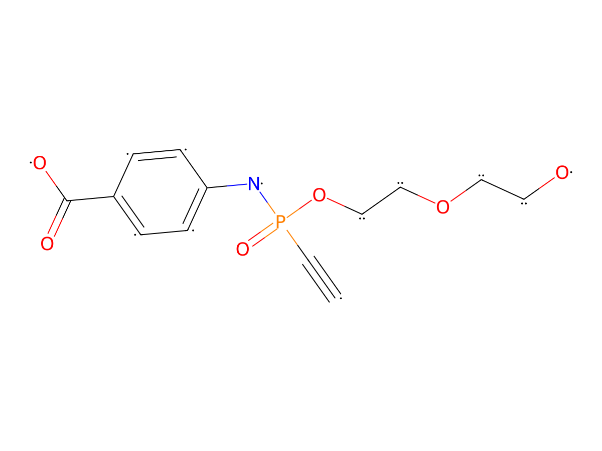
|
|||||
| Formula |
C33H56NO16P
|
|||||
| Isosmiles |
C#CP(NC1=CC=C(C(O)=O)C=C1)(OCCOCCOCCOCCOCCOCCOCCOCCOCCOCCOCCOCCO)=O
|
|||||
| InChI |
InChI=1S/C33H56NO16P/c1-2-51(38,34-32-5-3-31(4-6-32)33(36)37)50-30-29-49-28-27-48-26-25-47-24-23-46-22-21-45-20-19-44-18-17-43-16-15-42-14-13-41-12-11-40-10-9-39-8-7-35/h1,3-6,35H,7-30H2,(H,34,38)(H,36,37)
|
|||||
| InChIKey |
CPMPADBZTNSLLW-UHFFFAOYSA-N
|
|||||
| Pharmaceutical Properties |
Molecule Weight
|
753.776
|
Polar area
|
197.39
|
||
|
Complexity
|
970.2858391
|
xlogp Value
|
1.772
|
|||
|
Heavy Count
|
51
|
Rot Bonds
|
39
|
|||
|
Hbond acc
|
15
|
Hbond Donor
|
3
|
|||
Each Antibody-drug Conjugate Related to This Linker
Full Information of The Activity Data of The ADC(s) Related to This Linker
37828728 ADC 8 [Investigative]
Revealed Based on the Cell Line Data
| Experiment 1 Reporting the Activity Date of This ADC | [1] | ||||
| Efficacy Data | Half Maximal Effective Concentration (EC50) |
182.4 ng/mL
|
Positive HER2 expression (HER2+++/++) | ||
| Method Description |
To investigate the cytotoxicity of the unconjugated small-molecule TOP1 inhibitors and ADCs, respective cells were incubated for 4 days (small molecules) and 7 days (ADCs) with increasing concentrations of small molecules (0.015-1,000 nmol/L) and ADCs (0.05-3 ug/mL) to generate a dose-response curve. Killing was analyzed using resazurin cell viability dye at a final concentration of 55 umol/L (Merck) by dividing the fluorescence from control cells in medium by the fluorescence of ADC-treated cells. Fluorescence emission at 590 nmol/L was measured on a microplate reader Infinite 200 Pro (Tecan Group Ltd.).
Click to Show/Hide
|
||||
| In Vitro Model | Breast adenocarcinoma | SK-BR-3 cells | CVCL_0033 | ||
| Experiment 2 Reporting the Activity Date of This ADC | [1] | ||||
| Efficacy Data | Half Maximal Effective Concentration (EC50) | > 3000 ng/mL | Negative HER2 expression (HER2-) | ||
| Method Description |
To investigate the cytotoxicity of the unconjugated small-molecule TOP1 inhibitors and ADCs, respective cells were incubated for 4 days (small molecules) and 7 days (ADCs) with increasing concentrations of small molecules (0.015-1,000 nmol/L) and ADCs (0.05-3 ug/mL) to generate a dose-response curve. Killing was analyzed using resazurin cell viability dye at a final concentration of 55 umol/L (Merck) by dividing the fluorescence from control cells in medium by the fluorescence of ADC-treated cells. Fluorescence emission at 590 nmol/L was measured on a microplate reader Infinite 200 Pro (Tecan Group Ltd.).
Click to Show/Hide
|
||||
| In Vitro Model | Lung adenocarcinoma | HCC-78 cells | CVCL_2061 | ||
References