
Linker Information
General Information of This Linker (ID: LIN0OXUQL)
| Name |
Mc-Val-Ala-PABC
|
|||||
|---|---|---|---|---|---|---|
| Linker Type |
Cathepsin-cleavable linker
|
|||||
| Antibody-Linker Relation |
Cleavable
|
|||||
| Structure |
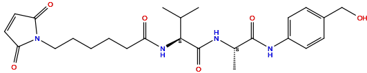
|
|||||
| Formula |
C25H34N4O6
|
|||||
| Isosmiles |
C[C@@H](C(=O)NC1=CC=C(C=C1)CO)NC(=O)[C@H](C(C)C)NC(=O)CCCCCN2C(=O)C=CC2=O
|
|||||
| PubChem CID | ||||||
| InChI |
InChI=1S/C25H34N4O6/c1-16(2)23(28-20(31)7-5-4-6-14-29-21(32)12-13-22(29)33)25(35)26-17(3)24(34)27-19-10-8-18(15-30)9-11-19/h8-13,16-17,23,30H,4-7,14-15H2,1-3H3,(H,26,35)(H,27,34)(H,28,31)/t17-,23-/m0/s1
|
|||||
| InChIKey |
DAMSURYLURICHN-SBUREZEXSA-N
|
|||||
| IUPAC Name |
6-(2,5-dioxopyrrol-1-yl)-N-[(2S)-1-[[(2S)-1-[4-(hydroxymethyl)anilino]-1-oxopropan-2-yl]amino]-3-methyl-1-oxobutan-2-yl]hexanamide
|
|||||
| Pharmaceutical Properties |
Molecule Weight
|
486.6
|
Polar area
|
145
|
||
|
Complexity
|
786
|
xlogp Value
|
0.9
|
|||
|
Heavy Count
|
35
|
Rot Bonds
|
13
|
|||
|
Hbond acc
|
6
|
Hbond Donor
|
4
|
|||
Each Antibody-drug Conjugate Related to This Linker
Full Information of The Activity Data of The ADC(s) Related to This Linker
Tamrintamab pamozirine [Phase 1 (discontinued)]
Identified from the Human Clinical Data
| Experiment 1 Reporting the Activity Date of This ADC | [1] | ||||
| Efficacy Data | Partial Response (PR) |
1.72%
|
|||
| Patients Enrolled |
Female patients (age 18 years) with EOC if they had evidence of progressive disease during or within 6 months of receiving a platinum regimen.
|
||||
| Administration Dosage |
1 of 6 dose levels (0.025-0.40 mg/kg) every 3 weeks (Q3W), utilizing a standard 3+3 design (dose-limiting toxicity [DLT] period: 21 days).
|
||||
| Related Clinical Trial | |||||
| NCT Number | NCT02539719 | Clinical Status | Phase 1 | ||
| Clinical Description |
A phase 1a/1b dose escalation and expansion study of SC-003 as a single-agent and in combination with ABBV-181 in subjects with platinum-resistant/ refractory ovarian cancer.
|
||||
| Primary Endpoint |
The MTD for the Q3W schedule was 0.30 mg/kg and the SC-003 doses selected for the dose-expansion phase of the study were 0.30 mg/kg and 0.20 mg/kg.
|
||||
| Other Endpoint |
ORR=5.17% (N=3/58), 3 patients achieved PR. All responses were observed at 0.20-0.30mg/kg. Responses were not durable, with only 1 PR confirmed on the follow-up 16-week scan.
|
||||
Cetuximab- (FGX16-11) [Investigative]
Discovered Using Cell Line-derived Xenograft Model
| Experiment 1 Reporting the Activity Date of This ADC | [2] | ||||
| Efficacy Data | Tumor Growth Inhibition value (TGI) | ≈ 42.79% | High EGFR expression (EGFR+++) | ||
| Method Description |
The inhibitory activity of Cetuximab-(FGX16-11) against cancer cell growth was evaluated in various human cancer cell lines in vivo. The cells were treated with 1 mg/kg.
|
||||
| In Vivo Model | Colon cancer CDX model | ||||
| In Vitro Model | Colon adenocarcinoma | SW48 cells | CVCL_1724 | ||
| Experiment 2 Reporting the Activity Date of This ADC | [2] | ||||
| Efficacy Data | Tumor Growth Inhibition value (TGI) | ≈ 63.01% | High EGFR expression (EGFR+++) | ||
| Method Description |
The inhibitory activity of Cetuximab-(FGX16-11) against cancer cell growth was evaluated in various human cancer cell lines in vivo. The cells were treated with 5 mg/kg.
|
||||
| In Vivo Model | Colon cancer CDX model | ||||
| In Vitro Model | Colon adenocarcinoma | SW48 cells | CVCL_1724 | ||
| Experiment 3 Reporting the Activity Date of This ADC | [2] | ||||
| Efficacy Data | Tumor Growth Inhibition value (TGI) | ≈ 92.22% | High EGFR expression (EGFR+++) | ||
| Method Description |
The inhibitory activity of Cetuximab-(FGX16-11) against cancer cell growth was evaluated in various human cancer cell lines in vivo. The cells were treated with 10 mg/kg.
|
||||
| In Vivo Model | Colon cancer CDX model | ||||
| In Vitro Model | Colon adenocarcinoma | SW48 cells | CVCL_1724 | ||
| Experiment 4 Reporting the Activity Date of This ADC | [2] | ||||
| Efficacy Data | Tumor Growth Inhibition value (TGI) | ≈ 99.98% | High EGFR expression (EGFR+++) | ||
| Method Description |
The inhibitory activity of Cetuximab-(FGX16-11) against cancer cell growth was evaluated in various human cancer cell lines in vivo. The cells were treated with 20 mg/kg.
|
||||
| In Vivo Model | Colon cancer CDX model | ||||
| In Vitro Model | Colon adenocarcinoma | SW48 cells | CVCL_1724 | ||
| Experiment 5 Reporting the Activity Date of This ADC | [2] | ||||
| Efficacy Data | Tumor Growth Inhibition value (TGI) | ≈ 100% | High EGFR expression (EGFR+++) | ||
| Method Description |
The inhibitory activity of Cetuximab-(FGX16-11) against cancer cell growth was evaluated in various human cancer cell lines in vivo. The cells were treated with 40 mg/kg.
|
||||
| In Vivo Model | Colon cancer CDX model | ||||
| In Vitro Model | Colon adenocarcinoma | SW48 cells | CVCL_1724 | ||
Anti-HER2-D265C-30.2371 [Investigative]
Revealed Based on the Cell Line Data
| Experiment 1 Reporting the Activity Date of This ADC | [3] | ||||
| Efficacy Data | Half Maximal Inhibitory Concentration (IC50) |
8 pM
|
Positive HER2 expression (HER2+++/++; HER2 MFI=1016) | ||
| Method Description |
The cytotoxic activity in vitro of ADCs, which are comprising an anti-HER2 antibody carrying a D265C mutation (T-D265C, anti-HER2-D265C) conjugated tostructurally different amanitin derivatives via its D265C residue was tested on JIMT-1 cells NCI-N87 cells and SKBR-3 cells.
|
||||
| In Vitro Model | Gastric tubular adenocarcinoma | NCI-N87 cells | CVCL_1603 | ||
| Experiment 2 Reporting the Activity Date of This ADC | [3] | ||||
| Efficacy Data | Half Maximal Inhibitory Concentration (IC50) |
10 pM
|
Positive HER2 expression (HER2 +++/++) | ||
| Method Description |
The cytotoxic activity in vitro of ADCs, which are comprising an anti-HER2 antibody carrying a D265C mutation (T-D265C, anti-HER2-D265C) conjugated tostructurally different amanitin derivatives via its D265C residue was tested on JIMT-1 cells NCI-N87 cells and SKBR-3 cells.
|
||||
| In Vitro Model | Breast adenocarcinoma | SK-BR-3 cells | CVCL_0033 | ||
| Experiment 3 Reporting the Activity Date of This ADC | [3] | ||||
| Efficacy Data | Half Maximal Inhibitory Concentration (IC50) |
71 pM
|
Moderate HER2 expression (HER2++) | ||
| Method Description |
The cytotoxic activity in vitro of ADCs, which are comprising an anti-HER2 antibody carrying a D265C mutation (T-D265C, anti-HER2-D265C) conjugated tostructurally different amanitin derivatives via its D265C residue was tested on JIMT-1 cells NCI-N87 cells and SKBR-3 cells.
|
||||
| In Vitro Model | Breast ductal carcinoma | JIMT-1 cells | CVCL_2077 | ||
Anti-PSMA-D265C-30.2371 [Investigative]
Revealed Based on the Cell Line Data
| Experiment 1 Reporting the Activity Date of This ADC | [3] | ||||
| Efficacy Data | Half Maximal Inhibitory Concentration (IC50) |
14 pM
|
Positive PSMA expression (PSMA +++/++) | ||
| Method Description |
The cytotoxic activity in vitro of ADCs, which are comprising an anti-PSMA antibody carrying a D265C mutation conjugated tostructurally different amanitin derivatives via its D265C residue was tested on LNCaP cells and 22RV1 cells.
|
||||
| In Vitro Model | Prostate carcinoma | LNCaP cells | CVCL_0395 | ||
| Experiment 2 Reporting the Activity Date of This ADC | [3] | ||||
| Efficacy Data | Half Maximal Inhibitory Concentration (IC50) |
71 pM
|
Positive PSMA expression (PSMA +++/++) | ||
| Method Description |
The cytotoxic activity in vitro of ADCs, which are comprising an anti-PSMA antibody carrying a D265C mutation conjugated tostructurally different amanitin derivatives via its D265C residue was tested on LNCaP cells and 22RV1 cells.
|
||||
| In Vitro Model | Prostate carcinoma | 22RV1 cells | CVCL_1045 | ||
Anti-HER2-D265C-30.1699 [Investigative]
Revealed Based on the Cell Line Data
| Experiment 1 Reporting the Activity Date of This ADC | [3] | ||||
| Efficacy Data | Half Maximal Inhibitory Concentration (IC50) |
17 pM
|
Positive HER2 expression (HER2 +++/++) | ||
| Method Description |
The cytotoxic activity in vitro of ADCs, which are comprising an anti-HER2 antibody carrying a D265C mutation (T-D265C, anti-HER2-D265C) conjugated tostructurally different amanitin derivatives via its D265C residue was tested on JIMT-1 cells NCI-N87 cells and SKBR-3 cells.
|
||||
| In Vitro Model | Breast adenocarcinoma | SK-BR-3 cells | CVCL_0033 | ||
| Experiment 2 Reporting the Activity Date of This ADC | [3] | ||||
| Efficacy Data | Half Maximal Inhibitory Concentration (IC50) |
18 pM
|
Positive HER2 expression (HER2+++/++; HER2 MFI=1016) | ||
| Method Description |
The cytotoxic activity in vitro of ADCs, which are comprising an anti-HER2 antibody carrying a D265C mutation (T-D265C, anti-HER2-D265C) conjugated tostructurally different amanitin derivatives via its D265C residue was tested on JIMT-1 cells NCI-N87 cells and SKBR-3 cells.
|
||||
| In Vitro Model | Gastric tubular adenocarcinoma | NCI-N87 cells | CVCL_1603 | ||
| Experiment 3 Reporting the Activity Date of This ADC | [3] | ||||
| Efficacy Data | Half Maximal Inhibitory Concentration (IC50) |
0.11 nM
|
Moderate HER2 expression (HER2++) | ||
| Method Description |
The cytotoxic activity in vitro of ADCs, which are comprising an anti-HER2 antibody carrying a D265C mutation (T-D265C, anti-HER2-D265C) conjugated tostructurally different amanitin derivatives via its D265C residue was tested on JIMT-1 cells NCI-N87 cells and SKBR-3 cells.
|
||||
| In Vitro Model | Breast ductal carcinoma | JIMT-1 cells | CVCL_2077 | ||
Anti-PSMA-D265C-30.1699 [Investigative]
Revealed Based on the Cell Line Data
| Experiment 1 Reporting the Activity Date of This ADC | [3] | ||||
| Efficacy Data | Half Maximal Inhibitory Concentration (IC50) |
22 pM
|
Positive PSMA expression (PSMA +++/++) | ||
| Method Description |
The cytotoxic activity in vitro of ADCs, which are comprising an anti-PSMA antibody carrying a D265C mutation conjugated tostructurally different amanitin derivatives via its D265C residue was tested on LNCaP cells and 22RV1 cells.
|
||||
| In Vitro Model | Prostate carcinoma | LNCaP cells | CVCL_0395 | ||
| Experiment 2 Reporting the Activity Date of This ADC | [3] | ||||
| Efficacy Data | Half Maximal Inhibitory Concentration (IC50) |
0.11 nM
|
Positive PSMA expression (PSMA +++/++) | ||
| Method Description |
The cytotoxic activity in vitro of ADCs, which are comprising an anti-PSMA antibody carrying a D265C mutation conjugated tostructurally different amanitin derivatives via its D265C residue was tested on LNCaP cells and 22RV1 cells.
|
||||
| In Vitro Model | Prostate carcinoma | 22RV1 cells | CVCL_1045 | ||
Anti-HER2-D265C-30.2115 [Investigative]
Revealed Based on the Cell Line Data
| Experiment 1 Reporting the Activity Date of This ADC | [3] | ||||
| Efficacy Data | Half Maximal Inhibitory Concentration (IC50) |
25 pM
|
Positive HER2 expression (HER2 +++/++) | ||
| Method Description |
The cytotoxic activity in vitro of ADCs, which are comprising an anti-HER2 antibody carrying a D265C mutation (T-D265C, anti-HER2-D265C) conjugated tostructurally different amanitin derivatives via its D265C residue was tested on JIMT-1 cells NCI-N87 cells and SKBR-3 cells.
|
||||
| In Vitro Model | Breast adenocarcinoma | SK-BR-3 cells | CVCL_0033 | ||
| Experiment 2 Reporting the Activity Date of This ADC | [3] | ||||
| Efficacy Data | Half Maximal Inhibitory Concentration (IC50) |
54 pM
|
Positive HER2 expression (HER2+++/++; HER2 MFI=1016) | ||
| Method Description |
The cytotoxic activity in vitro of ADCs, which are comprising an anti-HER2 antibody carrying a D265C mutation (T-D265C, anti-HER2-D265C) conjugated tostructurally different amanitin derivatives via its D265C residue was tested on JIMT-1 cells NCI-N87 cells and SKBR-3 cells.
|
||||
| In Vitro Model | Gastric tubular adenocarcinoma | NCI-N87 cells | CVCL_1603 | ||
| Experiment 3 Reporting the Activity Date of This ADC | [3] | ||||
| Efficacy Data | Half Maximal Inhibitory Concentration (IC50) |
0.28 nM
|
Moderate HER2 expression (HER2++) | ||
| Method Description |
The cytotoxic activity in vitro of ADCs, which are comprising an anti-HER2 antibody carrying a D265C mutation (T-D265C, anti-HER2-D265C) conjugated tostructurally different amanitin derivatives via its D265C residue was tested on JIMT-1 cells NCI-N87 cells and SKBR-3 cells.
|
||||
| In Vitro Model | Breast ductal carcinoma | JIMT-1 cells | CVCL_2077 | ||
Anti-HER2-D265C-30.2060 [Investigative]
Revealed Based on the Cell Line Data
| Experiment 1 Reporting the Activity Date of This ADC | [3] | ||||
| Efficacy Data | Half Maximal Inhibitory Concentration (IC50) |
31 pM
|
Positive HER2 expression (HER2 +++/++) | ||
| Method Description |
The cytotoxic activity in vitro of ADCs, which are comprising an anti-HER2 antibody carrying a D265C mutation (T-D265C, anti-HER2-D265C) conjugated tostructurally different amanitin derivatives via its D265C residue was tested on JIMT-1 cells NCI-N87 cells and SKBR-3 cells.
|
||||
| In Vitro Model | Breast adenocarcinoma | SK-BR-3 cells | CVCL_0033 | ||
| Experiment 2 Reporting the Activity Date of This ADC | [3] | ||||
| Efficacy Data | Half Maximal Inhibitory Concentration (IC50) |
32 pM
|
Positive HER2 expression (HER2+++/++; HER2 MFI=1016) | ||
| Method Description |
The cytotoxic activity in vitro of ADCs, which are comprising an anti-HER2 antibody carrying a D265C mutation (T-D265C, anti-HER2-D265C) conjugated tostructurally different amanitin derivatives via its D265C residue was tested on JIMT-1 cells NCI-N87 cells and SKBR-3 cells.
|
||||
| In Vitro Model | Gastric tubular adenocarcinoma | NCI-N87 cells | CVCL_1603 | ||
| Experiment 3 Reporting the Activity Date of This ADC | [3] | ||||
| Efficacy Data | Half Maximal Inhibitory Concentration (IC50) |
0.21 nM
|
Moderate HER2 expression (HER2++) | ||
| Method Description |
The cytotoxic activity in vitro of ADCs, which are comprising an anti-HER2 antibody carrying a D265C mutation (T-D265C, anti-HER2-D265C) conjugated tostructurally different amanitin derivatives via its D265C residue was tested on JIMT-1 cells NCI-N87 cells and SKBR-3 cells.
|
||||
| In Vitro Model | Breast ductal carcinoma | JIMT-1 cells | CVCL_2077 | ||
Anti-PSMA-D265C-30.2060 [Investigative]
Revealed Based on the Cell Line Data
| Experiment 1 Reporting the Activity Date of This ADC | [3] | ||||
| Efficacy Data | Half Maximal Inhibitory Concentration (IC50) |
32 pM
|
Positive PSMA expression (PSMA +++/++) | ||
| Method Description |
The cytotoxic activity in vitro of ADCs, which are comprising an anti-PSMA antibody carrying a D265C mutation conjugated tostructurally different amanitin derivatives via its D265C residue was tested on LNCaP cells and 22RV1 cells.
|
||||
| In Vitro Model | Prostate carcinoma | LNCaP cells | CVCL_0395 | ||
| Experiment 2 Reporting the Activity Date of This ADC | [3] | ||||
| Efficacy Data | Half Maximal Inhibitory Concentration (IC50) |
0.19 nM
|
Positive PSMA expression (PSMA +++/++) | ||
| Method Description |
The cytotoxic activity in vitro of ADCs, which are comprising an anti-PSMA antibody carrying a D265C mutation conjugated tostructurally different amanitin derivatives via its D265C residue was tested on LNCaP cells and 22RV1 cells.
|
||||
| In Vitro Model | Prostate carcinoma | 22RV1 cells | CVCL_1045 | ||
Anti-PSMA-D265C-30.2347 [Investigative]
Revealed Based on the Cell Line Data
| Experiment 1 Reporting the Activity Date of This ADC | [3] | ||||
| Efficacy Data | Half Maximal Inhibitory Concentration (IC50) |
43 pM
|
Positive PSMA expression (PSMA +++/++) | ||
| Method Description |
The cytotoxic activity in vitro of ADCs, which are comprising an anti-PSMA antibody carrying a D265C mutation conjugated tostructurally different amanitin derivatives via its D265C residue was tested on LNCaP cells and 22RV1 cells.
|
||||
| In Vitro Model | Prostate carcinoma | LNCaP cells | CVCL_0395 | ||
| Experiment 2 Reporting the Activity Date of This ADC | [3] | ||||
| Efficacy Data | Half Maximal Inhibitory Concentration (IC50) |
0.16 nM
|
Positive PSMA expression (PSMA +++/++) | ||
| Method Description |
The cytotoxic activity in vitro of ADCs, which are comprising an anti-PSMA antibody carrying a D265C mutation conjugated tostructurally different amanitin derivatives via its D265C residue was tested on LNCaP cells and 22RV1 cells.
|
||||
| In Vitro Model | Prostate carcinoma | 22RV1 cells | CVCL_1045 | ||
Anti-HER2-D265C-30.2347 [Investigative]
Revealed Based on the Cell Line Data
| Experiment 1 Reporting the Activity Date of This ADC | [3] | ||||
| Efficacy Data | Half Maximal Inhibitory Concentration (IC50) |
46 pM
|
Positive HER2 expression (HER2+++/++; HER2 MFI=1016) | ||
| Method Description |
The cytotoxic activity in vitro of ADCs, which are comprising an anti-HER2 antibody carrying a D265C mutation (T-D265C, anti-HER2-D265C) conjugated tostructurally different amanitin derivatives via its D265C residue was tested on JIMT-1 cells NCI-N87 cells and SKBR-3 cells.
|
||||
| In Vitro Model | Gastric tubular adenocarcinoma | NCI-N87 cells | CVCL_1603 | ||
| Experiment 2 Reporting the Activity Date of This ADC | [3] | ||||
| Efficacy Data | Half Maximal Inhibitory Concentration (IC50) |
59 pM
|
Positive HER2 expression (HER2 +++/++) | ||
| Method Description |
The cytotoxic activity in vitro of ADCs, which are comprising an anti-HER2 antibody carrying a D265C mutation (T-D265C, anti-HER2-D265C) conjugated tostructurally different amanitin derivatives via its D265C residue was tested on JIMT-1 cells NCI-N87 cells and SKBR-3 cells.
|
||||
| In Vitro Model | Breast adenocarcinoma | SK-BR-3 cells | CVCL_0033 | ||
| Experiment 3 Reporting the Activity Date of This ADC | [3] | ||||
| Efficacy Data | Half Maximal Inhibitory Concentration (IC50) |
0.13 nM
|
Moderate HER2 expression (HER2++) | ||
| Method Description |
The cytotoxic activity in vitro of ADCs, which are comprising an anti-HER2 antibody carrying a D265C mutation (T-D265C, anti-HER2-D265C) conjugated tostructurally different amanitin derivatives via its D265C residue was tested on JIMT-1 cells NCI-N87 cells and SKBR-3 cells.
|
||||
| In Vitro Model | Breast ductal carcinoma | JIMT-1 cells | CVCL_2077 | ||
Anti-PSMA-D265C-30.2115 [Investigative]
Revealed Based on the Cell Line Data
| Experiment 1 Reporting the Activity Date of This ADC | [3] | ||||
| Efficacy Data | Half Maximal Inhibitory Concentration (IC50) |
0.27 nM
|
Positive PSMA expression (PSMA +++/++) | ||
| Method Description |
The cytotoxic activity in vitro of ADCs, which are comprising an anti-PSMA antibody carrying a D265C mutation conjugated tostructurally different amanitin derivatives via its D265C residue was tested on LNCaP cells and 22RV1 cells.
|
||||
| In Vitro Model | Prostate carcinoma | LNCaP cells | CVCL_0395 | ||
| Experiment 2 Reporting the Activity Date of This ADC | [3] | ||||
| Efficacy Data | Half Maximal Inhibitory Concentration (IC50) |
0.36 nM
|
Positive PSMA expression (PSMA +++/++) | ||
| Method Description |
The cytotoxic activity in vitro of ADCs, which are comprising an anti-PSMA antibody carrying a D265C mutation conjugated tostructurally different amanitin derivatives via its D265C residue was tested on LNCaP cells and 22RV1 cells.
|
||||
| In Vitro Model | Prostate carcinoma | 22RV1 cells | CVCL_1045 | ||
Mil40-5 [Investigative]
Revealed Based on the Cell Line Data
| Experiment 1 Reporting the Activity Date of This ADC | [4] | ||||
| Efficacy Data | Half Maximal Inhibitory Concentration (IC50) |
14.5 nM
|
High HER2 expression (HER2+++) | ||
| Method Description |
SKOV-3, BT474 HerDR, MDA-MB-231 and MCF-7 cells were cultured under various concentrations of Mil40, SN-38 and ADCs for 10days, 9days, 6days, and 6days, respectively. Cytotoxicity assays were established using the CellTiter-Go assay kit (CTG).
|
||||
| In Vitro Model | Invasive breast carcinoma | BT474 HerDR cells | CVCL_0179 | ||
| Experiment 2 Reporting the Activity Date of This ADC | [4] | ||||
| Efficacy Data | Half Maximal Inhibitory Concentration (IC50) |
157.6 nM
|
High HER2 expression (HER2+++) | ||
| Method Description |
SKOV-3, BT474 HerDR, MDA-MB-231 and MCF-7 cells were cultured under various concentrations of Mil40, SN-38 and ADCs for 10days, 9days, 6days, and 6days, respectively. Cytotoxicity assays were established using the CellTiter-Go assay kit (CTG).
|
||||
| In Vitro Model | Ovarian serous cystadenocarcinoma | SK-OV-3 cells | CVCL_0532 | ||
| Experiment 3 Reporting the Activity Date of This ADC | [4] | ||||
| Efficacy Data | Half Maximal Inhibitory Concentration (IC50) | > 1000 nM | Negative HER2 expression (HER2-) | ||
| Method Description |
SKOV-3, BT474 HerDR, MDA-MB-231 and MCF-7 cells were cultured under various concentrations of Mil40, SN-38 and ADCs for 10days, 9days, 6days, and 6days, respectively. Cytotoxicity assays were established using the CellTiter-Go assay kit (CTG).
|
||||
| In Vitro Model | Invasive breast carcinoma | MCF-7 cells | CVCL_0031 | ||
| Experiment 4 Reporting the Activity Date of This ADC | [4] | ||||
| Efficacy Data | Half Maximal Inhibitory Concentration (IC50) | > 1000 nM | Negative HER2 expression (HER2-) | ||
| Method Description |
SKOV-3, BT474 HerDR, MDA-MB-231 and MCF-7 cells were cultured under various concentrations of Mil40, SN-38 and ADCs for 10days, 9days, 6days, and 6days, respectively. Cytotoxicity assays were established using the CellTiter-Go assay kit (CTG).
|
||||
| In Vitro Model | Breast adenocarcinoma | MDA-MB-231 cells | CVCL_0062 | ||
PD-L1 ADC 1 [Investigative]
Revealed Based on the Cell Line Data
| Experiment 1 Reporting the Activity Date of This ADC | [5] | ||||
| Efficacy Data | Half Maximal Inhibitory Concentration (IC50) |
265.3 nM
|
High PD-L1 expression (PD-L1+++) | ||
| Method Description |
The in vitro cytotoxicity of ADC 1 and ADC 2 was evaluated in three PD-L1-positive cell lines, i.e., Calu-1, MDA-MB-231, and SK-MES, and one PD-L1-negative cell line, i.e., AsPC-1.
|
||||
| In Vitro Model | Lung squamous cell carcinoma | SK-MES-1 cells | CVCL_0630 | ||
| Experiment 2 Reporting the Activity Date of This ADC | [5] | ||||
| Efficacy Data | Half Maximal Inhibitory Concentration (IC50) | > 663.08 nM | Negative PD-L1 expression (PD-L1-) | ||
| Method Description |
The in vitro cytotoxicity of ADC 1 and ADC 2 was evaluated in three PD-L1-positive cell lines, i.e., Calu-1, MDA-MB-231, and SK-MES, and one PD-L1-negative cell line, i.e., AsPC-1.
|
||||
| In Vitro Model | Pancreatic ductal adenocarcinoma | AsPC-1 cells | CVCL_0152 | ||
| Experiment 3 Reporting the Activity Date of This ADC | [5] | ||||
| Efficacy Data | Half Maximal Inhibitory Concentration (IC50) | > 663.08 nM | High PD-L1 expression (PD-L1+++) | ||
| Method Description |
The in vitro cytotoxicity of ADC 1 and ADC 2 was evaluated in three PD-L1-positive cell lines, i.e., Calu-1, MDA-MB-231, and SK-MES, and one PD-L1-negative cell line, i.e., AsPC-1.
|
||||
| In Vitro Model | Breast adenocarcinoma | MDA-MB-231 cells | CVCL_0062 | ||
| Experiment 4 Reporting the Activity Date of This ADC | [5] | ||||
| Efficacy Data | Half Maximal Inhibitory Concentration (IC50) | > 663.08 nM | High PD-L1 expression (PD-L1+++) | ||
| Method Description |
The in vitro cytotoxicity of ADC 1 and ADC 2 was evaluated in three PD-L1-positive cell lines, i.e., Calu-1, MDA-MB-231, and SK-MES, and one PD-L1-negative cell line, i.e., AsPC-1.
|
||||
| In Vitro Model | Lung squamous cell carcinoma | Calu-1 cells | CVCL_0608 | ||
PD-L1 ADC 3 [Investigative]
Revealed Based on the Cell Line Data
| Experiment 1 Reporting the Activity Date of This ADC | [5] | ||||
| Efficacy Data | Half Maximal Effective Concentration (EC50) |
9.75 nM
|
High PD-L1 expression (PD-L1+++) | ||
| Method Description |
The in vitro cytotoxicity of ADC 3 was quickly evaluated in three PD-L1-positive cell lines, ie, MDA-MB-231, PC 9, and A431, and one PD-L1-negative cell line, ie, Romas.
|
||||
| In Vitro Model | Lung adenocarcinoma | PC-9 cells | CVCL_B260 | ||
| Experiment 2 Reporting the Activity Date of This ADC | [5] | ||||
| Efficacy Data | Half Maximal Effective Concentration (EC50) |
10.33 nM
|
High PD-L1 expression (PD-L1+++) | ||
| Method Description |
The in vitro cytotoxicity of ADC 3 was quickly evaluated in three PD-L1-positive cell lines, ie, MDA-MB-231, PC 9, and A431, and one PD-L1-negative cell line, ie, Romas.
|
||||
| In Vitro Model | Breast adenocarcinoma | MDA-MB-231 cells | CVCL_0062 | ||
| Experiment 3 Reporting the Activity Date of This ADC | [5] | ||||
| Efficacy Data | Half Maximal Effective Concentration (EC50) |
11.94 nM
|
High PD-L1 expression (PD-L1+++) | ||
| Method Description |
The in vitro cytotoxicity of ADC 3 was quickly evaluated in three PD-L1-positive cell lines, ie, MDA-MB-231, PC 9, and A431, and one PD-L1-negative cell line, ie, Romas.
|
||||
| In Vitro Model | Skin squamous cell carcinoma | A431 cells | CVCL_0037 | ||
WO2024222841A1 ADC66 [Investigative]
Revealed Based on the Cell Line Data
| Experiment 1 Reporting the Activity Date of This ADC | [6] | ||||
| Efficacy Data | Half Maximal inhibitory Concentration (lC50) |
6.613 pM
|
High CD33 expression (CD33+++) | ||
| Method Description |
Cytotoxicity of ADC to MV4-11 cells
|
||||
| In Vitro Model | Childhood acute monocytic leukemia | MV4-11 cells | CVCL_0064 | ||
WO2024222841A1 ADC65 [Investigative]
Revealed Based on the Cell Line Data
| Experiment 1 Reporting the Activity Date of This ADC | [6] | ||||
| Efficacy Data | Half Maximal inhibitory Concentration (lC50) |
6.699 pM
|
High CD33 expression (CD33+++) | ||
| Method Description |
Cytotoxicity of ADC to MV4-11 cells
|
||||
| In Vitro Model | Childhood acute monocytic leukemia | MV4-11 cells | CVCL_0064 | ||
WO2024222841A1 ADC67 [Investigative]
Revealed Based on the Cell Line Data
| Experiment 1 Reporting the Activity Date of This ADC | [6] | ||||
| Efficacy Data | Half Maximal inhibitory Concentration (lC50) |
12.3 pM
|
High CD33 expression (CD33+++) | ||
| Method Description |
Cytotoxicity of ADC to MV4-11 cells
|
||||
| In Vitro Model | Childhood acute monocytic leukemia | MV4-11 cells | CVCL_0064 | ||
WO2024169913A1 ADC01-06-8 [Investigative]
Revealed Based on the Cell Line Data
| Experiment 1 Reporting the Activity Date of This ADC | [7] | ||||
| Efficacy Data | Half Maximal inhibitory Concentration (lC50) |
0.01116 nM
|
High HER2 expression (HER2+++) | ||
| Method Description |
Cytotoxicity of ADC to SK-BR-3 cells
|
||||
| In Vitro Model | Breast adenocarcinoma | SK-BR-3 cells | CVCL_0033 | ||
WO2024169913A1 ADC01-06-4 [Investigative]
Revealed Based on the Cell Line Data
| Experiment 1 Reporting the Activity Date of This ADC | [7] | ||||
| Efficacy Data | Half Maximal inhibitory Concentration (lC50) |
0.02107 nM
|
High HER2 expression (HER2+++) | ||
| Method Description |
Cytotoxicity of ADC to SK-BR-3 cells
|
||||
| In Vitro Model | Breast adenocarcinoma | SK-BR-3 cells | CVCL_0033 | ||
Su-C6-VA [Investigative]
Revealed Based on the Cell Line Data
| Experiment 1 Reporting the Activity Date of This ADC | [8] | ||||
| Efficacy Data | Half Maximal inhibitory Concentration (lC50) |
0.0231 nM
|
Positive HER2 expression (HER2+++/++) | ||
| Method Description |
HCC 1954, 1000cell/well was added to the designated 384-well cell culture plate in the appropriate growth media. The ADCs stock solution was diluted in series in a ratio of 1:3 to produce 10 diluted solutions with different concentrations (the compound stock solution was an L-His buffer salt solution with a concentration of about 2 mg/mL, which was diluted with PBS, and the initial maximum concentration at test point was about 500 mg/mL). 4ul of the compound diluted solutions with different concentrations was distributed into a 384-well plate. After 96 hours, the plate was taken from the incubator and equilibrated at room temperature for 10 minutes. 40uL of CellTiter-Glo reagent was added to each well to be tested (the ratio of CellTiter-Glo reagent to medium was 1:1).the plate was then placed at room temperature for 30 minutes, and then reading was carried out on an EnSpire reader for cell counting.
Click to Show/Hide
|
||||
| In Vitro Model | Breast ductal carcinoma | HCC1954 cells | CVCL_1259 | ||
| Experiment 2 Reporting the Activity Date of This ADC | [8] | ||||
| Efficacy Data | Half Maximal inhibitory Concentration (lC50) |
0.0563 nM
|
Positive HER2 expression (HER2+++/++) | ||
| Method Description |
NCI-N87, 1000cell/well was added to the designated 384-well cell culture plate in the appropriate growth media. The ADCs stock solution was diluted in series in a ratio of 1:3 to produce 10 diluted solutions with different concentrations (the compound stock solution was an L-His buffer salt solution with a concentration of about 2 mg/mL, which was diluted with PBS, and the initial maximum concentration at test point was about 500 mg/mL). 4ul of the compound diluted solutions with different concentrations was distributed into a 384-well plate. After 96 hours, the plate was taken from the incubator and equilibrated at room temperature for 10 minutes. 40uL of CellTiter-Glo reagent was added to each well to be tested (the ratio of CellTiter-Glo reagent to medium was 1:1).the plate was then placed at room temperature for 30 minutes, and then reading was carried out on an EnSpire reader for cell counting.
Click to Show/Hide
|
||||
| In Vitro Model | Gastric tubular adenocarcinoma | NCI-N87 cells | CVCL_1603 | ||
| Experiment 3 Reporting the Activity Date of This ADC | [8] | ||||
| Efficacy Data | Half Maximal inhibitory Concentration (lC50) |
0.0783 nM
|
Positive HER2 expression (HER2+++/++) | ||
| Method Description |
BT-474, 1000cell/well was added to the designated 384-well cell culture plate in the appropriate growth media. The ADCs stock solution was diluted in series in a ratio of 1:3 to produce 10 diluted solutions with different concentrations (the compound stock solution was an L-His buffer salt solution with a concentration of about 2 mg/mL, which was diluted with PBS, and the initial maximum concentration at test point was about 500 mg/mL). 4ul of the compound diluted solutions with different concentrations was distributed into a 384-well plate. After 96 hours, the plate was taken from the incubator and equilibrated at room temperature for 10 minutes. 40uL of CellTiter-Glo reagent was added to each well to be tested (the ratio of CellTiter-Glo reagent to medium was 1:1).the plate was then placed at room temperature for 30 minutes, and then reading was carried out on an EnSpire reader for cell counting.
Click to Show/Hide
|
||||
| In Vitro Model | Invasive breast carcinoma | BT-474 cells | CVCL_0179 | ||
| Experiment 4 Reporting the Activity Date of This ADC | [8] | ||||
| Efficacy Data | Half Maximal inhibitory Concentration (lC50) |
0.1518 nM
|
Positive HER2 expression (HER2+++/++) | ||
| Method Description |
SK-OV-3, 1000cell/well was added to the designated 384-well cell culture plate in the appropriate growth media. The ADCs stock solution was diluted in series in a ratio of 1:3 to produce 10 diluted solutions with different concentrations (the compound stock solution was an L-His buffer salt solution with a concentration of about 2 mg/mL, which was diluted with PBS, and the initial maximum concentration at test point was about 500 mg/mL). 4ul of the compound diluted solutions with different concentrations was distributed into a 384-well plate. After 96 hours, the plate was taken from the incubator and equilibrated at room temperature for 10 minutes. 40uL of CellTiter-Glo reagent was added to each well to be tested (the ratio of CellTiter-Glo reagent to medium was 1:1).the plate was then placed at room temperature for 30 minutes, and then reading was carried out on an EnSpire reader for cell counting.
Click to Show/Hide
|
||||
| In Vitro Model | Ovarian serous adenocarcinoma | SKOV-3 cells | CVCL_0532 | ||
| Experiment 5 Reporting the Activity Date of This ADC | [8] | ||||
| Efficacy Data | Half Maximal inhibitory Concentration (lC50) | > 1.6495 nM | Negative HER2 expression (HER2-) | ||
| Method Description |
MCF-7, 1000cell/well was added to the designated 384-well cell culture plate in the appropriate growth media. The ADCs stock solution was diluted in series in a ratio of 1:3 to produce 10 diluted solutions with different concentrations (the compound stock solution was an L-His buffer salt solution with a concentration of about 2 mg/mL, which was diluted with PBS, and the initial maximum concentration at test point was about 500 mg/mL). 4ul of the compound diluted solutions with different concentrations was distributed into a 384-well plate. After 96 hours, the plate was taken from the incubator and equilibrated at room temperature for 10 minutes. 40uL of CellTiter-Glo reagent was added to each well to be tested (the ratio of CellTiter-Glo reagent to medium was 1:1).the plate was then placed at room temperature for 30 minutes, and then reading was carried out on an EnSpire reader for cell counting.
Click to Show/Hide
|
||||
| In Vitro Model | Invasive breast carcinoma | MCF-7 cells | CVCL_0031 | ||
| Experiment 6 Reporting the Activity Date of This ADC | [8] | ||||
| Efficacy Data | Half Maximal inhibitory Concentration (lC50) | > 1.6495 nM | Negative HER2 expression (HER2-) | ||
| Method Description |
MDA-MB-468, 1000cell/well was added to the designated 384-well cell culture plate in the appropriate growth media. The ADCs stock solution was diluted in series in a ratio of 1:3 to produce 10 diluted solutions with different concentrations (the compound stock solution was an L-His buffer salt solution with a concentration of about 2 mg/mL, which was diluted with PBS, and the initial maximum concentration at test point was about 500 mg/mL). 4ul of the compound diluted solutions with different concentrations was distributed into a 384-well plate. After 96 hours, the plate was taken from the incubator and equilibrated at room temperature for 10 minutes. 40uL of CellTiter-Glo reagent was added to each well to be tested (the ratio of CellTiter-Glo reagent to medium was 1:1).the plate was then placed at room temperature for 30 minutes, and then reading was carried out on an EnSpire reader for cell counting.
Click to Show/Hide
|
||||
| In Vitro Model | Breast adenocarcinoma | MDA-MB-468 cells | CVCL_0419 | ||
WO2024169913A1 ADC04-07-8 [Investigative]
Revealed Based on the Cell Line Data
| Experiment 1 Reporting the Activity Date of This ADC | [7] | ||||
| Efficacy Data | Half Maximal inhibitory Concentration (lC50) |
0.04488 nM
|
High DLL3 expression (DLL3+++) | ||
| Method Description |
Cytotoxicity of ADC to NCI-H82 cells
|
||||
| In Vitro Model | Lung small cell carcinoma | NCI-H82 cells | CVCL_1591 | ||
FDA026-LE14 [Investigative]
Revealed Based on the Cell Line Data
| Experiment 1 Reporting the Activity Date of This ADC | [9] | ||||
| Efficacy Data | Half Maximal inhibitory Concentration (lC50) |
36.5 nM
|
|||
| Method Description |
SW620 (ATCC) cells were selected as the cell line for in vitro activity detection experiments. 2000 cells were inoculated into each well of a 96-well cell culture plate and cultured for 20-24 hours. The antibody-drug conjugate prepared according to the method in Example 3 was formulated with L15 cell culture medium containing 10% FBS into 11 concentration gradients of test solution: 1000, 166.7, 55.6, 18.6, 6.17, 2.06, 0.69, 0.23, 0.08, 0.008 and OnM. 100 ul of the diluted test solution per well was added to the culture plate containing the inoculated cells and incubated in a 37°C, 5% CO, incubator for 144 hours. Then, CellTiter-Glo® Luminescent Cell Viability Assay Reagent (50 ul/well) was added, and the mixture was shaken at 500 rpm at room temperature for 10 minutes. The data were read using a SpectraMaxL microplate reader (OD570nm, with 2-second interval readings), and the IC52 results were calculated.
Click to Show/Hide
|
||||
| In Vitro Model | Colon adenocarcinoma | SW620 cells | CVCL_0547 | ||
| Experiment 2 Reporting the Activity Date of This ADC | [9] | ||||
| Efficacy Data | Half Maximal inhibitory Concentration (lC50) |
37.5 nM
|
|||
| Method Description |
22Rv1 (ATCC) cells were selected as the cell line for in vitro activity detection experiments. 2000 cells were inoculated into each well of a 96-well cell culture plate and cultured for 20-24 hours. The antibody-drug conjugate prepared according to the method in Example 3 was formulated with L15 cell culture medium containing 10% FBS into 11 concentration gradients of test solution: 1000, 166.7, 55.6, 18.6, 6.17, 2.06, 0.69, 0.23, 0.08, 0.008 and OnM. 100 ul of the diluted test solution per well was added to the culture plate containing the inoculated cells and incubated in a 37°C, 5% CO, incubator for 144 hours. Then, CellTiter-Glo® Luminescent Cell Viability Assay Reagent (50 ul/well) was added, and the mixture was shaken at 500 rpm at room temperature for 10 minutes. The data were read using a SpectraMaxL microplate reader (OD570nm, with 2-second interval readings), and the IC50 results were calculated.
Click to Show/Hide
|
||||
| In Vitro Model | Prostate carcinoma | 22RV1 cells | CVCL_1045 | ||
| Experiment 3 Reporting the Activity Date of This ADC | [9] | ||||
| Efficacy Data | Half Maximal inhibitory Concentration (lC50) |
38.4 nM
|
|||
| Method Description |
NCI-H820 (ATCC) cells were selected as the cell line for in vitro activity detection experiments. 2000 cells were inoculated into each well of a 96-well cell culture plate and cultured for 20-24 hours. The antibody-drug conjugate prepared according to the method in Example 3 was formulated with L15 cell culture medium containing 10% FBS into 11 concentration gradients of test solution: 1000, 166.7, 55.6, 18.6, 6.17, 2.06, 0.69, 0.23, 0.08, 0.008 and OnM. 100 ul of the diluted test solution per well was added to the culture plate containing the inoculated cells and incubated in a 37°C, 5% CO, incubator for 144 hours. Then, CellTiter-Glo® Luminescent Cell Viability Assay Reagent (50 ul/well) was added, and the mixture was shaken at 500 rpm at room temperature for 10 minutes. The data were read using a SpectraMaxL microplate reader (OD570nm, with 2-second interval readings), and the IC50 results were calculated.
Click to Show/Hide
|
||||
| In Vitro Model | Lung papillary adenocarcinoma | NCI-H820 cells | CVCL_1592 | ||
| Experiment 4 Reporting the Activity Date of This ADC | [9] | ||||
| Efficacy Data | Half Maximal inhibitory Concentration (lC50) |
50.23 nM
|
|||
| Method Description |
OVCAR-8 (ATCC) cells were selected as the cell line for in vitro activity detection experiments. 2000 cells were inoculated into each well of a 96-well cell culture plate and cultured for 20-24 hours. The antibody-drug conjugate prepared according to the method in Example 3 was formulated with L15 cell culture medium containing 10% FBS into 11 concentration gradients of test solution: 1000, 166.7, 55.6, 18.6, 6.17, 2.06, 0.69, 0.23, 0.08, 0.008 and OnM. 100 ul of the diluted test solution per well was added to the culture plate containing the inoculated cells and incubated in a 37°C, 5% CO, incubator for 144 hours. Then, CellTiter-Glo® Luminescent Cell Viability Assay Reagent (50 ul/well) was added, and the mixture was shaken at 500 rpm at room temperature for 10 minutes. The data were read using a SpectraMaxL microplate reader (OD570nm, with 2-second interval readings), and the IC64 results were calculated.
Click to Show/Hide
|
||||
| In Vitro Model | High grade ovarian serous adenocarcinoma | OVCAR-8 cells | CVCL_1629 | ||
| Experiment 5 Reporting the Activity Date of This ADC | [9] | ||||
| Efficacy Data | Half Maximal inhibitory Concentration (lC50) |
62.38 nM
|
|||
| Method Description |
HCC827 (ATCC) cells were selected as the cell line for in vitro activity detection experiments. 2000 cells were inoculated into each well of a 96-well cell culture plate and cultured for 20-24 hours. The antibody-drug conjugate prepared according to the method in Example 3 was formulated with L15 cell culture medium containing 10% FBS into 11 concentration gradients of test solution: 1000, 166.7, 55.6, 18.6, 6.17, 2.06, 0.69, 0.23, 0.08, 0.008 and OnM. 100 ul of the diluted test solution per well was added to the culture plate containing the inoculated cells and incubated in a 37°C, 5% CO, incubator for 144 hours. Then, CellTiter-Glo® Luminescent Cell Viability Assay Reagent (50 ul/well) was added, and the mixture was shaken at 500 rpm at room temperature for 10 minutes. The data were read using a SpectraMaxL microplate reader (OD570nm, with 2-second interval readings), and the IC64 results were calculated.
Click to Show/Hide
|
||||
| In Vitro Model | Lung adenocarcinoma | HCC827 cells | CVCL_2063 | ||
| Experiment 6 Reporting the Activity Date of This ADC | [9] | ||||
| Efficacy Data | Half Maximal inhibitory Concentration (lC50) |
85.6 nM
|
|||
| Method Description |
LNCaP (ATCC) cells were selected as the cell line for in vitro activity detection experiments. 2000 cells were inoculated into each well of a 96-well cell culture plate and cultured for 20-24 hours. The antibody-drug conjugate prepared according to the method in Example 3 was formulated with L15 cell culture medium containing 10% FBS into 11 concentration gradients of test solution: 1000, 166.7, 55.6, 18.6, 6.17, 2.06, 0.69, 0.23, 0.08, 0.008 and OnM. 100 ul of the diluted test solution per well was added to the culture plate containing the inoculated cells and incubated in a 37°C, 5% CO, incubator for 144 hours. Then, CellTiter-Glo® Luminescent Cell Viability Assay Reagent (50 ul/well) was added, and the mixture was shaken at 500 rpm at room temperature for 10 minutes. The data were read using a SpectraMaxL microplate reader (OD570nm, with 2-second interval readings), and the IC50 results were calculated.
Click to Show/Hide
|
||||
| In Vitro Model | Prostate carcinoma | LNCaP cells | CVCL_0395 | ||
CN116916962A-ADC-B2 [Investigative]
Revealed Based on the Cell Line Data
| Experiment 1 Reporting the Activity Date of This ADC | [10] | ||||
| Efficacy Data | Half Maximal Effective Concentration (EC50) |
2.63 nM
|
High hCCR2 expression (hCCR2+++) | ||
| Method Description |
THP 1, 15,000cells/254uL per welld were plated at set in 384 well flat plate, in the appropriate growth media. The cell plates were dosed with 5uL of the hCCR2 targeting-ADC samples and then incubated at 37°C for 20 hours. At the end of the incubation 10uL/well of the QUANTJ-LucTM (InvivoGenftrep-qlc1) were added and luminescence was measured immediately using the LeadSeeker.
Click to Show/Hide
|
||||
| In Vitro Model | Acute monoblastic/monocytic leukemia, Childhood acute monocytic leukemia | THP1 cells | CVCL_0006 | ||
References