
Linker Information
General Information of This Linker (ID: LIN0MVTCK)
| Name |
PEG2-Val-Cit-PABC
|
|||||
|---|---|---|---|---|---|---|
| Linker Type |
Cathepsin-cleavable linker
|
|||||
| Antibody-Linker Relation |
Cleavable
|
|||||
| Structure |
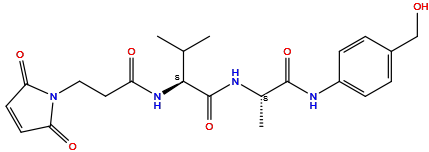
|
|||||
| Formula |
C22H28N4O6
|
|||||
| Isosmiles |
C[C@@H](C(=O)NC1=CC=C(C=C1)CO)NC(=O)[C@H](C(C)C)NC(=O)CCN2C(=O)C=CC2=O
|
|||||
| PubChem CID | ||||||
| InChI |
InChI=1S/C22H28N4O6/c1-13(2)20(25-17(28)10-11-26-18(29)8-9-19(26)30)22(32)23-14(3)21(31)24-16-6-4-15(12-27)5-7-16/h4-9,13-14,20,27H,10-12H2,1-3H3,(H,23,32)(H,24,31)(H,25,28)/t14-,20-/m0/s1
|
|||||
| InChIKey |
NSHZSEBCOJNZHE-XOBRGWDASA-N
|
|||||
| IUPAC Name |
(2S)-2-[3-(2,5-dioxopyrrol-1-yl)propanoylamino]-N-[(2S)-1-[4-(hydroxymethyl)anilino]-1-oxopropan-2-yl]-3-methylbutanamide
|
|||||
| Pharmaceutical Properties |
Molecule Weight
|
444.5
|
Polar area
|
145
|
||
|
Complexity
|
740
|
xlogp Value
|
-0.1
|
|||
|
Heavy Count
|
32
|
Rot Bonds
|
10
|
|||
|
Hbond acc
|
6
|
Hbond Donor
|
4
|
|||
Each Antibody-drug Conjugate Related to This Linker
Full Information of The Activity Data of The ADC(s) Related to This Linker
Anti-FucGM1-46 ADC [Investigative]
Discovered Using Cell Line-derived Xenograft Model
| Experiment 1 Reporting the Activity Date of This ADC | [1] | ||||
| Efficacy Data | Tumor Growth Inhibition value (TGI) | ≈ 61.24% | Positive FucGM1 expression (FucGM1+++/++) | ||
| Method Description |
Female CB-17 SCID mice were each inoculated in the flank area with the 5 million tumor cells. To examine the efficacy of anti-FucGM1 ADC-45, mice were dosed with a single IP administration of 0.005 mol/kg.
|
||||
| In Vivo Model | H187 CDX model | ||||
| In Vitro Model | Lung small cell carcinoma | NCI-H187 cells | CVCL_1501 | ||
| Experiment 2 Reporting the Activity Date of This ADC | [1] | ||||
| Efficacy Data | Tumor Growth Inhibition value (TGI) | ≈ 97.58% | Positive FucGM1 expression (FucGM1+++/++) | ||
| Method Description |
Female CB-17 SCID mice were each inoculated in the flank area with the 5 million tumor cells. To examine the efficacy of anti-FucGM1 ADC-45, mice were dosed with a single IP administration of 0.02 mol/kg.
|
||||
| In Vivo Model | H187 CDX model | ||||
| In Vitro Model | Lung small cell carcinoma | NCI-H187 cells | CVCL_1501 | ||
Revealed Based on the Cell Line Data
| Experiment 1 Reporting the Activity Date of This ADC | [1] | ||||
| Efficacy Data | Half Maximal Inhibitory Concentration (IC50) |
2.2 nM
|
Positive FucGM1 expression (FucGM1+++/++) | ||
| Method Description |
Cancer cell lines were incubated with compounds for 72 h. IC50 values were determined by quantitating viable cells using a CellTiter-Glo luminescent assay.
|
||||
| In Vitro Model | Lung small cell carcinoma | NCI-H187 cells | CVCL_1501 | ||
Anti-MSLN-46 ADC [Investigative]
Revealed Based on the Cell Line Data
| Experiment 1 Reporting the Activity Date of This ADC | [1] | ||||
| Efficacy Data | Half Maximal Inhibitory Concentration (IC50) |
0.02 nM
|
Positive MSLN expression (MSLN+++/++) | ||
| Method Description |
Cancer cell lines were incubated with compounds for 72 h. IC50 values were determined by quantitating viable cells using a CellTiter-Glo luminescent assay.
|
||||
| In Vitro Model | Gastric tubular adenocarcinoma | NCI-N87 cells | CVCL_1603 | ||
| Experiment 2 Reporting the Activity Date of This ADC | [1] | ||||
| Efficacy Data | Half Maximal Inhibitory Concentration (IC50) | > 100 nM | Negative MSLN expression (MSLN-) | ||
| Method Description |
Cancer cell lines were incubated with compounds for 72 h. IC50 values were determined by quantitating viable cells using a CellTiter-Glo luminescent assay.
|
||||
| In Vitro Model | Lung small cell carcinoma | NCI-H187 cells | CVCL_1501 | ||
Su-C3-VA [Investigative]
Revealed Based on the Cell Line Data
| Experiment 1 Reporting the Activity Date of This ADC | [2] | ||||
| Efficacy Data | Half Maximal inhibitory Concentration (lC50) |
0.0229 nM
|
Positive HER2 expression (HER2+++/++) | ||
| Method Description |
HCC 1954, 1000cell/well was added to the designated 384-well cell culture plate in the appropriate growth media. The ADCs stock solution was diluted in series in a ratio of 1:3 to produce 10 diluted solutions with different concentrations (the compound stock solution was an L-His buffer salt solution with a concentration of about 2 mg/mL, which was diluted with PBS, and the initial maximum concentration at test point was about 500 mg/mL). 4ul of the compound diluted solutions with different concentrations was distributed into a 384-well plate. After 96 hours, the plate was taken from the incubator and equilibrated at room temperature for 10 minutes. 40uL of CellTiter-Glo reagent was added to each well to be tested (the ratio of CellTiter-Glo reagent to medium was 1:1).the plate was then placed at room temperature for 30 minutes, and then reading was carried out on an EnSpire reader for cell counting.
Click to Show/Hide
|
||||
| In Vitro Model | Breast ductal carcinoma | HCC1954 cells | CVCL_1259 | ||
| Experiment 2 Reporting the Activity Date of This ADC | [2] | ||||
| Efficacy Data | Half Maximal inhibitory Concentration (lC50) |
0.0498 nM
|
Positive HER2 expression (HER2+++/++) | ||
| Method Description |
NCI-N87, 1000cell/well was added to the designated 384-well cell culture plate in the appropriate growth media. The ADCs stock solution was diluted in series in a ratio of 1:3 to produce 10 diluted solutions with different concentrations (the compound stock solution was an L-His buffer salt solution with a concentration of about 2 mg/mL, which was diluted with PBS, and the initial maximum concentration at test point was about 500 mg/mL). 4ul of the compound diluted solutions with different concentrations was distributed into a 384-well plate. After 96 hours, the plate was taken from the incubator and equilibrated at room temperature for 10 minutes. 40uL of CellTiter-Glo reagent was added to each well to be tested (the ratio of CellTiter-Glo reagent to medium was 1:1).the plate was then placed at room temperature for 30 minutes, and then reading was carried out on an EnSpire reader for cell counting.
Click to Show/Hide
|
||||
| In Vitro Model | Gastric tubular adenocarcinoma | NCI-N87 cells | CVCL_1603 | ||
| Experiment 3 Reporting the Activity Date of This ADC | [2] | ||||
| Efficacy Data | Half Maximal inhibitory Concentration (lC50) |
0.09 nM
|
Positive HER2 expression (HER2+++/++) | ||
| Method Description |
BT-474, 1000cell/well was added to the designated 384-well cell culture plate in the appropriate growth media. The ADCs stock solution was diluted in series in a ratio of 1:3 to produce 10 diluted solutions with different concentrations (the compound stock solution was an L-His buffer salt solution with a concentration of about 2 mg/mL, which was diluted with PBS, and the initial maximum concentration at test point was about 500 mg/mL). 4ul of the compound diluted solutions with different concentrations was distributed into a 384-well plate. After 96 hours, the plate was taken from the incubator and equilibrated at room temperature for 10 minutes. 40uL of CellTiter-Glo reagent was added to each well to be tested (the ratio of CellTiter-Glo reagent to medium was 1:1).the plate was then placed at room temperature for 30 minutes, and then reading was carried out on an EnSpire reader for cell counting.
Click to Show/Hide
|
||||
| In Vitro Model | Invasive breast carcinoma | BT-474 cells | CVCL_0179 | ||
| Experiment 4 Reporting the Activity Date of This ADC | [2] | ||||
| Efficacy Data | Half Maximal inhibitory Concentration (lC50) |
0.2181 nM
|
Positive HER2 expression (HER2+++/++) | ||
| Method Description |
SK-OV-3, 1000cell/well was added to the designated 384-well cell culture plate in the appropriate growth media. The ADCs stock solution was diluted in series in a ratio of 1:3 to produce 10 diluted solutions with different concentrations (the compound stock solution was an L-His buffer salt solution with a concentration of about 2 mg/mL, which was diluted with PBS, and the initial maximum concentration at test point was about 500 mg/mL). 4ul of the compound diluted solutions with different concentrations was distributed into a 384-well plate. After 96 hours, the plate was taken from the incubator and equilibrated at room temperature for 10 minutes. 40uL of CellTiter-Glo reagent was added to each well to be tested (the ratio of CellTiter-Glo reagent to medium was 1:1).the plate was then placed at room temperature for 30 minutes, and then reading was carried out on an EnSpire reader for cell counting.
Click to Show/Hide
|
||||
| In Vitro Model | Ovarian serous adenocarcinoma | SKOV-3 cells | CVCL_0532 | ||
| Experiment 5 Reporting the Activity Date of This ADC | [2] | ||||
| Efficacy Data | Half Maximal inhibitory Concentration (lC50) | > 1.6314 nM | Negative HER2 expression (HER2-) | ||
| Method Description |
MCF-7, 1000cell/well was added to the designated 384-well cell culture plate in the appropriate growth media. The ADCs stock solution was diluted in series in a ratio of 1:3 to produce 10 diluted solutions with different concentrations (the compound stock solution was an L-His buffer salt solution with a concentration of about 2 mg/mL, which was diluted with PBS, and the initial maximum concentration at test point was about 500 mg/mL). 4ul of the compound diluted solutions with different concentrations was distributed into a 384-well plate. After 96 hours, the plate was taken from the incubator and equilibrated at room temperature for 10 minutes. 40uL of CellTiter-Glo reagent was added to each well to be tested (the ratio of CellTiter-Glo reagent to medium was 1:1).the plate was then placed at room temperature for 30 minutes, and then reading was carried out on an EnSpire reader for cell counting.
Click to Show/Hide
|
||||
| In Vitro Model | Invasive breast carcinoma | MCF-7 cells | CVCL_0031 | ||
| Experiment 6 Reporting the Activity Date of This ADC | [2] | ||||
| Efficacy Data | Half Maximal inhibitory Concentration (lC50) | > 1.6314 nM | Negative HER2 expression (HER2-) | ||
| Method Description |
MDA-MB-468, 1000cell/well was added to the designated 384-well cell culture plate in the appropriate growth media. The ADCs stock solution was diluted in series in a ratio of 1:3 to produce 10 diluted solutions with different concentrations (the compound stock solution was an L-His buffer salt solution with a concentration of about 2 mg/mL, which was diluted with PBS, and the initial maximum concentration at test point was about 500 mg/mL). 4ul of the compound diluted solutions with different concentrations was distributed into a 384-well plate. After 96 hours, the plate was taken from the incubator and equilibrated at room temperature for 10 minutes. 40uL of CellTiter-Glo reagent was added to each well to be tested (the ratio of CellTiter-Glo reagent to medium was 1:1).the plate was then placed at room temperature for 30 minutes, and then reading was carried out on an EnSpire reader for cell counting.
Click to Show/Hide
|
||||
| In Vitro Model | Breast adenocarcinoma | MDA-MB-468 cells | CVCL_0419 | ||
References