Linker Information
General Information of This Linker
| Linker ID |
LIN0KDOJZ
|
|||||
|---|---|---|---|---|---|---|
| Linker Name |
Mal-PEG2
|
|||||
| Linker Type |
Flexible reactive (thiol) linker
|
|||||
| Antibody-Linker Relation |
Uncleavable
|
|||||
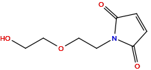
| Structure |

|
|||||
| Formula |
C8H11NO4
|
|||||
| Isosmiles |
C1=CC(=O)N(C1=O)CCOCCO
|
|||||
| PubChem CID | ||||||
| InChI |
InChI=1S/C8H11NO4/c10-4-6-13-5-3-9-7(11)1-2-8(9)12/h1-2,10H,3-6H2
|
|||||
| InChIKey |
IIYLRYHFPWHINS-UHFFFAOYSA-N
|
|||||
| IUPAC Name |
1-[2-(2-hydroxyethoxy)ethyl]pyrrole-2,5-dione
|
|||||
| Pharmaceutical Properties |
Molecule Weight
|
185.18
|
Polar area
|
66.8
|
||
|
Complexity
|
218
|
xlogp Value
|
-0.2
|
|||
|
Heavy Count
|
13
|
Rot Bonds
|
5
|
|||
|
Hbond acc
|
4
|
Hbond Donor
|
1
|
|||
Each Antibody-drug Conjugate Related to This Linker
Full Information of The Activity Data of The ADC(s) Related to This Linker
Anti-CD22- (LC:K149C)-SN36248 [Investigative]
Discovered Using Cell Line-derived Xenograft Model
| Experiment 1 Reporting the Activity Date of This ADC | [1] | ||||
| Efficacy Data | Tumor Growth Inhibition value (TGI) | ≈ 95.70% (Day 10) | Positive CD22 expression (CD22+++/++) | ||
| Method Description |
The inhibitory activity of thio-hu anti-CD22-(LC:K149C)-SN36248 against cancer cell growth was compared with a Pinatuzumab vedotin (pina) and polatuzumab vedotin (pola) against various human cancer cell lines in vitro.
|
||||
| In Vitro Model | EBV-related Burkitt lymphoma | Raji cells | CVCL_0511 | ||
| Experiment 2 Reporting the Activity Date of This ADC | [1] | ||||
| Efficacy Data | Tumor Growth Inhibition value (TGI) | ≈ 96.00% (Day 15) | Positive CD22 expression (CD22+++/++) | ||
| Method Description |
The inhibitory activity of thio-hu anti-CD22-(LC:K149C)-SN36248 against cancer cell growth was compared with a Pinatuzumab vedotin (pina) and polatuzumab vedotin (pola) against various human cancer cell lines in vitro.
|
||||
| In Vitro Model | Diffuse large B-cell lymphoma | WSU-DLCL2 cells | CVCL_1902 | ||
| Experiment 3 Reporting the Activity Date of This ADC | [1] | ||||
| Efficacy Data | Tumor Growth Inhibition value (TGI) | ≈ 96.70% (Day 14) | Positive CD22 expression (CD22+++/++) | ||
| Method Description |
The inhibitory activity of thio-hu anti-CD22-(LC:K149C)-SN36248 against cancer cell growth was compared with a Pinatuzumab vedotin (pina) and polatuzumab vedotin (pola) against various human cancer cell lines in vitro.
|
||||
| In Vitro Model | Burkitt lymphoma | BJAB.Luc-22R1.2 cells | CVCL_5711 | ||
| Experiment 4 Reporting the Activity Date of This ADC | [1] | ||||
| Efficacy Data | Tumor Growth Inhibition value (TGI) | ≈ 99.20% (Day 15) | Positive CD22 expression (CD22+++/++) | ||
| Method Description |
The inhibitory activity of thio-hu anti-CD22-(LC:K149C)-SN36248 against cancer cell growth was compared with a Pinatuzumab vedotin (pina) and polatuzumab vedotin (pola) against various human cancer cell lines in vitro.
|
||||
| In Vitro Model | Burkitt lymphoma | BJAB.Luc cells | CVCL_5711 | ||
Alpha-CD22-LC-K149C-11 [Investigative]
Revealed Based on the Cell Line Data
| Experiment 1 Reporting the Activity Date of This ADC | [2] | ||||
| Efficacy Data | Half Maximal Inhibitory Concentration (IC50) |
0.08 nM
|
Positive CD22 expression (CD22+++/++) | ||
| Method Description |
Seco-CBI-Dimer TDCs Exhibit Potent Antigen-Dependent Antiproliferation Effects in Vitro. All cell-based assay results are reported as the arithmetic mean of at least three separate runs (n = 3).
|
||||
| In Vitro Model | Diffuse large B-cell lymphoma | WSU-DLCL2 cells | CVCL_1902 | ||
| Experiment 2 Reporting the Activity Date of This ADC | [2] | ||||
| Efficacy Data | Half Maximal Inhibitory Concentration (IC50) |
0.25 nM
|
Positive CD22 expression (CD22+++/++) | ||
| Method Description |
Seco-CBI-Dimer TDCs Exhibit Potent Antigen-Dependent Antiproliferation Effects in Vitro. All cell-based assay results are reported as the arithmetic mean of at least three separate runs (n = 3).
|
||||
| In Vitro Model | Burkitt lymphoma | BJAB cells | CVCL_5711 | ||
| Experiment 3 Reporting the Activity Date of This ADC | [2] | ||||
| Efficacy Data | Half Maximal Inhibitory Concentration (IC50) |
24.00 nM
|
Positive CD22 expression (CD22+++/++) | ||
| Method Description |
Seco-CBI-Dimer TDCs Exhibit Potent Antigen-Dependent Antiproliferation Effects in Vitro. All cell-based assay results are reported as the arithmetic mean of at least three separate runs (n = 3).
|
||||
| In Vitro Model | T acute lymphoblastic leukemia | Jurkat cells | CVCL_0065 | ||
FOLR1-Mal-PEG2-Eribulin [Investigative]
Revealed Based on the Cell Line Data
| Experiment 1 Reporting the Activity Date of This ADC | [3] | ||||
| Efficacy Data | Half Maximal Inhibitory Concentration (IC50) |
0.33 nM
|
High FOLR1 expression(FOLR1+++) | ||
| Method Description |
Cells were sub-cultured and seeded at 5,000 cells/well in complete growth medium in 96-well tissue culture plates, and incubated at 37°C, 5% CO2 overnight. Test reagents were serially diluted and added to the cell plates (initial concentration of 100 nM). Plates were incubated at 37°C, 5% CO2 for an additional 3 d.
|
||||
| In Vitro Model | Ovarian endometrioid adenocarcinoma | IGROV-1 cells | CVCL_1304 | ||
| Experiment 2 Reporting the Activity Date of This ADC | [3] | ||||
| Efficacy Data | Half Maximal Inhibitory Concentration (IC50) |
38.00 nM
|
Moderate FOLR1 expression(FOLR1++) | ||
| Method Description |
Cells were sub-cultured and seeded at 5,000 cells/well in complete growth medium in 96-well tissue culture plates, and incubated at 37°C, 5% CO2 overnight. Test reagents were serially diluted and added to the cell plates (initial concentration of 100 nM). Plates were incubated at 37°C, 5% CO2 for an additional 3 d.
|
||||
| In Vitro Model | Lung non-small cell carcinoma | NCI-H2110 cells | CVCL_1530 | ||
| Experiment 3 Reporting the Activity Date of This ADC | [3] | ||||
| Efficacy Data | Half Maximal Inhibitory Concentration (IC50) | > 100 nM | Low FOLR1 expression(FOLR1+) | ||
| Method Description |
Cells were sub-cultured and seeded at 5,000 cells/well in complete growth medium in 96-well tissue culture plates, and incubated at 37°C, 5% CO2 overnight. Test reagents were serially diluted and added to the cell plates (initial concentration of 100 nM). Plates were incubated at 37°C, 5% CO2 for an additional 3 d.
|
||||
| In Vitro Model | Skin squamous cell carcinoma | A431 cells | CVCL_0037 | ||
| Experiment 4 Reporting the Activity Date of This ADC | [3] | ||||
| Efficacy Data | Half Maximal Inhibitory Concentration (IC50) | > 100 nM | Negative FOLR1 expression(FOLR1-) | ||
| Method Description |
Cells were sub-cultured and seeded at 5,000 cells/well in complete growth medium in 96-well tissue culture plates, and incubated at 37°C, 5% CO2 overnight. Test reagents were serially diluted and added to the cell plates (initial concentration of 100 nM). Plates were incubated at 37°C, 5% CO2 for an additional 3 d.
|
||||
| In Vitro Model | Osteosarcoma | SJSA-1 cells | CVCL_1697 | ||
Alpha-Ly6E-LC-K149C-11 [Investigative]
Revealed Based on the Cell Line Data
| Experiment 1 Reporting the Activity Date of This ADC | [2] | ||||
| Efficacy Data | Half Maximal Inhibitory Concentration (IC50) |
9.00 nM
|
Positive CD22 expression (CD22+++/++) | ||
| Method Description |
Seco-CBI-Dimer TDCs Exhibit Potent Antigen-Dependent Antiproliferation Effects in Vitro. All cell-based assay results are reported as the arithmetic mean of at least three separate runs (n = 3).
|
||||
| In Vitro Model | Diffuse large B-cell lymphoma | WSU-DLCL2 cells | CVCL_1902 | ||
| Experiment 2 Reporting the Activity Date of This ADC | [2] | ||||
| Efficacy Data | Half Maximal Inhibitory Concentration (IC50) |
15.00 nM
|
Positive CD22 expression (CD22+++/++) | ||
| Method Description |
Seco-CBI-Dimer TDCs Exhibit Potent Antigen-Dependent Antiproliferation Effects in Vitro. All cell-based assay results are reported as the arithmetic mean of at least three separate runs (n = 3).
|
||||
| In Vitro Model | T acute lymphoblastic leukemia | Jurkat cells | CVCL_0065 | ||
| Experiment 3 Reporting the Activity Date of This ADC | [2] | ||||
| Efficacy Data | Half Maximal Inhibitory Concentration (IC50) |
61.00 nM
|
Positive CD22 expression (CD22+++/++) | ||
| Method Description |
Seco-CBI-Dimer TDCs Exhibit Potent Antigen-Dependent Antiproliferation Effects in Vitro. All cell-based assay results are reported as the arithmetic mean of at least three separate runs (n = 3).
|
||||
| In Vitro Model | Burkitt lymphoma | BJAB cells | CVCL_5711 | ||
Alpha-NaPi2b-LC-K149C-11 [Investigative]
Revealed Based on the Cell Line Data
| Experiment 1 Reporting the Activity Date of This ADC | [2] | ||||
| Efficacy Data | Half Maximal Inhibitory Concentration (IC50) |
9.60 nM
|
Positive CD22 expression (CD22+++/++) | ||
| Method Description |
Seco-CBI-Dimer TDCs Exhibit Potent Antigen-Dependent Antiproliferation Effects in Vitro. All cell-based assay results are reported as the arithmetic mean of at least three separate runs (n = 3).
|
||||
| In Vitro Model | Diffuse large B-cell lymphoma | WSU-DLCL2 cells | CVCL_1902 | ||
| Experiment 2 Reporting the Activity Date of This ADC | [2] | ||||
| Efficacy Data | Half Maximal Inhibitory Concentration (IC50) |
17.00 nM
|
Positive CD22 expression (CD22+++/++) | ||
| Method Description |
Seco-CBI-Dimer TDCs Exhibit Potent Antigen-Dependent Antiproliferation Effects in Vitro. All cell-based assay results are reported as the arithmetic mean of at least three separate runs (n = 3).
|
||||
| In Vitro Model | T acute lymphoblastic leukemia | Jurkat cells | CVCL_0065 | ||
| Experiment 3 Reporting the Activity Date of This ADC | [2] | ||||
| Efficacy Data | Half Maximal Inhibitory Concentration (IC50) |
51.00 nM
|
Positive CD22 expression (CD22+++/++) | ||
| Method Description |
Seco-CBI-Dimer TDCs Exhibit Potent Antigen-Dependent Antiproliferation Effects in Vitro. All cell-based assay results are reported as the arithmetic mean of at least three separate runs (n = 3).
|
||||
| In Vitro Model | Burkitt lymphoma | BJAB cells | CVCL_5711 | ||
MSLN-Mal-PEG2-Eribulin [Investigative]
Revealed Based on the Cell Line Data
| Experiment 1 Reporting the Activity Date of This ADC | [3] | ||||
| Efficacy Data | Half Maximal Inhibitory Concentration (IC50) |
43.00 nM
|
Negative MSLN expression(MSLN-) | ||
| Method Description |
Cells were sub-cultured and seeded at 5,000 cells/well in complete growth medium in 96-well tissue culture plates, and incubated at 37°C, 5% CO2 overnight. Test reagents were serially diluted and added to the cell plates (initial concentration of 100 nM). Plates were incubated at 37°C, 5% CO2 for an additional 3 d.
|
||||
| In Vitro Model | Ovarian endometrioid adenocarcinoma | IGROV-1 cells | CVCL_1304 | ||
| Experiment 2 Reporting the Activity Date of This ADC | [3] | ||||
| Efficacy Data | Half Maximal Inhibitory Concentration (IC50) |
50.00 nM
|
Moderate MSLN expression(MSLN++) | ||
| Method Description |
Cells were sub-cultured and seeded at 5,000 cells/well in complete growth medium in 96-well tissue culture plates, and incubated at 37°C, 5% CO2 overnight. Test reagents were serially diluted and added to the cell plates (initial concentration of 100 nM). Plates were incubated at 37°C, 5% CO2 for an additional 3 d.
|
||||
| In Vitro Model | Lung non-small cell carcinoma | NCI-H2110 cells | CVCL_1530 | ||
| Experiment 3 Reporting the Activity Date of This ADC | [3] | ||||
| Efficacy Data | Half Maximal Inhibitory Concentration (IC50) | > 100 nM | Negative MSLN expression(MSLN-) | ||
| Method Description |
Cells were sub-cultured and seeded at 5,000 cells/well in complete growth medium in 96-well tissue culture plates, and incubated at 37°C, 5% CO2 overnight. Test reagents were serially diluted and added to the cell plates (initial concentration of 100 nM). Plates were incubated at 37°C, 5% CO2 for an additional 3 d.
|
||||
| In Vitro Model | Skin squamous cell carcinoma | A431 cells | CVCL_0037 | ||
| Experiment 4 Reporting the Activity Date of This ADC | [3] | ||||
| Efficacy Data | Half Maximal Inhibitory Concentration (IC50) | > 100 nM | Negative MSLN expression(MSLN-) | ||
| Method Description |
Cells were sub-cultured and seeded at 5,000 cells/well in complete growth medium in 96-well tissue culture plates, and incubated at 37°C, 5% CO2 overnight. Test reagents were serially diluted and added to the cell plates (initial concentration of 100 nM). Plates were incubated at 37°C, 5% CO2 for an additional 3 d.
|
||||
| In Vitro Model | Osteosarcoma | SJSA-1 cells | CVCL_1697 | ||
References
