
Linker Information
General Information of This Linker (ID: LIN0JQWNO)
| Name |
MC-K (Ac)VC
|
|||||
|---|---|---|---|---|---|---|
| Linker Type |
Cathepsin-cleavable linker
|
|||||
| Antibody-Linker Relation |
Cleavable
|
|||||
| Structure |
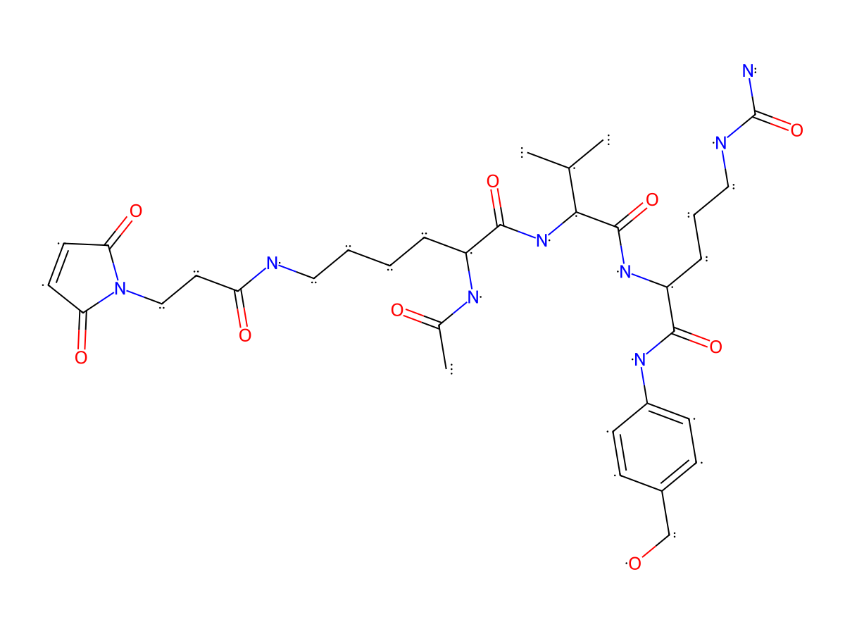
|
|||||
| Formula |
C33H48N8O9
|
|||||
| Isosmiles |
O=C(NCCCC[C@H](NC(C)=O)C(N[C@H](C(N[C@@H](CCCNC(N)=O)C(NC1=CC=C(CO)C=C1)=O)=O)C(C)C)=O)CCN2C(C=CC2=O)=O
|
|||||
| InChI |
InChI=1S/C33H48N8O9/c1-20(2)29(32(49)39-25(8-6-17-36-33(34)50)30(47)38-23-11-9-22(19-42)10-12-23)40-31(48)24(37-21(3)43)7-4-5-16-35-26(44)15-18-41-27(45)13-14-28(41)46/h9-14,20,24-25,29,42H,4-8,15-19H2,1-3H3,(H,35,44)(H,37,43)(H,38,47)(H,39,49)(H,40,48)(H3,34,36,50)/t24-,25-,29-/m0/s1
|
|||||
| InChIKey |
YALRGPACELLQOY-QEMZJVQQSA-N
|
|||||
| Pharmaceutical Properties |
Molecule Weight
|
700.794
|
Polar area
|
258.23
|
||
|
Complexity
|
1332.864752
|
xlogp Value
|
-0.7022
|
|||
|
Heavy Count
|
50
|
Rot Bonds
|
21
|
|||
|
Hbond acc
|
9
|
Hbond Donor
|
8
|
|||
Each Antibody-drug Conjugate Related to This Linker
Full Information of The Activity Data of The ADC(s) Related to This Linker
aCEA5-K (Ac)-exatecan [Investigative]
Revealed Based on the Cell Line Data
| Experiment 1 Reporting the Activity Date of This ADC | [1] | ||||
| Efficacy Data | Half Maximal inhibitory Concentration (lC50) |
0.2۪.04 nM
|
High CEACAM5 expression (CEACAM5 +++) | ||
| Method Description |
Human cancer cell lines were obtained from the American Type Culture Collection (CEA5-positive: SK-CO-1, MKN-45, LS174T; CEA5-negative MDA-MB-231) and maintained according to standard culture conditions (SK-CO-1, MKN-45: 37 °C, 5% CO2, 95% humidity; LS174T, MDA-MB-231: 37 °C, 10% CO2, 95% humidity). SK-CO-1 cells were cultured in Minimum Essential Medium Eagle (Sigma-Aldrich, FG0325) including l-alanylglutamine supplemented with 10% fetal bovine serum (FBS) and 1 mM sodium pyruvate. LS174T cells were cultured in Dulbecco's modified Eagle's medium (DMEM) (Sigma-Aldrich, FG0435) including l-alanyl-glutamine supplemented with 10% FBS, 1 mM sodium pyruvate, and 1% (v/v) nonessential amino acid 100× mix (Sigma-Aldrich, M7145). MDA-MBA-231 cells were cultured in DMEM including l-alanylglutamine supplemented with 10% FBS and 1 mM sodium pyruvate. MKN-45 cells were cultured in Roswell Park Memorial Institute (RPMI) 1640 (Sigma-Aldrich, FG1215) medium including l-alanylglutamine supplemented with 10% FBS and 1 mM sodium pyruvate. For subculturing, adherent cells were detached by the addition of TrypLE Express (1×) (Gibco, 12604-013), diluted with fresh medium, and transferred into a new culturing flask. For the evaluation of exatecan and exatecan conjugates, 90 uL of viable cell (vc) suspension was seeded into 96-well plates (SK-CO-1 2,500 vc/well, MKN-45 500 vc/well, LS174T 2,500 vc/well, MDA-MB-231 1,500 vc/well) followed by incubation (37 °C, 5 or 10% CO2) in a humid chamber overnight. Test compounds were formulated at a 10-fold starting concentration, prepared in serial dilution (1:4), and 10 uL was added to cells for each dilution in triplicates. The cell viability was determined after 6 days using the CellTiter-Glo reagent (Promega, G7573) according to the manufacturer's instructions. Luminescence values were measured by using a Varioskan plate reader (Thermo Fisher) and normalized to the luminescence of nontreated cells. Dose-response curves were fitted using the log (inhibitor) vs response variable slope 4-parameter fitting function of GraphPad Prism (GraphPad Software, Inc.). Data are shown as % relative cell viability versus molar compound concentration. Error bars indicate the standard deviation (SD) of triplicates. Geometric mean values of IC50's derived from multiple experiments were calculated.
Click to Show/Hide
|
||||
| In Vitro Model | Colon adenocarcinoma | SK-CO-1 cells | CVCL_0626 | ||
| Experiment 2 Reporting the Activity Date of This ADC | [1] | ||||
| Efficacy Data | Half Maximal inhibitory Concentration (lC50) |
0.28۪.05 nM
|
High CEACAM5 expression (CEACAM5 +++) | ||
| Method Description |
Human cancer cell lines were obtained from the American Type Culture Collection (CEA5-positive: SK-CO-1, MKN-45, LS174T; CEA5-negative MDA-MB-231) and maintained according to standard culture conditions (SK-CO-1, MKN-45: 37 °C, 5% CO2, 95% humidity; LS174T, MDA-MB-231: 37 °C, 10% CO2, 95% humidity). SK-CO-1 cells were cultured in Minimum Essential Medium Eagle (Sigma-Aldrich, FG0325) including l-alanylglutamine supplemented with 10% fetal bovine serum (FBS) and 1 mM sodium pyruvate. LS174T cells were cultured in Dulbecco's modified Eagle's medium (DMEM) (Sigma-Aldrich, FG0435) including l-alanyl-glutamine supplemented with 10% FBS, 1 mM sodium pyruvate, and 1% (v/v) nonessential amino acid 100× mix (Sigma-Aldrich, M7145). MDA-MBA-231 cells were cultured in DMEM including l-alanylglutamine supplemented with 10% FBS and 1 mM sodium pyruvate. MKN-45 cells were cultured in Roswell Park Memorial Institute (RPMI) 1640 (Sigma-Aldrich, FG1215) medium including l-alanylglutamine supplemented with 10% FBS and 1 mM sodium pyruvate. For subculturing, adherent cells were detached by the addition of TrypLE Express (1×) (Gibco, 12604-013), diluted with fresh medium, and transferred into a new culturing flask. For the evaluation of exatecan and exatecan conjugates, 90 uL of viable cell (vc) suspension was seeded into 96-well plates (SK-CO-1 2,500 vc/well, MKN-45 500 vc/well, LS174T 2,500 vc/well, MDA-MB-231 1,500 vc/well) followed by incubation (37 °C, 5 or 10% CO2) in a humid chamber overnight. Test compounds were formulated at a 10-fold starting concentration, prepared in serial dilution (1:4), and 10 uL was added to cells for each dilution in triplicates. The cell viability was determined after 6 days using the CellTiter-Glo reagent (Promega, G7573) according to the manufacturer's instructions. Luminescence values were measured by using a Varioskan plate reader (Thermo Fisher) and normalized to the luminescence of nontreated cells. Dose-response curves were fitted using the log (inhibitor) vs response variable slope 4-parameter fitting function of GraphPad Prism (GraphPad Software, Inc.). Data are shown as % relative cell viability versus molar compound concentration. Error bars indicate the standard deviation (SD) of triplicates. Geometric mean values of IC50's derived from multiple experiments were calculated.
Click to Show/Hide
|
||||
| In Vitro Model | Colon adenocarcinoma | LS174T cells | CVCL_1384 | ||
| Experiment 3 Reporting the Activity Date of This ADC | [1] | ||||
| Efficacy Data | Half Maximal inhibitory Concentration (lC50) |
0.53۪.09 nM
|
Negative CEACAM5 expression (CEACAM5-) | ||
| Method Description |
Human cancer cell lines were obtained from the American Type Culture Collection (CEA5-positive: SK-CO-1, MKN-45, LS174T; CEA5-negative MDA-MB-231) and maintained according to standard culture conditions (SK-CO-1, MKN-45: 37 °C, 5% CO2, 95% humidity; LS174T, MDA-MB-231: 37 °C, 10% CO2, 95% humidity). SK-CO-1 cells were cultured in Minimum Essential Medium Eagle (Sigma-Aldrich, FG0325) including l-alanylglutamine supplemented with 10% fetal bovine serum (FBS) and 1 mM sodium pyruvate. LS174T cells were cultured in Dulbecco's modified Eagle's medium (DMEM) (Sigma-Aldrich, FG0435) including l-alanyl-glutamine supplemented with 10% FBS, 1 mM sodium pyruvate, and 1% (v/v) nonessential amino acid 100× mix (Sigma-Aldrich, M7145). MDA-MBA-231 cells were cultured in DMEM including l-alanylglutamine supplemented with 10% FBS and 1 mM sodium pyruvate. MKN-45 cells were cultured in Roswell Park Memorial Institute (RPMI) 1640 (Sigma-Aldrich, FG1215) medium including l-alanylglutamine supplemented with 10% FBS and 1 mM sodium pyruvate. For subculturing, adherent cells were detached by the addition of TrypLE Express (1×) (Gibco, 12604-013), diluted with fresh medium, and transferred into a new culturing flask. For the evaluation of exatecan and exatecan conjugates, 90 uL of viable cell (vc) suspension was seeded into 96-well plates (SK-CO-1 2,500 vc/well, MKN-45 500 vc/well, LS174T 2,500 vc/well, MDA-MB-231 1,500 vc/well) followed by incubation (37 °C, 5 or 10% CO2) in a humid chamber overnight. Test compounds were formulated at a 10-fold starting concentration, prepared in serial dilution (1:4), and 10 uL was added to cells for each dilution in triplicates. The cell viability was determined after 6 days using the CellTiter-Glo reagent (Promega, G7573) according to the manufacturer's instructions. Luminescence values were measured by using a Varioskan plate reader (Thermo Fisher) and normalized to the luminescence of nontreated cells. Dose-response curves were fitted using the log (inhibitor) vs response variable slope 4-parameter fitting function of GraphPad Prism (GraphPad Software, Inc.). Data are shown as % relative cell viability versus molar compound concentration. Error bars indicate the standard deviation (SD) of triplicates. Geometric mean values of IC50's derived from multiple experiments were calculated.
Click to Show/Hide
|
||||
| In Vitro Model | Breast adenocarcinoma | MDA-MB-231 cells | CVCL_0062 | ||
| Experiment 4 Reporting the Activity Date of This ADC | [1] | ||||
| Efficacy Data | Half Maximal inhibitory Concentration (lC50) |
0.7۪.24 nM
|
High CEACAM5 expression (CEACAM5 +++) | ||
| Method Description |
Human cancer cell lines were obtained from the American Type Culture Collection (CEA5-positive: SK-CO-1, MKN-45, LS174T; CEA5-negative MDA-MB-231) and maintained according to standard culture conditions (SK-CO-1, MKN-45: 37 °C, 5% CO2, 95% humidity; LS174T, MDA-MB-231: 37 °C, 10% CO2, 95% humidity). SK-CO-1 cells were cultured in Minimum Essential Medium Eagle (Sigma-Aldrich, FG0325) including l-alanylglutamine supplemented with 10% fetal bovine serum (FBS) and 1 mM sodium pyruvate. LS174T cells were cultured in Dulbecco's modified Eagle's medium (DMEM) (Sigma-Aldrich, FG0435) including l-alanyl-glutamine supplemented with 10% FBS, 1 mM sodium pyruvate, and 1% (v/v) nonessential amino acid 100× mix (Sigma-Aldrich, M7145). MDA-MBA-231 cells were cultured in DMEM including l-alanylglutamine supplemented with 10% FBS and 1 mM sodium pyruvate. MKN-45 cells were cultured in Roswell Park Memorial Institute (RPMI) 1640 (Sigma-Aldrich, FG1215) medium including l-alanylglutamine supplemented with 10% FBS and 1 mM sodium pyruvate. For subculturing, adherent cells were detached by the addition of TrypLE Express (1×) (Gibco, 12604-013), diluted with fresh medium, and transferred into a new culturing flask. For the evaluation of exatecan and exatecan conjugates, 90 uL of viable cell (vc) suspension was seeded into 96-well plates (SK-CO-1 2,500 vc/well, MKN-45 500 vc/well, LS174T 2,500 vc/well, MDA-MB-231 1,500 vc/well) followed by incubation (37 °C, 5 or 10% CO2) in a humid chamber overnight. Test compounds were formulated at a 10-fold starting concentration, prepared in serial dilution (1:4), and 10 uL was added to cells for each dilution in triplicates. The cell viability was determined after 6 days using the CellTiter-Glo reagent (Promega, G7573) according to the manufacturer's instructions. Luminescence values were measured by using a Varioskan plate reader (Thermo Fisher) and normalized to the luminescence of nontreated cells. Dose-response curves were fitted using the log (inhibitor) vs response variable slope 4-parameter fitting function of GraphPad Prism (GraphPad Software, Inc.). Data are shown as % relative cell viability versus molar compound concentration. Error bars indicate the standard deviation (SD) of triplicates. Geometric mean values of IC50's derived from multiple experiments were calculated.
Click to Show/Hide
|
||||
| In Vitro Model | Gastric adenocarcinoma | MKN-45 cells | CVCL_0434 | ||