
Linker Information
General Information of This Linker (ID: LIN0JFJEO)
| Name |
FDA026-LE17 linker
|
|||||
|---|---|---|---|---|---|---|
| Linker Type |
Cathepsin-cleavable linker
|
|||||
| Antibody-Linker Relation |
Cleavable
|
|||||
| Structure |
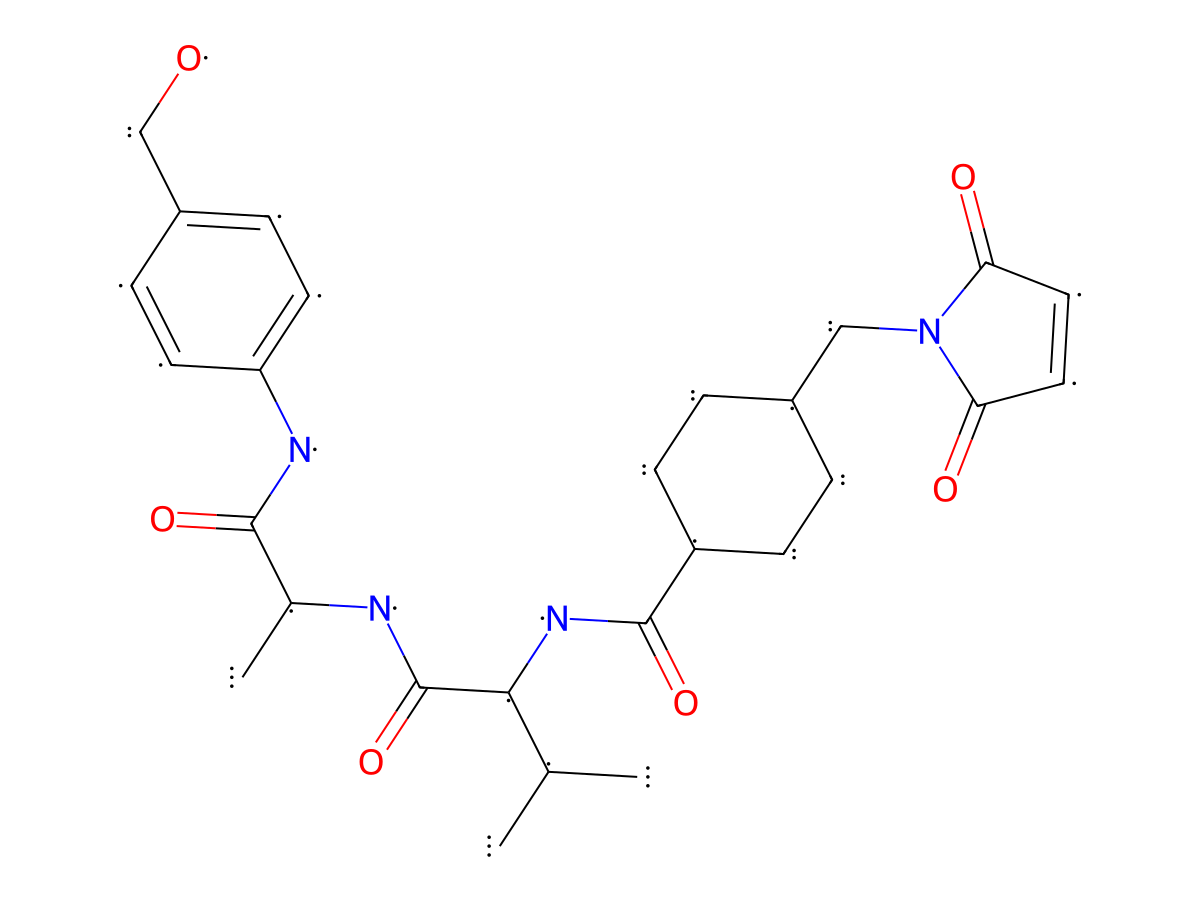
|
|||||
| Formula |
C27H36N4O6
|
|||||
| Isosmiles |
C[C@H](NC([C@@H](NC(C(CC1)CCC1CN2C(C=CC2=O)=O)=O)C(C)C)=O)C(NC3=CC=C(CO)C=C3)=O
|
|||||
| PubChem CID | ||||||
| InChI |
InChI=1S/C27H36N4O6/c1-16(2)24(27(37)28-17(3)25(35)29-21-10-6-19(15-32)7-11-21)30-26(36)20-8-4-18(5-9-20)14-31-22(33)12-13-23(31)34/h6-7,10-13,16-18,20,24,32H,4-5,8-9,14-15H2,1-3H3,(H,28,37)(H,29,35)(H,30,36)/t17-,18?,20?,24-/m0/s1
|
|||||
| InChIKey |
WGEPEKUWUWACJU-NZKRWOFHSA-N
|
|||||
| Pharmaceutical Properties |
Molecule Weight
|
512.607
|
Polar area
|
144.91
|
||
|
Complexity
|
984.6229671
|
xlogp Value
|
1.4943
|
|||
|
Heavy Count
|
37
|
Rot Bonds
|
10
|
|||
|
Hbond acc
|
6
|
Hbond Donor
|
4
|
|||
Each Antibody-drug Conjugate Related to This Linker
Full Information of The Activity Data of The ADC(s) Related to This Linker
FDA026-LE17 [Investigative]
Revealed Based on the Cell Line Data
| Experiment 1 Reporting the Activity Date of This ADC | [1] | ||||
| Efficacy Data | Half Maximal inhibitory Concentration (lC50) |
40.36 nM
|
|||
| Method Description |
SK-BR-3 (ATCC) cells were selected as the cell line for in vitro activity detection experiments. 2000 cells were inoculated into each well of a 96-well cell culture plate and cultured for 20-24 hours. The antibody-drug conjugate prepared according to the method in Example 3 was formulated with L15 cell culture medium containing 10% FBS into 11 concentration gradients of test solution: 1000, 166.7, 55.6, 18.6, 6.17, 2.06, 0.69, 0.23, 0.08, 0.008 and OnM. 100 ul of the diluted test solution per well was added to the culture plate containing the inoculated cells and incubated in a 37°C, 5% CO, incubator for 144 hours. Then, CellTiter-Glo® Luminescent Cell Viability Assay Reagent (50 ul/well) was added, and the mixture was shaken at 500 rpm at room temperature for 10 minutes. The data were read using a SpectraMaxL microplate reader (OD570nm, with 2-second interval readings), and the IC50 results were calculated.
Click to Show/Hide
|
||||
| In Vitro Model | Breast adenocarcinoma | SK-BR-3 cells | CVCL_0033 | ||
| Experiment 2 Reporting the Activity Date of This ADC | [1] | ||||
| Efficacy Data | Half Maximal inhibitory Concentration (lC50) |
42.38 nM
|
|||
| Method Description |
SW620 (ATCC) cells were selected as the cell line for in vitro activity detection experiments. 2000 cells were inoculated into each well of a 96-well cell culture plate and cultured for 20-24 hours. The antibody-drug conjugate prepared according to the method in Example 3 was formulated with L15 cell culture medium containing 10% FBS into 11 concentration gradients of test solution: 1000, 166.7, 55.6, 18.6, 6.17, 2.06, 0.69, 0.23, 0.08, 0.008 and OnM. 100 ul of the diluted test solution per well was added to the culture plate containing the inoculated cells and incubated in a 37°C, 5% CO, incubator for 144 hours. Then, CellTiter-Glo® Luminescent Cell Viability Assay Reagent (50 ul/well) was added, and the mixture was shaken at 500 rpm at room temperature for 10 minutes. The data were read using a SpectraMaxL microplate reader (OD570nm, with 2-second interval readings), and the IC55 results were calculated.
Click to Show/Hide
|
||||
| In Vitro Model | Colon adenocarcinoma | SW620 cells | CVCL_0547 | ||
FDA026-LE16 [Investigative]
Revealed Based on the Cell Line Data
| Experiment 1 Reporting the Activity Date of This ADC | [1] | ||||
| Efficacy Data | Half Maximal inhibitory Concentration (lC50) |
46.18 nM
|
|||
| Method Description |
SK-BR-3 (ATCC) cells were selected as the cell line for in vitro activity detection experiments. 2000 cells were inoculated into each well of a 96-well cell culture plate and cultured for 20-24 hours. The antibody-drug conjugate prepared according to the method in Example 3 was formulated with L15 cell culture medium containing 10% FBS into 11 concentration gradients of test solution: 1000, 166.7, 55.6, 18.6, 6.17, 2.06, 0.69, 0.23, 0.08, 0.008 and OnM. 100 ul of the diluted test solution per well was added to the culture plate containing the inoculated cells and incubated in a 37°C, 5% CO, incubator for 144 hours. Then, CellTiter-Glo® Luminescent Cell Viability Assay Reagent (50 ul/well) was added, and the mixture was shaken at 500 rpm at room temperature for 10 minutes. The data were read using a SpectraMaxL microplate reader (OD570nm, with 2-second interval readings), and the IC50 results were calculated.
Click to Show/Hide
|
||||
| In Vitro Model | Breast adenocarcinoma | SK-BR-3 cells | CVCL_0033 | ||
| Experiment 2 Reporting the Activity Date of This ADC | [1] | ||||
| Efficacy Data | Half Maximal inhibitory Concentration (lC50) |
53.12 nM
|
|||
| Method Description |
SW620 (ATCC) cells were selected as the cell line for in vitro activity detection experiments. 2000 cells were inoculated into each well of a 96-well cell culture plate and cultured for 20-24 hours. The antibody-drug conjugate prepared according to the method in Example 3 was formulated with L15 cell culture medium containing 10% FBS into 11 concentration gradients of test solution: 1000, 166.7, 55.6, 18.6, 6.17, 2.06, 0.69, 0.23, 0.08, 0.008 and OnM. 100 ul of the diluted test solution per well was added to the culture plate containing the inoculated cells and incubated in a 37°C, 5% CO, incubator for 144 hours. Then, CellTiter-Glo® Luminescent Cell Viability Assay Reagent (50 ul/well) was added, and the mixture was shaken at 500 rpm at room temperature for 10 minutes. The data were read using a SpectraMaxL microplate reader (OD570nm, with 2-second interval readings), and the IC54 results were calculated.
Click to Show/Hide
|
||||
| In Vitro Model | Colon adenocarcinoma | SW620 cells | CVCL_0547 | ||